Dr BSA Medical College Harassment Case: Health Dept asked to take disciplinary action against AP Pharmacology

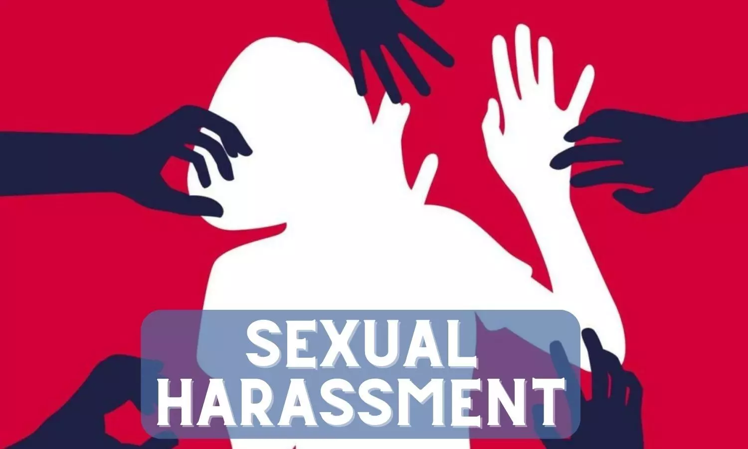
New Delhi: Days after 13 female MBBS students of Dr Baba Saheb Ambedkar Medical College and Hospital, Delhi have accused an Assistant Professor of Pharmacology of sexual harassment, the Vigilance department has asked the Health department to send a proposal for initiating disciplinary action against the accused.

According to PTI, the 22-year-old medical student alleged that her professor “sexually harassed” her during a viva examination, police said on Monday.

A case has been registered under sections 354A (assault or criminal force to woman with intent to outrage her modesty) and 509 (word, gesture or act intended to insult the modesty of a woman) of the Indian Penal Code (IPC) against the accused, police said.

Medical Dialogues team recently reported that based on the complaint by the students, a case has been registered by the police. Meanwhile, soon after the matter came to light, a demonstration was organized by a women’s group and the protest soon manifested as a massive Public Outrage outside the college premises. Protesters were seen holding placards denouncing the accused professor and demanding justice.

Writing to the Principal of the medical college, a student belonging to the 2021 MBBS batch stated that the alleged incident took place during a practical exam in the pharmacology department.

In a formal letter addressed to the Secretary of the Health and Family Welfare Department, the Vigilance Directorate highlighted the significance of media reports regarding the incident.

“From the media, it is also observed that at least two students have recorded their statement under Section 164 of Cr. P.C. It is also reported in the media that the accused was arrested and was booked under sections 354(A) and 509 of the IPC,” said the directorate letter, news agency PTI reported.

“It is requested to send the proposal for initiating necessary disciplinary action against the accused professor to the Directorate of Vigilance, Delhi government, for placing before the Competent Authority as per extant rules,” it said.

The Health Department has so far not sent any file for taking disciplinary action against the accused, it added.

Powered by WPeMatico

AIIMS Raipur inks MoU with Defence Ministry for health services to ex-servicemen

New Delhi: In a significant development aimed at enhancing healthcare services for ex-servicemen, a Memorandum of Understanding (MoU) was signed between the All India Institute of Medical Sciences (AIIMS) Raipur and the Ex-servicemen Contributory Health Scheme (ECHS) wing of the Ministry of Defence. 

The agreement, inked during a ceremony at AIIMS Raipur, aims to facilitate ex-servicemen in availing health benefits at the facility in a “capless and cashless manner”.

According to PTI, It was signed between the AIIMS-Raipur and the ECHS wing of Ministry of Defence, the senior official said. 

Also Read:Lt Gen Ashok Kumar Jindal appointed as new Executive Director and CEO of AIIMS Raipur

AIIMS Raipur becomes only the second AIIMS hospital to be empanelled with ECHS, following AIIMS Bhopal, underscoring the growing collaboration between premier medical institutions and the armed forces to cater to the healthcare needs of veterans.

This MoU will allow ex-servicemen to avail health benefits at the facility in a “capless and cashless manner”, a senior official said.

Lt Gen. Padam Singh Shekhawat, General Officer Commanding, Madhya Pradesh Area, expressed his happiness at empathising with AIIMS-R for ECHS. ”There are more than 9000 servicemen in Chhattisgarh and more than 21,000 dependents. With the empanelment of AIIMS-R, the ECHS has extended its services for all 30,000 beneficiaries in Chhattisgarh. AIIMS is an institute of national importance equipped with state-of-the-art medical facilities. Ex-servicemen will certainly get treatment with empathy in AIIMS from a dedicated medical fraternity, Now, there are 14 empaneled hospitals in Chhattisgarh, he said, news agency ANI reported.

The signing ceremony witnessed the presence of esteemed dignitaries including Lt Gen Padam Singh Shekhawat, General Officer Commanding, Madhya Bharat Area; Lt Gen (retd) Ashok Kumar Jindal, Executive Director AIIMS; Brigadier Jitendra Singh, Deputy Managing Director, ECHS; and Brig Aman Anand, Commander, Chhattisgarh and Odisha Sub Area, reaffirming the collective commitment to serving the healthcare needs of ex-servicemen.

Also Read:Union Minister Mandaviya lays foundation stone of Rs 100 crore critical care health block at AIIMS Raipur

Powered by WPeMatico

Rheumatic diseases linked to childbearing problems, finds study

A new study published in the journal Rheumatology, published by Oxford University Press, found that rheumatic diseases can lead to reproductive problems, though some conditions have more detrimental effects than others.

Immune-mediated diseases encompass a diverse range of conditions, all characterized by abnormal immune system activity. While some, like type 1 diabetes, typically manifest before reproductive age, others emerge later in life. Scientists have investigated systemic lupus erythematosus for its effects on reproductive health as the condition increases the risk for some adverse pregnancy outcomes, including pre-eclampsia, preterm delivery, C-sections, and low birth weight. However, the impact of other autoimmune diseases—such as psoriasis—on fertility and pregnancy is unclear.

In the study, researchers used data from Finnish nationwide health registers to study the impact of immune-mediated diseases on reproductive health measures such as reproductive success, and for women, ever having experienced adverse maternal and perinatal outcomes. Out of all people born in Finland between 1964 and 1984, 7.9% of the women and 7.8% of the men had an autoimmune disease diagnosed before or during reproductive years.

The findings highlighted that many immune-mediated diseases had little impact on the number of children. However, women with selected immune-mediated diseases experience a higher prevalence of childlessness, with the top three diseases being Addison’s disease (23.9% more childlessness), juvenile idiopathic arthritis (9.3%), and vitamin B12 deficiency anaemia (8.6%). Several of the rheumatic diseases—particularly systemic lupus erythematosus and juvenile idiopathic arthritis—lead to higher rates of childlessness and fewer children.

The risks for pre-eclampsia, low birth weight, preterm delivery, non-elective C-sections and need of neonatal intensive care were increased for many conditions. Systemic lupus erythematosus and type 1 diabetes showed over two-fold risks for some of these outcomes. However, the risk of gestational diabetes was not higher for patients with any of the rheumatic diseases compared to the population.

“Despite seeing an elevated risk for diverse childbearing problems in rheumatic and other immune-mediated diseases, many of the complications are still fairly rare,” said Anne Kerola, the lead author of the study. “Family planning should actively be discussed between patients, both men and women, with rheumatic diseases and their healthcare providers. Pregnancies in women with rheumatic diseases are carefully followed up to tailor medications appropriately, which helps reduce risks.”

Reference: Anne M Kerola, Antti Palomäki, Hannele Laivuori, Tarja Laitinen, Martti Färkkilä, Kari K Eklund, Samuli Ripatti, Markus Perola, Andrea Ganna, Joni V Lindbohm, Nina Mars; Patterns of reproductive health in inflammatory rheumatic diseases and other immune-mediated diseases: a nationwide registry study; Journal: Rheumatology; DOI: 10.1093/rheumatology/keae122

Powered by WPeMatico

Do glucose levels affect cognitive performance in people with type 1 diabetes? Study finds out

A new study led by researchers at McLean Hospital (a member of Mass General Brigham) and Washington State University have demonstrated that naturally occurring glucose fluctuations impact cognitive function in people with Type 1 Diabetes (T1D).

The study published in the journal npj Digital Medicine revealed that cognition was slower in moments when glucose was atypical – that is, considerably higher or lower than someone’s usual glucose level. However, some people were more susceptible to the cognitive effects of large glucose fluctuations than others.

T1D is an autoimmune disease characterized by glucose variability. Previous laboratory studies have shown that very low and very high glucose levels impair cognitive function. However, technological limitations made it difficult to study the impact of naturally occurring glucose fluctuations on cognition outside of the laboratory, preventing researchers from obtaining repeated, high-frequency measurements within the same individuals over time. High-frequency measurements are necessary to understand whether glucose fluctuations impact cognition similarly for everyone.

In the study, researchers collected high-frequency glucose and cognitive data from 200 individuals with T1D using digital glucose sensors and smartphone-based tests. Glucose levels were measured every five minutes, and cognitive assessments were conducted three times daily for fifteen days. This data collection allowed researchers to analyze the cognitive effects of natural glucose fluctuations. Utilizing machine learning, they investigated individual differences in the impact of glucose on cognition.

The results showed that cognitive function was impaired when glucose was considerably higher or lower than usual. The researchers also found that people differed from each other in terms of how much glucose fluctuations impacted their cognitive speed, and some people – including older adults and adults with certain health conditions – were much more impacted by glucose fluctuations than others.

“Our results demonstrate that people can differ a lot from one another in how their brains are impacted by glucose. We found that minimizing glucose fluctuations in daily life is important for optimizing processing speed, and this is especially true for people who are older or have other diabetes-related health conditions,” said Laura Germine, PhD, co-senior author of the paper and director of McLean’s Laboratory for Brain and Cognitive Health Technology.

Reference: Z. W. Hawks, E. D. Beck, L. Jung, L. M. Fonseca, M. J. Sliwinski, R. S. Weinstock, E. Grinspoon, I. Xu, R. W. Strong, S. Singh, H. P. A. Van Dongen, M. R. Frumkin, J. Bulger, M. J. Cleveland, K. Janess, Y. C. Kudva, R. Pratley, M. R. Rickels, S. R. Rizvi, N. S. Chaytor & L. T. Germine; Dynamic associations between glucose and ecological momentary cognition in Type 1 Diabetes; Journal: npj Digital Medicine; DOI: 10.1038/s41746-024-01036-5

Powered by WPeMatico

Primary dengue infections can also lead to severe disease among kids, study questions common belief

India: A collaborative study in India has questioned the currently widely held belief that severe dengue is associated predominantly with secondary infections and stresses the importance of developing treatments or vaccines to protect dengue-naive populations.

According to the study, primary infections can also lead to severe disease, including dengue shock syndrome or dengue hemorrhagic fever challenging the belief that only secondary dengue infections are severe. The study findings, published in Nature Medicine, call for a reevaluation of dengue understanding and strategies in vaccine intervention research.

It is a common belief that dengue gets severe when one gets affected for the second time. The new study from the International Centre for Genetic Engineering and Biotechnology (ICGEB, New Delhi) showed that a substantial number of primary dengue infections also carry severe disease conditions.

Dengue is a global epidemic that has led to over 100 million cases annually. The clinical symptoms vary from mild fever to severe haemorrhage and shock, including some fatalities. The current paradigm is that these severe dengue cases mostly occur during secondary infections due to antibody-dependent enhancement after infection with a different dengue virus serotype.

India has the highest dengue burden worldwide, but not much is known about the disease severity and its link with primary and secondary dengue infections. To address this issue, Charu Aggarwal, International Centre for Genetic Engineering and Biotechnology, New Delhi, India, and her colleagues examined 619 children with febrile dengue-confirmed infection from three hospitals in different regions of India.

They classified primary and secondary infections based on IgM: IgG ratios using a dengue-specific enzyme-linked immunosorbent assay according to the World Health Organization (WHO) guidelines.

The main findings of the study were as follows:

  • Primary dengue infections accounted for more than half of total clinical cases (344 of 619), severe dengue cases (112 of 202) and fatalities (5 of 7).
  • Consistent with the classification based on binding antibody data, dengue-neutralizing antibody titers were also significantly lower in primary infections compared to secondary infections.

To conclude, dengue virus infection is a huge public health problem in India. Many patients develop severe diseases that can also be sometimes fatal. However, much of the ongoing vaccine intervention research is focused on the currently widely held global belief that primary dengue infections are not usually dangerous and that severe dengue diseases are mainly due to secondary dengue infections.

The study questions this widely held belief and shows that primary infections constitute a substantial fraction of severe disease cases and fatalities.

Reference:

Aggarwal, C., Ahmed, H., Sharma, P., Reddy, E. S., Nayak, K., Singla, M., Maheshwari, D., Chawla, Y. M., Panda, H., Rai, R. C., Gunisetty, S., Priyamvada, L., Bhaumik, S. K., Ahamed, S. F., Vivek, R., Bhatnagar, P., Singh, P., Kaur, M., Dixit, K., . . . Chandele, A. (2024). Severe disease during both primary and secondary dengue virus infections in pediatric populations. Nature Medicine, 1-5. https://doi.org/10.1038/s41591-024-02798-x

Powered by WPeMatico

Breast arterial calcification on mammography could identify women at high risk of CVD in future: Study

USA: Breast arterial calcifications (BAC) on a screening mammogram may identify women at potentially increased risk of future cardiovascular disease (CVD) without radiation exposure and additional cost, a recent study published in Clinical Imaging has found.

In the cross-sectional study of women who underwent both screening mammography and cardiac computed tomographic angiography, BAC was found in 18% of women.

Researchers found links between such calcifications and older age, younger age at first birth, diabetes, parity, and hypertension in women who underwent both screening mammography and cardiac CT angiography (CCTA).

Additionally, they found that when neither BAC nor coronary arterial calcifications were present, the estimated 10-year risk of developing atherosclerotic CVD was lowest, whereas, the highest risk was observed when both calcifications were present.

Breast arterial calcifications are incidentally observed on mammograms, yet there is no clarity on their implications. Shadi Azam, Department of Population Health Sciences, Weill Cornell Medicine, New York, USA, and colleagues investigated reproductive, lifestyle, and cardiovascular determinants of BAC in women undergoing mammography screening. Further, they investigated the relationship between BAC, coronary arterial calcifications (CAC), and estimated 10-year atherosclerotic CVD (ASCVD) risk.

In the cross-sectional study, the researchers obtained reproductive history and CVD risk factors from 215 women aged 18 or above who underwent mammography and CCTA within 2 years between 2007 and 2017 at a hospital.

BAC was divided as binary (present/absent) and semi-quantitatively (mild, moderate, severe). CAC was investigated using the Agatston method and recorded as binary (present/absent).

Adjusted odds ratios (ORs) were calculated, accounting for age as a confounding factor. ASCVD risk over ten years was calculated using the Pooled Cohort Risk Equations.

The researchers reported the following findings:

  • Older age, systolic and diastolic blood pressures, higher parity, and younger age at the first birth (≤28 years) were significantly associated with greater odds of BAC.
  • Women with both BAC and CAC had the highest estimated 10-year risk of ASCVD (13.30 %).
  • Those with only BAC (8.80 %), only CAC (5.80 %), and no BAC or CAC (4.40 %) had lower estimated 10-year risks of ASCVD.
  • The researchers detected no association between the presence of BAC and CAC.

The researchers highlighted that finding links between the mammographic presence of BAC and cardiovascular disease risk could help better manage patients without needing additional exposure to radiation and could help save on costs.

“There is a need for further research in large cohorts with longer follow-up periods to delineate better the association between [calcification] burden and CVD outcomes and to establish the clinical utility of reporting breast arterial calcifications detected through mammography screening,” the researchers wrote.

Reference:

Azam, S., Tamimi, R. M., Drotman, M. B., Babagbemi, K., Levy, A. D., & Peña, J. M. (2024). Assessing breast arterial calcification in mammograms and its implications for atherosclerotic cardiovascular disease risk. Clinical Imaging, 110129. https://doi.org/10.1016/j.clinimag.2024.110129

Powered by WPeMatico

Smoking, number of Implants Placed and implant location linked to early implant failure in new study

Smoking, number of Implants Placed and implant location Linked to Early implant failure in a new study published in The International Journal of Oral & Maxillofacial Implants.

A study was done to examine whether patient-level disease diagnoses, drug treatments, and dental clinical factors are correlates of early implant failure. This historical, nested case-control study used electronic medical records to retrieve patient-level data on individuals with implant failure occurring up to 6 months after implantation, including demographics, clinical information (number, location, and complicated status of implants), underlying diseases (osteoporosis, diabetes, hypertension, inflammatory bowel disease [IBD], myocardial infarction [MI]), and drug purchases (chemotherapy, bisphosphonates, selective serotonin inhibitors, antihypertensive medications, proton pump inhibitors [PPIs], disease-modifying antirheumatic drugs, corticosteroids, and nonsteroidal anti-inflammatory drugs) among adult members of a large, state-mandated health provider in Israel between 2015 and 2020. Individuals with implant extraction occurring up to 6 months after implantation were matched 1:1 to controls. Univariate and adjusted multivariate conditional logistic regression models were used to examine the odds ratios (ORs) of all dental implant risk factors among the patients. Results: A total of 585 individuals with implant failure (mean age, 53 years [42,62], 51.3% females and 49.7% males) were identified among 29,378 eligible patients. Compared to controls, patients who experienced implant failure (ie, cases) were more likely to have smoked in their lifetime (18% vs 11.5%, P = .009), experienced IBD (1.5% vs 0.02%, P = .059), and consumed PPIs within 6 months of the implant surgery (40.0% vs 32.5%, P = .028). Clinical factors, including the number of implants per person (individuals with two implants, OR = 1.53; individuals with at least four implants, OR = 3.33; P < .01), location of implant (maxilla; OR = 1.59; 95% CI: 1.2–2.04), and smoking (OR = 1.57; 95% CI: 1.09–2.26) were significant correlates of early implant failure among the included cases. Crohn’s disease, MI, and osteoporosis were found to be borderline significant correlates of early dental implant failure. Conclusions: Among the factors examined, the number of implants, the location of implants, and smoking history were significant correlates of early implant failure, while Crohn’s disease, MI, and osteoporosis were found to not be significant. Larger patient-level studies are needed to examine the individual and combined effects of diseases, medications, and clinical factors on early implant failure.

Reference:

Basson, A. A., Mann, J., Findler, M., & Chodick, G. (Year). Correlates of Early Dental Implant Failure: A Retrospective Study. Journal of Oral Implantology, Volume(Issue), 897-906. DOI: 10.11607/jomi.10199

Keywords:

Smoking, number of implants, implant location, early implant failure, new study, The International Journal of Oral & Maxillofacial Implants, dental implant failure, smoking, Crohn’s disease, myocardial infarction, osteoporosis, maxillary implants, Basson, A. A., Mann, J., Findler, M., & Chodick, G

Powered by WPeMatico

Surgical Helmet System hoods in joint arthroplasty may not completely sterile after it has been applied: study

The incidence of prosthetic joint infection (PJI) is increasing, coincident with the rising volume of joint arthroplasty being performed. With recent controversy regarding the efficacy of surgical helmet systems (SHS) in pre venting infection, the focus has turned to the correct donning techniques and usage of surgical hoods. McAleese et al conducted a study to compare the bacterial contamination of the operating surgeon’s gloves after two common donning techniques of SHS hoods.

The study was conducted at ‘Tallaght University Hospital, Dublin, Ireland’. It has been published in ‘Arthroplasty’ journal.

The bacterial contamination rate was quantified using colony-forming units (CFUs), with 50 trials per formed per donning technique. Samples were cultured on 5% Columbia blood agar in ambient air at 37 °C for 48 h and all subsequent bacterial growth was identified using a MALDI-TOF mass spectrometer. In Group 1, the operating surgeon donned their colleague’s hood. In Group 2, the operating surgeon had their hood applied by a non-scrubbed colleague. After each trial, the operating surgeon immediately inoculated their gloves onto an agar plate. The immediate sterility of 50 SHS hoods was assessed at two separate zones—the screen (Zone 1) and the neckline (Zone 2).

Key findings of the study were:

• There was no significant difference in contamination rates between the two techniques (3% vs. 2%, P=0.99) or between right and left glove contamination rates.

• Immediately after donning, 6/50 (12%) of SHS hoods cultured an organism.

• Contamination rates at both the face shield and neckline zones were equivalent.

• The majority of bacteria cultured were Bacillus species.

The authors concluded that – “We found no statistically significant difference between the bacterial contamination rates of the operating surgeon’s gloves using two common techniques of SHS hood application. These trials were performed under laminar airflow with late fan activation. We advise that extreme care should be exercised when the operating surgeon assists donning a surgical colleague to negate the risk of inadvertent contamination. Importantly, based on our results, the SHS hood should not be presumed to be completely sterile after it has been applied and we recommend against adjusting it intraoperatively. Further studies examining the clinical significance of these results are warranted. Research comparing different donning techniques using different brands of gloves and sterile hoods may further enlighten this area.”

Further reading:

Surgical helmet systems in total joint arthroplasty: assessment of hood sterility and donning technique

McAleese et al

Arthroplasty (2023) 5:53

https://doi.org/10.1186/s42836-023-00212-4

Powered by WPeMatico

Hospitals with high mitral surgical volumes yield best M-TEER outcomes at one year: Circulation

USA: New data suggests that transcatheter Edge-to-Edge Repair (TEER) of the mitral valve for mitral regurgitation, can be safely performed in centers with low mitral valve (MV) repair volumes. However, 1-year mortality and heart failure readmission are superior at centers with higher surgical MV repair (MVr) volume.

“While there was no difference in the risk of death in the hospital or at 30 days with transcatheter edge-to-edge repair for primary mitral regurgitation when done at low-, intermediate-, and high-volume surgical centers, that equivalence didn’t hold up at one year,” the researchers reported.

The findings were published online in Circulation: Cardiovascular Interventions on March 4, 2024.

Transcatheter edge-to-edge mitral valve repair is an effective treatment for patients with primary mitral regurgitation at prohibitive risk for surgical MV repair. High-volume TEER centers and high-volume MVr centers have better outcomes than low-volume centers. However, it is unknown whether MVr volume predicts TEER outcomes.

Against the above background, Paul A. Grayburn, Baylor Scott, and White Heart Hospital Plano, TX, and colleagues hypothesized that high-volume MV surgical centers would have superior risk-adjusted outcomes for TEER than low-volume centers.

To test the hypothesis, the researchers combined data from the Society of Thoracic Surgeons adult cardiac surgery database and the American College of Cardiology/Society of Thoracic Surgeons Transcatheter Valve Therapy registry.

Surgical MV repair was defined as leaflet resection or artificial chords with or without annuloplasty and was evaluated as a continuous variable and as predefined categories (<25, 25-49, and ≥50 MV repairs/year). A generalized linear mixed model assessed risk-adjusted in-hospital/30-day mortality, TEER success (mitral regurgitation ≤2+ and gradient <5 mm Hg), and 30-day heart failure readmission.

The study revealed the following findings:

  • The study included 41,834 patients from 500 sites of which 66.4% were low, 20.4% intermediate, and 13.2% high-volume surgical centers.
  • TEER success was 54.6% and was not statistically significantly different across MV surgical site volumes.
  • TEER mortality at 30 days was 3.5%, with no significant difference across MVr volume on unadjusted or adjusted analysis of volume as a continuous variable.
  • One-year mortality was 15.0% and was lower for higher MVr volume centers when adjusted for clinical and demographic variables.
  • Heart failure readmission at one year was 9.4% and statistically significantly lower in high-volume centers on both unadjusted and adjusted analysis.

In conclusion, TEER can be performed safely in centers with low volumes of MV repair. However, hospitals with more surgical experience see better M-TEER outcomes in 1 year.

Reference:

Grayburn PA, Mack MJ, Manandhar P, Kosinski AS, Sannino A, Smith RL 2nd, Szerlip M, Vemulapalli S. Comparison of Transcatheter Edge-to-Edge Mitral Valve Repair for Primary Mitral Regurgitation Outcomes to Hospital Volumes of Surgical Mitral Valve Repair. Circ Cardiovasc Interv. 2024 Mar 4:e013581. doi: 10.1161/CIRCINTERVENTIONS.123.013581. Epub ahead of print. PMID: 38436084.

Powered by WPeMatico

High ligation with endovenous laser therapy for varicosis of great saphenous vein found safe in diabetes: Study

High ligation with endovenous laser therapy for varicosis of great saphenous vein found safe in diabetes patients,  suggests a new study published in the journal Medicine.

A study was done to analyze the effects of high ligation plus endovenous laser therapy on intraoperative blood loss, postoperative visual analogue scale (VAS) score, and complications in patients with varicosis of great saphenous vein and type 2 diabetes. About 61 patients with varicosis of great saphenous vein and type 2 diabetes treated at our hospital were included. About 32 patients (37 affected limbs) receiving conventional surgery were included in control group, while 29 patients (34 affected limbs) receiving high ligation plus endovenous laser therapy were included in study group. The intraoperative blood loss, operation time, length of postoperative hospital stay, postoperative VAS scores, blood glucose levels, Venous Clinical Severity Score (VCSS), and incidence of complications were compared between the two groups. Compared with the control group, there were less intraoperative blood loss (P < .05), shorter operation time (P < .05), and shorter length of postoperative hospital stay in the study group (P < .05). Besides, there was also lower VAS scores at different time points after surgery in the study group than in the control group (P < .05). Blood glucose levels were decreased in both groups after surgery (P < .05). Moreover, VCSS was decreased in either group at 3 months after surgery (P < .05). Blood glucose levels of patients in the study group were lower than those of the control group after surgery (P < .05). The VCSS was also lower in the study group at 3 months after surgery than in the control group (P < .05). High ligation with endovenous laser therapy for varicosis of great saphenous vein in patients with type 2 diabetes was safe and feasible.

Reference:

Yang, Zheng, et al. “Effects of High Ligation Plus Endovenous Laser Therapy in Patients With Varicosis of Great Saphenous Vein and Type 2 Diabetes.” Medicine, vol. 103, no. 11, 2024, pp. E37508.

Keywords:

High ligation, endovenous, laser, therapy, varicosis, great saphenous vein, diabetes, Yang, Zheng, Medicine (Baltimore)

Powered by WPeMatico