Daily aspirin can significantly reduce liver fat content in metabolic dysfunction-associated steatotic liver disease: Study

USA: A recent clinical trial revealed that six months of daily low-dose aspirin significantly reduced hepatic fat quantity compared with placebo in patients with metabolic dysfunction-associated steatotic liver disease (MASLD). 

The most common chronic liver disease is metabolic dysfunction-associated steatotic liver disease characterized by an increased buildup of fat in the liver due to factors such as obesity and type 2 diabetes.

Such elevated fat poses serious health risks, but the clinical trial published in JAMA and conducted by investigators from Massachusetts General Hospital, a founding member of the Mass General Brigham healthcare system, reveals that daily aspirin can significantly reduce liver fat content.

“Since MASLD is estimated to affect up to a third of U.S. adults, aspirin represents an attractive potential low-cost option to prevent progression to cirrhosis or liver cancer, the most feared complications of MASLD,” said senior author Andrew T. Chan, MD, MPH, a gastroenterologist and chief of the Clinical and Translational Epidemiology Unit at Massachusetts General Hospital.

Chan and his colleagues tested aspirin’s potential because the drug reduces inflammation and affects fat metabolism.

In their phase 2 trial, 80 adults with MASLD were randomized to receive daily low-dose aspirin (81 mg) or placebo for six months.

At the end of the trial, the average change in liver fat content was -6.6% with aspirin versus +3.6% with placebo, indicating that low-dose aspirin reduced the average liver fat content by 10.2% compared with placebo. Low-dose aspirin was found to be safe and well-tolerated.

Aspirin also improved various markers of liver health. “Multiple non-invasive blood and imaging-based tests for liver fat, inflammation, and fibrosis all showed a similar direction of benefit that favored aspirin treatment,” said lead author and Principal Investigator Tracey G. Simon, MD, MPH, a hepatologist in the Division of Gastroenterology at Massachusetts General Hospital. “Together, these data support the potential for aspirin to provide benefits for patients with MASLD.”

References: Tracey G. Simon, MD, MPH1,2,3; Robert M. Wilechansky, MD1,2,4; Stefania Stoyanova, BA4; et al JAMA. 2024;331(11):920-929. doi:10.1001/jama.2024.1215

Powered by WPeMatico

Adding ribociclib to hormone therapy reduces the risk of breast cancer recurrence: NEJM

A new treatment approach that combines a targeted therapy drug with hormone therapy significantly increased the amount of time a person with stage 2 or 3 HR-positive, HER2-negative early breast cancer lives without the cancer returning, according to a new study co-led by UCLA Health Jonsson Comprehensive Cancer Center investigators.

The team found adding ribociclib, a drug that belongs to a class of CDK4/6 inhibitors, to standard hormone therapy not only improved invasive-free survival in women with this type of early-stage breast cancer, but also improves distant disease-free survival and recurrence-free survival.

The results were published today in the New England Journal of Medicine and findings were presented last year at the American Society of Clinical Oncology Annual Meeting in Chicago.

“We found that adding ribociclib to the standard hormone therapy resulted in a relative reduction in the recurrence rate by as much as 25%,” said first author of the study Dr. Dennis Slamon, chair of hematology-oncology at the David Geffen School of Medicine at UCLA and director of clinical and translational research at the UCLA Health Jonsson Comprehensive Cancer Center. “And that’s huge for this the group of patients, who make up 70% to 75% of breast cancer cases.”

Many patients with this type of breast cancer are treated with surgery, and in some cases with radiation and chemotherapy, followed by endocrine therapy for up to 10 years to help reduce their risk of recurrence.

While endocrine therapy improves outcomes, there is still a risk of the cancer coming back years later after the initial diagnosis. For patients with stage 2 disease, there is a 27% to 37% risk of the cancer returning and for stage 3 disease there’s a 46% to 57% change of the cancer coming back.

To help fill this unmet need, researchers looked at adding ribociclib to endocrine therapy to see if it can improve outcomes in the early breast cancer setting.

Previously, Slamon and researchers at the UCLA Health Jonsson Comprehensive Cancer Center demonstrated this combination approach improves overall survival in both premenopausal and postmenopausal women with metastatic HR-positive, HER2-negative breast cancer.

Building on this past research, the team opened a clinical trial, called NATALEE, which enrolled 5101 patients with stage 2 or 3 HR positive, HER2 negative early breast cancer. Participants were randomly assigned to either receive ribociclib plus endocrine therapy (2549 patients), consisting of a nonsteroidal aromatase inhibitor or to receive endocrine therapy alone (2552 patients).

The median duration on study follow-up was 34 months, with three-year and two-year duration of ribociclib completed by 20% and 57% patients respectively.

At the three-year mark, the invasive disease-free survival rates were 90.4% for the combination arm, compared to 87.1% for women who were treated with only hormone therapy.

The study’s secondary endpoint, distant disease-free survival and recurrence-free survival, also favored treatment with ribociclib and endocrine therapy. The distant-free survival rates were 90.8% for the combination arm, compared to 88.6% for endocrine therapy alone. Patients on the combination had a 91.7% recurrence-free survival compared to 88.6% for endocrine therapy alone.

The side effects were similar in both groups, with the most common issues being neutropenia, arthralgia and liver-related events.

“Overall, the NATALEE trial supports support ribociclib plus endocrine therapy as a new treatment option for a much larger population of patients with HR-positive, HER2-negative early breast cancer,” said Slamon. “These findings should change how we evaluate and treat patients.”

Reference:

Dennis Slamon, Oleg Lipatov, Zbigniew Nowecki, Nicholas McAndrew, Bozena Kukielka-Budny, Daniil Stroyakovskiy, Denise A. Yardley, Chiun-Sheng Huang, Peter A. Fasching, John Crown, Aditya Bardia, Ribociclib plus Endocrine Therapy in Early Breast Cancer, New England Journal of Medicine, DOI: 10.1056/NEJMoa2305488.

Powered by WPeMatico
Statins could help calm gum disease by altering behavior of macrophages: Study

USA: A recent study has suggested that statins could improve gum health and reduce the risk of heart disease. 

Could taking statins benefit your mouth in addition to your arteries? The study conducted in cell cultures showed that cholesterol-lowering drugs help to dampen the inflammation associated with periodontal disease by altering the behavior of macrophages, a type of immune cell.

Statins are the most common type of prescription medication in the United States today, taken by over 40 million Americans to lower cholesterol. The study suggests these drugs improve gum health and reduce the risk of heart disease.

Subramanya Pandruvada, an assistant professor in the College of Dental Medicine at the Medical University of South Carolina, oversaw the work.

“During our study, we replicated specific conditions in periodontal disease and demonstrated that introducing statins to our in vitro model modifies macrophage response,” Pandruvada said. “This allowed us to explore how medication like statins can help us treat inflammatory conditions such as periodontal disease.”

Pandruvada will present the new research at Discover BMB, the annual meeting of the American Society for Biochemistry and Molecular Biology, which is being held March 23–26 in San Antonio. The study’s lead authors are Waleed Alkakhan, a graduate dental resident in periodontology, and Nico Farrar, a dental student at the Medical University of South Carolina.

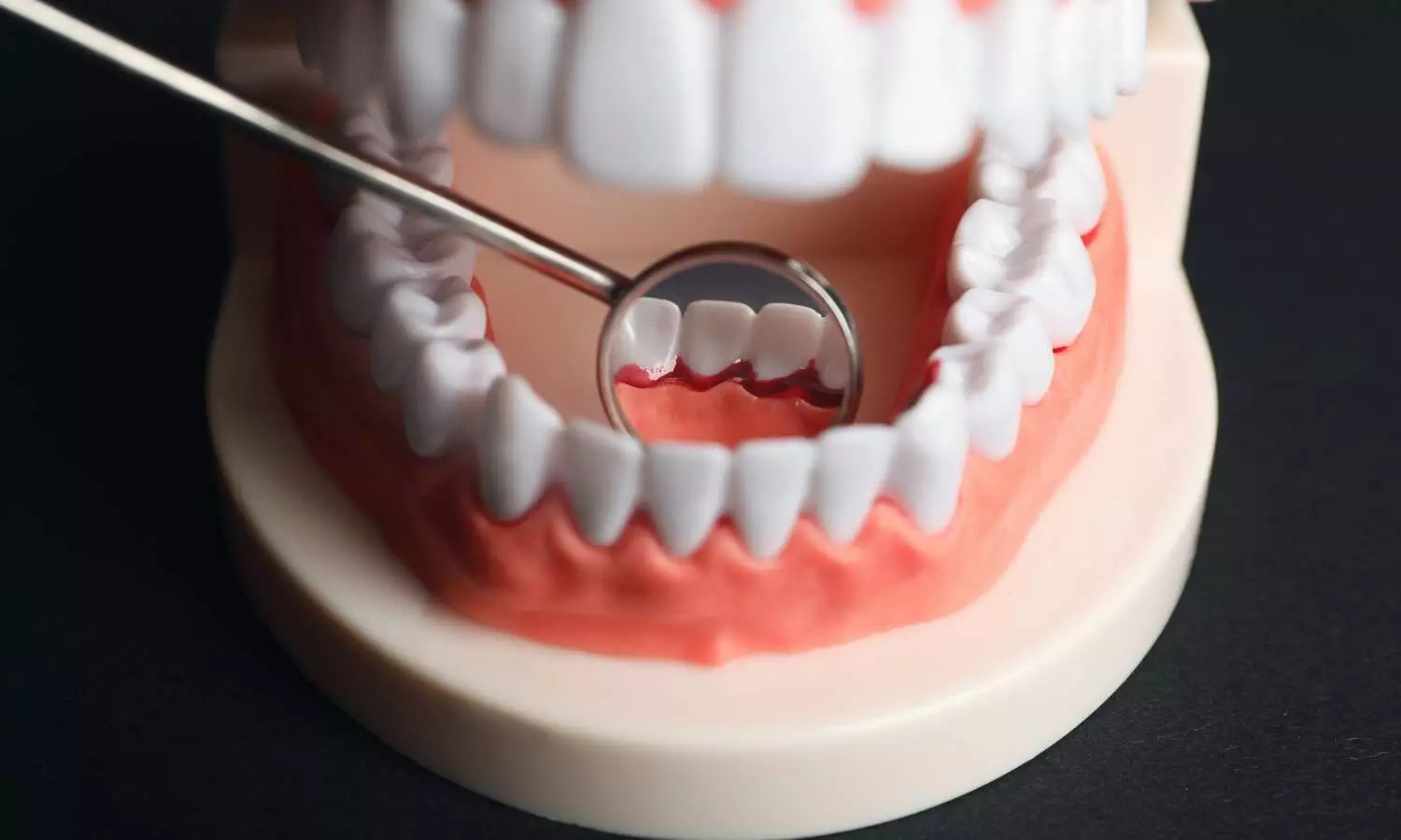
Periodontal disease occurs when the growth of bacteria in the gums causes the immune system to mount an inflammatory response, contributing to symptoms such as swelling, bleeding, and bone degradation. Untreated, it can lead to tooth loss. Nearly half of adults over age 30 have some form of periodontal disease, according to the U.S. Centers for Disease Control and Prevention.

Current treatments for advanced periodontal disease include antibiotics, deep cleanings of tooth and root surfaces, and various surgical procedures. Researchers have sought new ways to calm gum disease through less invasive treatment strategies.

Some previous studies have shown that people taking statins tend to show fewer signs of periodontitis than people who do not take statins. The new study is the first to trace the biochemical pathways through which statins appear to reduce periodontal inflammation.

“Recent periodontal literature has shown the beneficial effects of statins when used with traditional periodontal therapy,” Pandruvada said. “However, our study highlights a novel approach in which statins affect macrophages specifically, which, through this mechanism, can help treat periodontal disease.”

Macrophages play an important role in helping the body fight infections; however, they can also worsen inflammation depending on the form they take at different phases of the immune response. The researchers grew macrophages and gum cells together for the study and exposed them to various conditions. They found that exposure to simvastatin, a common statin drug, suppressed the macrophage inflammatory response.

As a next step, the researchers plan to study the impacts of statins on periodontal disease in animal models, a step toward determining whether this strategy might be a safe and effective approach for future periodontal therapies.

The new findings build upon the group’s initial results, which were published last year in the journal Cells.

Powered by WPeMatico
Low exposure to antibiotics in newborns may not increase risk of early-onset sepsis: JAMA

In a nationwide study conducted in Sweden, researchers have
discovered that administering antibiotics to newborns during their first week
of life does not substantially heighten the risk of early-onset sepsis (EOS) or
associated mortality. This finding comes as a significant reassurance to
healthcare providers and parents, amid growing concerns regarding the overuse
of antibiotics and the development of antibiotic resistance.


The study results were published in the journal JAMA Network
Open.

Antibiotic management is a boon in saving the lives of
newborns with EOS. However irrational use may develop resistance and adverse
outcomes in the development. Antibiotic prescriptions have to be scrutinized to
optimize treatment strategies. Hence researchers from Sweden conducted a nationwide
observational study titled the Sweden Neonatal Antibiotic Use
study and examined data from over 1 million newborns born between 2012 and 2020
across various neonatal units. The study evaluated the relationship between
antibiotic usage and the incidence of EOS among late-preterm and full-term
neonates.


By including All hospital live births from 34 weeks’ gestation, the
study was carried out among the neonatal intensive care during the first week
of life to identify the usage of intravenous antibiotics during the
first week of life, the duration of antibiotic therapy, the rate of
culture-proven EOS, and mortality associated with EOS.


Findings:

  • The study found that only a small percentage (1.88%) of
    newborns received antibiotics during their first week of life.
  • Of these, about 3.4% were diagnosed with EOS.

    Antibiotic treatment duration for newborns without EOS had a
    median of 5 days, totaling 113 antibiotic days per 1000 live births.


  • Despite no significant change in neonatal antibiotic exposure
    or antibiotic days per 1000 live births over the study period, EOS incidence
    decreased significantly from 0.74 in 2012 to 0.34 in 2020.

  • EOS-associated mortality remained stable at 1.39%.

  • Remarkably, the researchers observed no significant increase
    in the incidence of EOS or associated mortality over the study period, despite
    the relatively low antibiotic exposure.

  • The incidence of EOS was 0.63 per 1000 live births.

Unnecessary antibiotic use not only contributes to the
emergence of antibiotic-resistant bacteria but also poses potential risks to
newborns, including adverse effects and disruptions to the developing
microbiome. Moving forward, the findings of this study may guide healthcare
providers in optimizing antibiotic treatment strategies for newborns, striking
a delicate balance between effective infection management and antimicrobial
stewardship.

Further reading: Gyllensvärd J, Studahl M, Gustavsson L, et al. Antibiotic Use in Late Preterm and Full-Term Newborns. JAMA Netw Open. 2024;7(3):e243362. doi:10.1001/jamanetworkopen.2024.3362

Powered by WPeMatico

Intravascular Imaging Increases Cost-Effectiveness of Coronary Artery Procedures: AHA

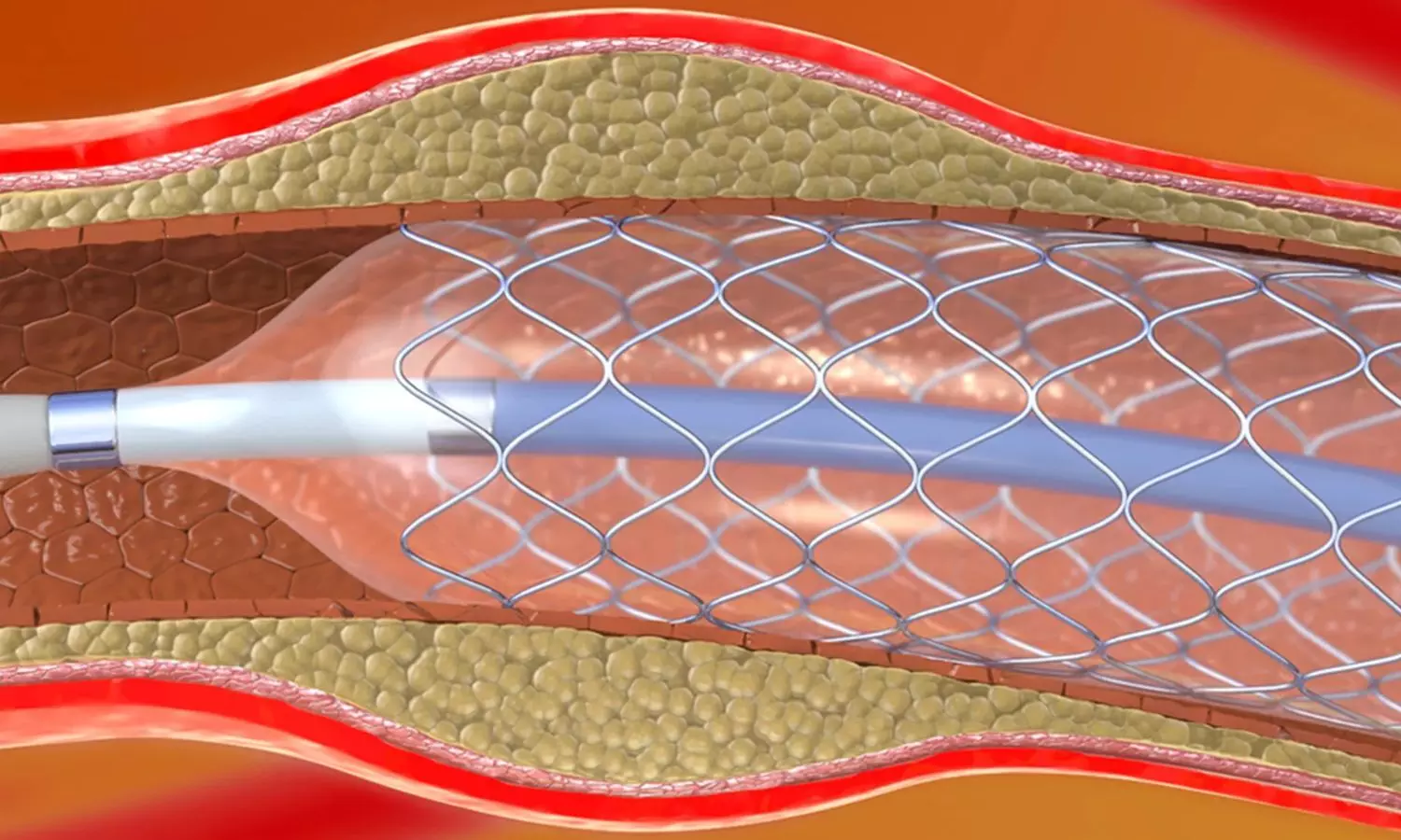
A recent study unveiled the cost-effectiveness of intravascular imaging-guided percutaneous coronary intervention (PCI) compared to conventional angiography-guided procedures. The findings were published in Circulation: Cardiovascular Quality and Outcomes reveal significant benefits for patients with complex coronary artery lesions.

The RENOVATE-COMPLEX-PCI trial was conducted in Korea from May 2018 to May 2021 and examined the clinical and economic outcomes of employing intravascular imaging during PCI procedures. This randomized controlled trial involved a total of 1639 patients and utilized a Markov model to simulate various health states over a 3-year and lifetime horizon.

The results from this research showed compelling evidence in favor of intravascular imaging-guided PCI. Over the 3-year follow-up period, the patients who underwent this advanced technique experienced higher quality-adjusted life years (QALY) and incurred slightly greater medical costs when compared to the individuals who underwent angiography-guided PCI. The incremental cost-effectiveness ratio (ICER) within the trial data stood at $57,040 per QALY gained that fell within the threshold of $35,000 per QALY.

Despite initial higher costs, the long-term analysis revealed a reversal in cumulative medical expenses between the two groups. The patients who underwent intravascular imaging-guided PCI enjoyed consistently higher QALY and also incurred lower total cumulative medical costs that resulted in a dominant incremental cost-effectiveness ratio.

Overall, this study suggests that intravascular imaging-guided PCI is not only clinically beneficial but also more cost-effective in the management of complex coronary artery lesions over the long term. This advanced imaging technology optimizes patient outcomes while minimizing healthcare expenditures by reducing medical costs and improving quality of life.

Reference:

Hong, D., Lee, J., Lee, H., Cho, J., Guallar, E., Choi, K. H., Lee, S. H., Shin, D., Lee, J.-Y., Lee, S.-J., Lee, S. Y., Kim, S. M., Yun, K. H., Cho, J. Y., Kim, C. J., Ahn, H.-S., Nam, C.-W., Yoon, H.-J., … Park, Y. H. (2024). Cost-Effectiveness of Intravascular Imaging-Guided Complex PCI: Prespecified Analysis of RENOVATE-COMPLEX-PCI Trial. In Circulation: Cardiovascular Quality and Outcomes. Ovid Technologies (Wolters Kluwer Health). https://doi.org/10.1161/circoutcomes.123.010230

Powered by WPeMatico

Invasive Angiography Adds Nothing to CCTA Alone for risk stratification of patients with stable CAD: ISCHEMIA

Canada: An analysis of the ISCHEMIA trial revealed that both invasive coronary angiography and coronary CT angiography (CCTA) add incremental information over baseline characteristics for predicting risk in stable coronary artery disease (CAD) patients.

The findings, published in JACC: Cardiovascular Imaging, also show that invasive angiography addition on top of CCTA does not provide any more prognostic information over the less invasive imaging test alone.

The data reaffirm the prognostic usefulness of assessing coronary anatomy with CCTA compared to invasive angiography and should reassure physicians who are apprehensive about using CT in certain patients, such as those with a high pretest likelihood of CAD.

According to the researchers, “With modern CT, with good image quality and experienced readers, we can do very well, even in complex CAD. We are not saying you other tests should not be considered in high-risk patients, but you shouldn’t necessarily avoid doing CT because you think it’s not going to have value.” 

The new study, published as a research letter, builds on prior work from the ISCHEMIA trial that revealed that CCTA has excellent anatomical agreement with invasive angiography for the identification of angiographically significant CAD. The analysis was performed by Jonathon Leipsic, Department of Imaging St Paul’s Hospital, Vancouver, British Columbia, Canada, and colleagues.

Invasive angiography has been the gold standard for the diagnosis of coronary disease, but CCTA has emerged as a less invasive test with similar diagnostic utility. The current US chest pain guidelines recommend CCTA for CAD diagnosis to aid in risk stratification and to guide treatment in stable patients at intermediate-to-high risk.

To date, no head-to-head studies have compared invasive angiography with CCTA for risk stratification, but the ISCHEMIA trial data allowed researchers to evaluate whether invasive angiography added anything to CCTA for predicting the risk of all-cause mortality and myocardial infarction (MI).

The analysis included 1,418 patients (median age 64.2 years; 19.5% female) randomized to the invasive strategy with both an interpretable CCTA and an invasive coronary angiogram.

The number of disease vessels, defined as stenosis of 50%, on invasive angiography and CCTA added incremental value to risk prediction over baseline characteristics alone. However, the number of diseased vessels on invasive angiography—defined as a stenosis of 50% or 70%—did not add anything to the CCTA data for predicting all-cause mortality or MI.

The use of the anatomical Duke Jeopardy Score, which assesses both lesion severity and location, did go beyond the CCTA-derived segment stenosis and segment involvement scores.

Reference:

Leipsic J, Ben Zekry S, Tzimas G, Broderick S, Mancini GBJ, Hague CJ, Budoff MJ, Rockhold FW, Chaitman BR, Kirby R, Stone GW, Ali ZA, Min JK, Hochman JS, Maron DJ, Reynolds HR; ISCHEMIA Research Group. Comparative Prognostic Utility of Coronary CT and Invasive Angiography: Insights From the ISCHEMIA Trial. JACC Cardiovasc Imaging. 2024 Mar 4:S1936-878X(24)00067-6. doi: 10.1016/j.jcmg.2023.11.015. Epub ahead of print. PMID: 38483421.

Powered by WPeMatico
Inflammation-reducing drug shows no benefit for dry age-related macular degeneration in NIH trial

The drug minocycline, an antibiotic that also decreases inflammation, failed to slow vision loss or expansion of geographic atrophy in people with dry age-related macular degeneration (AMD), according to a phase II clinical study at the National Eye Institute (NEI), part of the National Institutes of Health.

Dry AMD affects the macula, the part of the eye’s retina that allows for clear central vision. In people with dry AMD, patches of light-sensing photoreceptors and their nearby support cells begin to die off, leaving regions known as geographic atrophy. Over time, these regions expand, causing people to lose more and more of their central vision. Microglia, immune cells that help maintain tissue and clear up debris, are present at higher levels around damaged retinal regions in people with dry AMD than in people without AMD. Scientists have suggested that inflammation-and particularly microglia-may be driving the expansion of geographic atrophy regions.

This study, led by Tiarnan Keenan, M.D., Ph.D., a Stadtman Tenure-Track Investigator at the NEI’s Division of Epidemiology and Clinical Applications, tested whether inhibiting microglia with minocycline might help slow geographic atrophy expansion and its corresponding vision loss. The trial enrolled 37 participants at the NIH Clinical Center in Bethesda, Maryland, and at the Bristol Eye Hospital, United Kingdom. After a nine-month period where the researchers tracked each participant’s rate of geographic atrophy expansion, the participants took twice-daily doses of minocycline for two years. The researchers compared each participant’s rate of geographic atrophy expansion while taking minocycline to their baseline rate, and found there was no difference in geographic atrophy expansion rate or vision loss with minocycline.

Previous studies have shown that minocycline can help reduce inflammation and microglial activity in the eye, including the retina. The drug has shown beneficial effects for conditions such as diabetic retinopathy, but has not previously been tested for dry AMD.

Reference:

Keenan TDL, Bailey C, Abraham M, Orndahl C, Menezes S, Bellur S, Arunachalam T, Kangale-Whitney C, Srinivas S, Karamat A, Nittala M, Cunningham D, Jeffrey BG, Wiley HE, Thavikulwat AT, Sadda S, Cukras CA, Chew EY, Wong WT. Phase 2 Trial Evaluating Minocycline for Geographic Atrophy in Age-Related Macular Degeneration: A Nonrandomized Controlled Trial. JAMA Ophthalmol. 2024 Mar 14. doi: 10.1001/jamaophthalmol.2024.0118.

Powered by WPeMatico

Switching to integrase inhibitor may accelerate weight gain during menopause inHIV patients: Study

Integrase strand transfer inhibitors (INSTIs) are associated with more significant weight gain in women with HIV (WWH) compared to men with HIV. Menopausal transition leads to changes in body composition.

A finding published in CROI, Conference on Retroviruses and Opportunistic Infections, mentioned that switching to INSTIs during the late peri- and postmenopausal phase is associated with increased waist circumference (WC) and BMI. It was found that following this switch, peak effects occurred around 41 months.
More data is needed on whether INSTI initiation during the menopausal transition affects WC and BMI trajectories.
This study included 1159 women with HIV who were non-pregnant and virally suppressed between 2006 and 2019. Four hundred twenty-four women switched to an INSTI during the study period, while 735 did not. Nine hundred-four women without HIV (WWOH) from the Women’s Interagency HIV Study were included. The visit at which they reported switching was defined as the index visit. The researchers used mixed effect models with quadratic terms to analyze changes in waist circumference and BMI by menopausal phase, as determined by anti-Müllerian hormone levels at the index visit. This hormone is a biomarker of ovarian reserve. The models were adjusted for demographic, baseline measurements, behavioural and comorbidity, and HIV-related factors.
Key findings from the study are:
  • 66% identified as Black, 28% were premenopausal, 10% were early peri-, 28% were late peri-, and 34% were postmenopausal at the index. INSTI+ were older than INSTI- and WWOH, with median BMIs of 30 vs 29 vs 31.5kg/m2, respectively.
  • 64% of INSTI+ and 79% of INSTI- were on tenofovir DF before the index visit.
  • In premenopausal women, INSTI+ and INSTI—were associated with a 0.06cm per 6 months and 0.08cm per 6 months faster linear increase in WC, respectively, compared to WWOH; INSTI+ had a 0.05cm per 6 months faster increase than INSTI-.
  • When compared to WWOH, In late perimenopausal women, INSTI+ had faster increases in WC, which peaked at 41mo and then declined.
  • INSTI—had smaller increases in WC, while INSTI+ had a 0.39cm per 6mo faster linear increase in WC than INSTI-.
  • In postmenopausal women, INSTI+ was associated with faster WC increases up to 39mo, then declines compared to INSTI-.
  • BMI trajectories were similar for late peri- and postmenopausal women.
Concluding further, Switching to an INSTI-based regimen during late peri- and postmenopause is tied to early accelerated increases in Waist circumference and BMI when compared to women who did not switch.
The results of this study suggest that menopausal status should be considered when switching to an INSTI.

Powered by WPeMatico

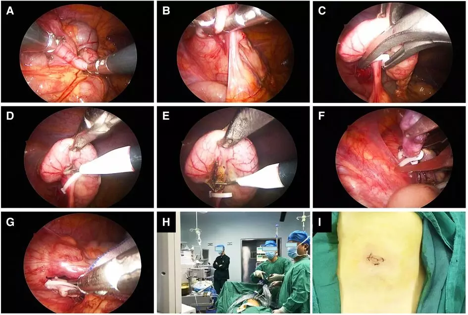
No advatage of Laparoscopic surgery over open surgery in reducing risk of bowel obstruction in complicated appendicitis: Study

No advantage of Laparoscopic surgery over open surgery in reducing the risk of bowel obstruction in complicated appendicitis suggests a new study published in the Journal of Pediatric Surgery. Complicated appendicitis is associated with a higher risk of postoperative complications, including adhesive bowel obstruction. This meta-analysis aims to investigate the difference in rates of postoperative bowel obstruction in paediatric patients with complicated versus simple appendicitis and whether the surgical approach influences this. A systematic literature search following PRISMA guidelines was conducted using MEDLINE, Embase and Cochrane Library for studies that analysed the incidence of adhesive bowel obstruction in paediatric patients after appendicectomy. Studies from 1998 – 2022 were included in the analysis. Results: A pooled analysis of 6 studies with low risk of bias and adequate follow-up periods, considering 58,962 cases of appendicectomy, revealed complex appendicitis was associated with a near two-fold increase in the incidence of SBO (pooled odds ratio 2.02 (95% CI 1.35 – 2.69)).

Interestingly, a similar pooled analysis of 10 studies, considering 62,433 cases of appendicectomy, revealed no significant difference between open and laparoscopic management of complex appendicitis (pooled odds ratio 0.93 (95% CI 0.24 to 1.62)). Complex appendicitis is associated with a two-fold increase in the rates of adhesive bowel obstruction. Whilst there are cosmetic advantages of a laparoscopic approach, surgical expertise should be favoured in decision-making relating to the surgical approach (laparoscopic versus open) as the evidence for a laparoscopic approach reducing risks of adhesive bowel obstruction is not convincing.

Reference:

Neel Doshi, Soham Bandyopadhyay, Madeline Green, Edward Richardson, Ahmad Komber, Si Emma Chen, Rahul Shah, Kokila Lakhoo. The risk of adhesive Bowel obstruction in children with appendicitis: A systematic review, Journal of Pediatric Surgery, 2024, ISSN 0022-3468. https://doi.org/10.1016/j.jpedsurg.2024.03.021.

(https://www.sciencedirect.com/science/article/pii/S0022346824001738)

Keywords:

No advantage, Laparoscopic surgery, open surgery, risk, bowel obstruction, complicated appendicitis, Journal of Pediatric Surgery, Simple; Complex; Appendicitis; Adhesive; Bowel; Obstruction

Powered by WPeMatico

Which are effective treatments for Reducing Nocturia among Women with Overactive Bladder?

In
a recent study, researchers have shed light on the efficacy of various
treatments for a common symptom of overactive bladder (OAB) known as nocturia.
The secondary analysis study found that for women with nocturia of at least 2 voids per night, treatment with anticholinergic
medication, onabotulinum
toxin A (BTX)
(either 100 or 200 units), or sacral neuromodulation (SNM) resulted in a
significant decrease in the number of voids per night at the 6-month follow-up.


The study results were published in the journal
Urogynecology.

Nocturia,
characterized by waking up multiple times during the night to urinate, can
significantly impact the quality of life of those affected. The prevalence of nocturia among
individuals with overactive bladder underscores its significance as a
distressing symptom, yet there remains a notable gap in our understanding of
how various treatments for overactive bladder affect this nocturnal urgency.
The study, conducted by analyzing data from the ABC and ROSETTA trials,
aimed to compare the effectiveness of anticholinergic medication, onabotulinum
toxin A (BTX), and sacral neuromodulation (SNM) in reducing nocturia.

A
team of researchers from MedStar
Washington Hospital Center, Washington D.C., by utilizing the
National Institutes of Health Data and Specimen Hub database, conducted a study
that included 197 female patients who reported a mean of at least 2 voids per
night on a 3-day diary. These patients were divided into cohorts based on the
treatment they received: anticholinergic medication, BTX 100 units, BTX 200
units, or SNM. The main outcome was to measure the change in mean voids/night on 3-day diary from baseline to
6 months.

Findings:

  • One
    of the key findings of the study was that all treatment cohorts demonstrated a
    significant reduction in the mean number of voids per night at the 6-month
    mark.

  • This
    reduction was a substantial 41% in mean voids per night, marking a significant
    improvement in the nocturia symptoms experienced by the participants.

  • Importantly,
    this improvement was observed across all treatment modalities.


  • Prior
    to treatment initiation, there were no significant differences noted among the
    cohorts in terms of the number of voids per night, demographic factors, or
    urodynamic values.
  • However,
    after 6 months of treatment, the results showed consistent and substantial
    improvements across the board, regardless of the treatment method utilized.


Notably,
the study found no significant differences in the effectiveness of the various
treatments in reducing nocturia. Whether patients received anticholinergic
medication, BTX (either 100 or 200 units), or SNM, the reduction in the mean
number of voids per night was comparable across all groups. This suggests that
multiple treatment options are equally effective in addressing the bothersome
symptoms of nocturia in women with OAB.


Further reading: Comparing Impact of Overactive Bladder Therapies on Nocturia. DOI: 10.1097/SPV.0000000000001465

Powered by WPeMatico