Shared digital NHS prescribing record could avoid nearly 1 million annual drug errors

Implementing a single shared digital prescribing record across the NHS in England could avoid nearly 1 million drug errors every year, stopping up to 16,000 fewer patients from being harmed and saving up to 22 lives every year, suggests a modeling study published online in BMJ Quality & Safety.

Powered by WPeMatico

Consistently exercising 2–3 times a week over the long term linked to lower current insomnia risk

Consistently exercising 2–3 times a week over the long term is linked to a lower current risk of insomnia as well as the ability to clock up the recommended 6–9 hours of shut-eye every night, suggests an international 10-year study published in the open access journal BMJ Open.

Powered by WPeMatico

AI-based analysis identifies two plant extracts with potential as GLP-1 agonist weight loss pills

Two plant compounds with potential as GLP-1 agonist weight loss pills have been identified in an AI (artificial intelligence)-based study, the European Congress on Obesity (ECO 2024) (Venice 12-15 May) will hear.

Powered by WPeMatico

Optimizing electronic health records: Study reveals improvements in departmental productivity

In a study published in the Annals of Family Medicine, researchers at the Marshall University Joan C. Edwards School of Medicine identify transformative effects of electronic health record (EHR) optimization on departmental productivity. With the universal implementation of EHR systems, the study sheds light on the importance of collaborative efforts between clinicians and information technology (IT) experts in maximizing the potential of these digital tools.

Powered by WPeMatico

Merck gets USFDA nod for WINREVAIR (sotatercept-csrk) for adults with pulmonary arterial hypertension

Rahway: Merck, known as MSD outside of the United States and Canada, has announced that the U.S. Food and Drug Administration (FDA) has approved sotatercept-csrk (U.S. Brand Name: WINREVAIR, for injection, 45mg, 60mg) for the treatment of adults with pulmonary arterial hypertension (PAH, World Health Organization [WHO] Group 1) to increase exercise capacity, improve WHO functional class (FC), and reduce the risk of clinical worsening events.
WINREVAIR was previously granted Breakthrough Therapy Designation by the FDA.
“WINREVAIR is the first FDA-approved activin signaling inhibitor therapy for PAH, representing a new class of therapy that works by improving the balance between pro- and anti-proliferative signaling to regulate vascular cell proliferation underlying PAH,” the release stated.

“Pulmonary arterial hypertension is a rare, progressive and ultimately life-threatening disease in which blood vessels in the lungs thicken and narrow, causing significant strain on the heart,” said Dr. Marc Humbert, Professor of Medicine and Director of the Pulmonary Hypertension Reference Center at the Université Paris-Saclay and investigator on the Phase 3 STELLAR study. “Based on the Phase 3 STELLAR trial, adding WINREVAIR to background PAH therapy demonstrated significant clinical benefits compared to background PAH therapy alone. This approval is an important milestone, as it offers healthcare providers a novel therapeutic option that targets a new PAH treatment pathway.”

The approval is based on the Phase 3 STELLAR trial, which compared WINREVAIR (n=163) to placebo (n=160), both in combination with background standard of care therapies in adult patients with PAH (WHO Group 1 FC II or III). Results showed adding WINREVAIR to background therapy increased six-minute walk distance from baseline by 41 meters (95% CI: 28, 54; p<0.001; placebo-adjusted) at Week 24 and significantly improved multiple important secondary outcome measures, including reducing the risk of death from any cause or PAH clinical worsening events by 84% versus background therapy alone (number of events: 9 vs 42, hazard ratio=0.16; 95% CI: 0.08, 0.35; p<0.001).

“Healthcare providers should monitor hemoglobin and platelets before each dose of WINREVAIR for the first 5 doses, or longer if values are unstable, and periodically thereafter to determine if dose adjustments are required. WINREVAIR may increase hemoglobin and may lead to erythrocytosis, which if severe may increase the risk of thromboembolic events or hyperviscosity syndrome. WINREVAIR also may decrease platelet count and lead to severe thrombocytopenia, which may increase the risk of bleeding; thrombocytopenia occurred more frequently in patients also receiving prostacyclin infusion. Treatment should not be initiated if platelet count is <50,000/mm³,” the release added.

“The Pulmonary Hypertension Association welcomes the development of new therapies for those with PAH,” said Matt Granato, president and chief executive officer, Pulmonary Hypertension Association. “A diagnosis of PAH is a life-changing experience for patients and families due to its chronic, progressive nature. Patients with PAH experience limiting symptoms such as shortness of breath and fatigue. We are excited to see industry research leading to a better understanding of PAH and the development of a medicine in a novel treatment pathway that expands options for the patient community.”

“New treatment options continue to be needed for patients with pulmonary arterial hypertension that support important clinical goals, including increasing exercise capacity and improving functional class,” said Dr. Aaron Waxman, Executive Director of the Center for Pulmonary Heart Diseases at Brigham and Women’s Hospital and investigator on the Phase 3 STELLAR study. “Sotatercept added to background therapy has the potential to become a new standard of care option for patients with pulmonary arterial hypertension.”

WINREVAIR is given once every three weeks by subcutaneous injection and may be administered by appropriate patients or caregivers with guidance, training and follow-up from a healthcare provider. Healthcare providers and patients/caregivers should refer to the Instructions for Use for information on the proper preparation and administration of WINREVAIR. Merck estimates that WINREVAIR will be available for dispensing by select specialty pharmacies in the U.S. by the end of April.

“PAH remains a debilitating disease with high morbidity and mortality,” said Dr. Eliav Barr, senior vice president and head of global clinical development, chief medical officer, Merck Research Laboratories. “This approval of WINREVAIR is an important milestone and a testament to our science-led strategy and focus on the development of innovations that can help people affected by rare diseases like PAH. We are proud to bring this novel medicine to patients.”

The primary efficacy endpoint in the STELLAR trial was the change from baseline at Week 24 in 6-Minute Walk Distance (6MWD). In the WINREVAIR treatment group, the placebo-adjusted median increase in 6MWD was 41 meters (95% CI: 28, 54; p<0.001, Hodges-Lehmann estimate). Additionally, patients who added WINREVAIR had statistically significantly greater improvements compared to placebo across multiple secondary endpoints:

  • Treatment with WINREVAIR led to an improvement in FC from baseline at Week 24 in 29% of patients compared to 14% of patients treated with placebo (p<0.001).
  • Treatment with WINREVAIR resulted in an 84% reduction in the occurrence of death or PAH clinical worsening events compared to placebo (median duration of exposure 33.6 weeks; number of events: 9/163 vs 42/160, hazard ratio=0.16; 95% CI: 0.08, 0.35; p<0.001).
  • Treatment with WINREVAIR led to an improvement from baseline in pulmonary vascular resistance (PVR). The median treatment difference in PVR between WINREVAIR and placebo was -235 dynes*sec/cm5 (95% CI: -288, -181; p<0.001).
  • Treatment with WINREVAIR led to an improvement from baseline in N-terminal pro-B-type natriuretic peptide (NT-proBNP) levels. The median treatment difference in NT-proBNP between WINREVAIR and placebo was -442 pg/mL (95% CI: -574, -310; p<0.001).

The STELLAR study (NCT04576988) was a global, double-blind, placebo-controlled, multicenter, parallel-group clinical trial in which 323 patients with PAH (WHO Group 1 FC II or III) were randomized 1:1 to WINREVAIR (target dose 0.7 mg/kg) (n=163) or placebo (n=160) plus stable background therapy administered subcutaneously once every 3 weeks.

The most common PAH etiologies were idiopathic PAH (59%), heritable PAH (18%), and PAH associated with connective tissue diseases (CTD) (15%). Most participants were receiving either three (61%) or two (35%) background drugs for PAH, and 40% were receiving prostacyclin infusions. The mean time from PAH diagnosis was 8.8 years. Patients had a WHO FC II (49%) or III (51%) at baseline.

WINREVAIR is FDA-approved for the treatment of adults with pulmonary arterial hypertension (PAH, WHO Group 1) to increase exercise capacity, improve WHO functional class (FC) and reduce the risk of clinical worsening events. WINREVAIR is the first activin signaling inhibitor therapy approved to treat PAH. WINREVAIR improves the balance between pro-proliferative and anti-proliferative signaling to modulate vascular proliferation. In preclinical models, WINREVAIR induced cellular changes that were associated with thinner vessel walls, partial reversal of right ventricular remodeling, and improved hemodynamics.

WINREVAIR is the subject of a licensing agreement with Bristol Myers Squibb.

Read also: Merck plans to conduct clinical trials of Multi-Valent HPV vaccine, single-dose regimen for GARDASIL9

Powered by WPeMatico

PHFI bags second place in global ranking of public health institutions

Hyderabad: In a significant milestone for India’s healthcare landscape, the Public Health Foundation of India (PHFI) has secured the prestigious second position among 26 global public health institutions in the inaugural rankings. Notably, PHFI stands as the lone Indian representative among this esteemed cohort of global public health institutions. 

This remarkable achievement has seen PHFI surpassing renowned institutions like Harvard TH Chan School of Public Health, which stands in the third spot now.
According to The Hindu, the rankings, unveiled in the International Journal of Public Health, result from a pioneering pilot project, conducted by an independent consortium of researchers, including Margaret Chan, former director-general of the World Health Organisation (WHO). Unlike conventional rankings that merely quantify research output, this initiative emphasizes the quality and impact of the institutions’ research endeavours.
Organizations like the London School of Hygiene and Tropical Medicine, PHFI, Harvard TH Chan School of Public Health, Swiss School of Public Health, and Johns Hopkins Bloomberg School of Public Health are leading the way internationally in this regard.
PHFI’s ascension to the second position underscores its unwavering commitment to advancing public health initiatives in India and beyond. Founded as a collaborative endeavour between public and private entities, PHFI has evolved through extensive consultations with diverse stakeholders, including academia, government bodies, and civil society organizations, both domestically and internationally. Focusing on a broad spectrum of public health services encompassing promotive, preventive, and therapeutic measures, PHFI addresses critical gaps in policy planning and public perception.
One of PHFI’s flagship initiatives includes the establishment of five Indian Institutes of Public Health (IIPHs) strategically located across India. These institutes serve as hubs for cutting-edge research and education, driving innovation and excellence in public health practices nationwide, reports The Daily. 
President of the PHFI K Srinath Reddy, “In today’s globalized world, international collaborations are essential for producing research that holds global relevance. The team utilized a total of 11 indicators distributed across four categories: productivity, quality, accessibility for readers, and international collaborations. These indicators were selected because they are pertinent to the discipline of Public Health.”
PHFI’s meteoric rise in the global rankings underscores India’s growing prowess in the field of public health. It reinforces its status as a key player in shaping the future of healthcare on the global stage. As PHFI continues its trajectory of excellence, it remains steadfast in its mission to catalyze positive change and foster healthier, more resilient communities worldwide. Mr Srinath said the ranking made PHFI assert its position as the public and policymaker. “This acknowledgement validates our mission and performance, lending credibility not only internationally but also nationally, where both Central and state governments will recognize our value in the field of public health. Additionally, prospective students applying to our courses can now have confidence in studying at a globally ranked institution,” he added. 

Powered by WPeMatico

Demolition of Historic Women Hospital at PMCH sparks outcry

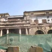
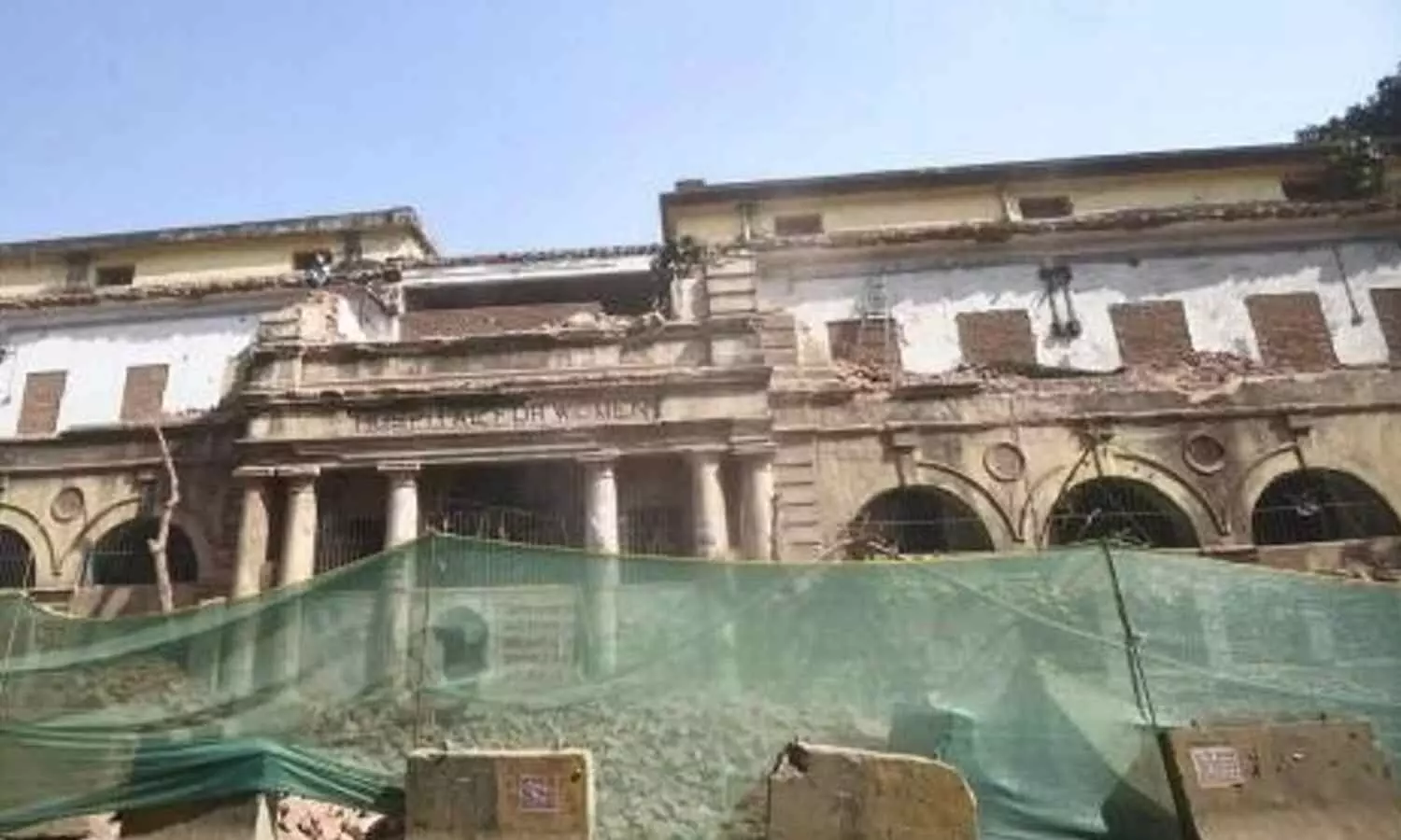
Patna: The demolition of the entire frontal structure of the more than 90-year-old Women Hospital at the iconic Patna Medical College and Hospital (PMCH) has stirred anguish among heritage enthusiasts and former students of the esteemed institution. 

The PMCH was established in 1925 as Bihar and Orissa province’s first medical college, it holds significant historical importance, having been founded in the early years following the region’s separation from the Bengal Presidency in 1912.

On March 22, coinciding with the 112th anniversary of Bihar’s modern statehood, laborers were observed dismantling the remaining frontal structures of the Women Hospital building situated within the PMCH campus along the historic Ashok Rajpath.

Also Read:Save heritage landmarks of PMCH: Alumni urges Bihar Govt

Once boasting a distinguished European architectural design featuring Doric pillars at its entrance, the old two-storey building now stands bruised, starkly contrasting its former handsome facade.

Officials said a segment of the front portion of the Women Hospital building has been demolished to make way for infrastructure projects. They said the demolition work started earlier this month, news agency PTI reported.

Chief Minister Nitish Kumar on February 27 inaugurated the first phase of the PMCH redevelopment project.

As part of the mega infrastructure revamp plan, a 5,462-bed hospital complex will come up at the old PMCH site for Rs 5,540 crore. The project is expected to be completed in seven years. The foundation stone of the mega project was laid by Kumar on February 8, 2021.

The ongoing redevelopment initiative, which began with the demolition of select structures in 2021 to make room for modern high-rise facilities, aims to revitalize the PMCH campus, originally established as the Prince of Wales Medical College in 1925.

This was Bihar and Orissa province’s first medical college. The college, renamed PMCH after Independence, was in turn born out of the Temple Medical School, established in 1874 in Bankipore.

Bihar and Orissa as a separate province was carved out of the Bengal presidency in 1912, with the capital at Patna. On April 1, 1936, Orissa (now Odisha) became a separate state.

Bihar government celebrates March 22 as Bihar Diwas, marking the formation of the state.

Several old buildings of PMCH, including the old medical superintendent’s bungalow, prison ward, and nurses hostel, have been demolished as part of the revamp project. Its alumni had earlier made a fervent appeal to the authorities to spare the core heritage structures which tell the stories of the famed institution’s inception.

According to PTI, PMCH Alumni Association president Satyajeet Kumar Singh reiterated his appeal to at least spare the historic old Bankipore General Hospital building and Administrative Block to “let future generations see the legacy of the institution in a tangible form”.

The partial demolition of the Women’s Hospital, set up in 1930, has also sparked an outcry among heritage lovers.

Aman Lal, 20, a student of Patna College said, “The government is slowly dismantling all key heritage buildings of the city which gave our state and Patna its identity.”

“On Bihar Diwas I was on my way to college when I saw labourers pulling down the remnants of the front portion of the iconic Women’s Hospital building. It broke my heart. Instead of celebrating our built heritage on Bihar Diwas, the government razed it. It’s a shame,” he said.

Prateek Nishant, a PMCH alumnus whose great grandfather, Tarini Prasad Sinha, was among the first graduating batch in 1927, said, “Heritage and development can co-exist and it needs sensitive planning”.

“As a former student, I feel sad. The heritage buildings of PMCH, which turns 100 next year, should have been saved for posterity. The new blocks of PMCH should have been built elsewhere in the city,” he said.

Phase one of the redevelopment project includes a G+9 multi-utility building, housing the OPD, blood banks, surgical store, medical store, blood bank, among other facilities, the senior official said.

Partial demolition of the Women Hospital has been carried out to also accommodate the alignment of an under-construction double-decker flyover along a section of the Ashok Rajpath from Kargil Chowk to NIT More, the official said.

Many educational and other institutions located along the Ashok Rajpath have been heavily impacted due to the double-decker flyover project and the ongoing construction of a corridor of Patna Metro.

The foundation stone of the little over 2 km flyover was laid on September 4, 2021, by the chief minister.

While PMCH’s main gate has been closed since July 2022 for the construction of a metro station, a significant portion of the front of the historic cancer ward building was demolished by early 2023 to build the metro corridor.

In September 2022, official sources told PTI that the old Women Hospital of the PMCH shall not be affected by the revised alignment of the Patna Metro along Ashok Rajpath.

Powered by WPeMatico

Adjuvant hyperbaric oxygen Therapy may alleviate inflammation and benefit diabetic foot patients, claims study

Adjuvant hyperbaric oxygen Therapy may alleviate inflammation and benefit diabetic foot patients suggests a new study published in the Medicine.

Diabetes Mellitus (DM) is a metabolic disease with a high morbidity and mortality and increasing in prevalence all over the world. Due to the hypoxic, ischemic, inflammatory, and infective environment in DM, diabetic foot ulcers have been treated with medico-surgical interventions and adjuvant hyperbaric oxygen Therapy (HBOT). The purpose of this study was to evaluate the effects of HBOT on haematological indices and biochemical parameters in patients with diabetic foot. The study group was formed from the file records of 103 male patients who applied to Yunus Emre State Hospital HBOT Center between September 1, 2016, and December 31, 2020, and were treated with HBOT with a multidisciplinary approach. RESULTS: There were negative low correlations between number of HBOT sessions and Mean Corpuscular Hemoglobin (MCH) (P = .037, r = -0.207) and Blood Urea Nitrogen (BUN) (P = .037, r = -0.222). White Blood Cell Count (WBC), Neutrophils (NEU), Monocytes (MON), Platelet Count (PLT), and Plateletcrit (PTC) parameters were found to be decreased.

An increase in lymphocytes (LYM), Eosinophils (EOS), Mean Corpuscular Hemoglobin Concentration (MCHC), and Red Cell Distribution Width (RDW) parameters were detected after the treatments (P < .05). Again, after the treatment, glucose (Glu), C-Reactive Protein (CRP), direct bilirubin, and total protein (TP) levels were decreased. Uric acid (UA) levels increased (P < .05). HBOT improved haematological indices in patients and had a beneficial effect on biochemical parameters, particularly Glu and CRP levels. Adjuvant HBOT alleviates diabetic inflammation and has a beneficial effect on diabetic patient treatment.

Reference:

Ercan, Erdinç, et al. “The Effect of Hyperbaric Oxygen Therapy On Hematological Indices and Biochemical Parameters in Patients With Diabetic Foot.” Medicine, vol. 103, no. 12, 2024, pp. e37493.

Powered by WPeMatico

MRI and lumbar puncture not necessarily required to manage CAR T-cell therapy complications: Study

Magnetic resonance imaging (MRI) and lumbar puncture (LP) may not always be necessary for diagnosing and managing a serious neurological complication associated with CAR T-cell therapy, according to a new Blood Advances study. Findings further validated the use of the electroencephalogram (EEG) – a noninvasive test measuring electrical activity in the brain-in managing this neurotoxicity.

“When treating patients for CAR T-cell associated toxicities, we typically follow pretty rigid guidelines based on phase one and two studies, and there is little to no clinical evidence to validate these,” explains Dr. Guillaume Manson, a hematologist at the University Hospital of Rennes in Rennes, France, and the study’s senior author. “Some of these tests, like a lumbar puncture, can be extremely taxing and invasive for patients. Here, we wanted to get a better sense of when these interventions are necessary versus when we could do without them.”

Authors sought to evaluate the necessity of three diagnostic tests used to manage care for recipients of CAR T-cell therapy experiencing immune effector cell-associated neurotoxicity syndrome (ICANS). While ICANS’ underlying mechanisms are not entirely understood, patients with ICANS may experience a range of neurological symptoms, including confusion, tremors, seizures, and, in rare cases, serious brain swelling and comas.

National and international diagnostic guidelines recommend that health care providers perform an MRI, a LP, and and/or an EEG, based on a complication’s severity, before beginning ICANS treatment. These interventions are expensive, can be invasive, and require extensive hospital resources. Further, these interventions are typically used to rule out other conditions and treatments are seldom modified by the results of these tests.

The study authors collected data from 190 patients treated with CAR T-cells at the University Hospital Centre of Rennes from August of 2018 to January of 2023. Out of all the patients in the study, about 62% were male, and the average age was 64 years old, with ages ranging from 15 to 81. Most of the patients were being treated for a type of lymphoma called refractory/relapsed diffuse large B cell lymphoma (DLBCL) (around 73%). During treatment, roughly 48% of patients developed ICANS.

All study participants met with a neurologist and underwent an MRI scan as a baseline assessment before receiving CAR T-cell infusions. Among those who experienced ICANS, their treatment protocol depended on their unique case and physician recommendation, abiding by the existing guidelines. Most patients with ICANS (80%) underwent at least one intervention, and a third (34%) underwent all three (MRI, LP, and EEG).

Researchers assessed how the different interventions affected patient treatment, such as how medications, for example, antibiotics, and anti-seizure therapy, were prescribed based on abnormal findings and how these treatment courses changed patient outcomes.

Of the 78% of patients who underwent MRI scans, 80% of scans showed normal results. Only 4% of those with abnormal findings had a change in treatment plans. Approximately 47% of patients underwent lumbar punctures, and no cases identified active infections, but 7% of patients had treatment change based on suspected infections. Among the 56% of patients who received EEGs, only 18% of these scans showed normal results. In 45% of patients, EEGs detected brain dysfunction, and in some cases, signs of seizures, even in those with no prior symptoms. As a result, 16% of treatment plans were altered based on abnormal EEG findings.

Results ultimately revealed that abnormal results were more common in patients with more severe ICANS. MRI results were often normal and while LP and EEG often showed abnormalities, it was more frequent with more severe cases of ICANS.

When it came to treatment decisions, MRI rarely led to changes, LP sometimes led to unnecessary treatments for suspected infections, and EEG often resulted in adjustments to antiepileptic medications.

This study is limited by its relatively small sample size and requires validation by further clinical studies. However, these findings suggest that LP and MRI might not always be necessary for all patients with ICANS, as they often did not influence treatment. However, EEG often led to adjustments in medications, suggesting it remains a necessary diagnostic intervention.

Ultimately, Dr. Manson conveys that these results represent a call to action to update existing guidelines for managing ICANS based on clinical evidence. He emphasizes that these results do not imply that practitioners should never perform MRIs or LPs, only that they may not be needed in all cases. If further evidence confirms that MRIs and LPs are not necessary in all instances of ICANs, revised guidelines could potentially save patients time, money, and spare them from unnecessary medical procedures.

“Every patient’s case is different, and these findings certainly do not say that certain tests should or should not be performed,” said Dr. Manson. “We did this research to generate clinical evidence to inform guidelines that support physicians in making clinical decisions when treating patients with these complex, and sometimes severe conditions.”

Reference:

Mauget, M., et al. (2024) Impact of diagnostic investigations in the management of CAR T-cell-associated neurotoxicity. Blood Advances. doi.org/10.1182/bloodadvances.2023011669.

Powered by WPeMatico

Long-term intake of reused deep-fried oil tied to increased neurodegeneration: Study

USA: Deep-frying at high temperatures has been linked with several metabolic disorders, but there have been no long-term investigations on the influence of deep-fried oil consumption and its detrimental effects on health. 

A recent study is the first to report long-term deep-fried oil supplementation increases neurodegeneration in the first-generation offspring.

The study found higher levels of neurodegeneration in rats that consumed reused deep-fried cooking oils and their offspring compared to rats on a normal diet. Deep frying, which involves completely submerging food in hot oil, is a common method of food preparation around the world.

Results from the study also suggest that the increased neurodegeneration is tied to the oil’s effects on the bidirectional communication network between the liver, gut and brain. The liver–gut–brain axis plays a crucial role in regulating various physiological functions, and its dysregulation has been associated with neurological disorders.

Kathiresan Shanmugam, an associate professor from Central University of Tamil Nadu in Thiruvarur, led the research team.

“Deep-frying at high temperatures has been linked with several metabolic disorders, but there have been no long-term investigations on the influence of deep-fried oil consumption and its detrimental effects on health,” said Shanmugam, formerly at Madurai Kamaraj University, Madurai. “To our knowledge, we are first to report long-term deep-fried oil supplementation increases neurodegeneration in the first-generation offspring.”

Sugasini Dhavamani, a research collaborator from the University of Illinois at Chicago, will present the research at Discover BMB, the annual meeting of the American Society for Biochemistry and Molecular Biology, which will be held March 23–26 in San Antonio.

Deep frying food not only adds calories; reusing the same oil for frying, a common practice in both homes and restaurants, removes many of the oil’s natural antioxidants and health benefits. Oil that is reused also can contain harmful components such as acrylamide, trans fat, peroxides and polar compounds.

To explore the long-term effects of reused deep-fried frying oil, the researchers divided female rats into five groups that each received either standard chow alone or standard chow with 0.1 ml per day of unheated sesame oil, unheated sunflower oil, reheated sesame oil or reheated sunflower oil for 30 days. The reheated oils simulated reused frying oil.

Compared with the other groups, the rats that consumed reheated sesame or sunflower oil showed increased oxidative stress and inflammation in the liver. These rats also showed significant damage in the colon that brought on changes in endotoxins and lipopolysaccharides — toxins released from certain bacteria. “As a result, liver lipid metabolism was significantly altered, and the transport of the important brain omega-3 fatty acid DHA was decreased. This, in turn, resulted in neurodegeneration, which was seen in the brain histology of the rats consuming the reheated oil as well as their offspring.”

Additional studies in which MSG was used to induce neurotoxicity in the offspring showed that the offspring that consumed the reheated oils were more likely to show neuronal damage than the control group receiving no oil or those that received unheated oil.

Although more studies are needed, the researchers say that supplementation with omega-3 fatty acids and nutraceuticals such as curcumin and oryzanol might be helpful in reducing liver inflammation and neurodegeneration. They added that clinical studies in humans are needed to evaluate the adverse effects of eating fried foods, especially those made with oil that is used repeatedly.

As a next step, the researchers would like to study the effects of deep-frying oil on neurodegenerative diseases such as Alzheimer’s and Parkinson’s as well as on anxiety, depression and neuroinflammation. They would also like to further explore the relationship between gut microbiota and the brain to identify potential new ways to prevent or treat neurodegeneration and neuroinflammation.

Powered by WPeMatico