Quinoa-incorporated bread falls under low-GI foods and improves glycemic control, lipid profiles: Study

India: A recent in vivo study published in Frontiers in Nutrition has shed light on the effect of quinoa (Chenopodium quinoa W.) flour supplementation in breads on the lipid profile and glycemic index (GI).

The study showed that quinoa-incorporated bread possessed a low glycemic index (42.00 ± 0.83) compared to the control (69.20 ± 1.84) and long-term consumption proved to contain functional efficacies, in terms of hypolipidemic effect.

“Incorporating quinoa into bread formulations not only enhances their nutritional content but also offers potential health benefits, particularly in managing cardiovascular health and blood sugar levels,” the researchers wrote. The findings highlight the positive effects of quinoa flour supplementation in bread on lipid profile and glycemic index.

Quinoa has recently gained significant popularity as a nutritious and versatile grain alternative. Beyond its use as a standalone dish, researchers have been exploring its potential as a functional ingredient in various food products. One such avenue of investigation is its incorporation into bread, a staple food consumed worldwide.

Quinoa (Chenopodium quinoa W.) is a pseudo-cereal rich in protein, vitamins, dietary fiber, and minerals. Its nutritional profile makes it an attractive candidate for improving the health properties of baked goods like bread.

Against the above background, Natasha R. Marak, Central Agricultural University, Tura, Meghalaya, India, and colleagues aimed to investigate the effects of quinoa bread on physical, chemical, and bioactive components, glycaemic index, and biochemical parameters.

For this purpose, the researchers fed human subjects aged between 20 and 50 years with the absence of morbid factors daily with quinoa bread for 3 months to study its pre-and post-treatment effects on lipid profile, glycosylated hemoglobin, and blood glucose.

The effort was made to incorporate the maximum amount of quinoa into the bread without compromising the bread’s acceptability. Of the 14 formulations, TQ13, containing 20% quinoa flour with 3% wheat bran, was selected for further analysis.

Based on the study, the researchers reported the following findings:

  • The GI study revealed that quinoa bread peaked at 45 min with a gradual increase after ingestion of the bread and a steady decline thereafter.
  • Before and after supplementation with quinoa-incorporated bread, the observed value for blood glucose levels was 86.96 ± 15.32 mg/dL and 84.25 ± 18.26 mg/dL, respectively.
  • There was a statistically significant decrease in levels of triglycerides, total cholesterol, low-density lipoprotein (LDL), and very-LDL (VLDL) levels before and after supplementation.
  • Non-significant changes were observed for high-density lipoprotein levels from the pre-and post-treatment with the quinoa-incorporated bread.

According to the study, these benefits have proved crucial in the dietary treatment of diabetes mellitus by resulting in improved glycaemic control and several metabolic parameters, such as improved blood lipid levels.

Based on the data obtained in the study, the researchers concluded that quinoa incorporation into wheat bread results in essential health benefits.

High consumption of phenolic compounds has long been associated with reduced risk of cardiovascular diseases and certain cancers. “Current trends in the enhancement of the antioxidant capacity of wheat bread by quinoa flour addition rich in phenolic compounds might be beneficial in the health status of a population,” the researchers concluded. “The results obtained in the present study corroborate the data reported in the literature, indicating that quinoa bread can be used in glycemic control and plasma lipids.”

Reference:

Marak, N. R., Das, P., Das Purkayastha, M., & Baruah, L. D. (2024). Effect of quinoa (Chenopodium quinoa W.) flour supplementation in breads on the lipid profile and glycemic index: An in vivo study. Frontiers in Nutrition, 11, 1341539. https://doi.org/10.3389/fnut.2024.1341539

Powered by WPeMatico

FeNO helps in diagnosis of chest tightness-variant asthma in children, claims study

A recent research identified a potential breakthrough in diagnosing a novel form of asthma in children known as Chest Tightness-Variant Asthma (CTVA). CTVA is characterized by chest tightness as the primary symptom and presents a diagnostic challenge due to its atypical nature. However, a recent study published in the recent edition of Respiratory Medicine journal illuminates the promise of this diagnostic approach.

The study was conducted on a total of 95 children aged 6 to 14 years with chest tightness as their only symptom for more than four weeks. Tengteng Zhang and team evaluated the efficacy of combining bronchial provocation testing with fractional exhaled nitric oxide (FeNO) measurement for the diagnosis of CTVA.

The results revealed that while lung function remained normal in both CTVA and non-CTVA groups, the FeNO levels significantly differed. The children in the CTVA group expressed FeNO levels averaging 22.35 parts per billion (ppb) that was markedly higher than the average of 14.85 ppb in the control group. This discrepancy in FeNO levels proved statistically significant (P < 0.05).

Utilizing the receiver operating characteristic (ROC) curves, further analysis demonstrated that FeNO could help in diagnosing CTVA, with an optimal cutoff point of 18.5 ppb. At this threshold, FeNO expressed a sensitivity of 60.3% and specificity of 77.8%. Additionally, a negative correlation was observed between FeNO levels and bronchial hyperresponsiveness parameters such as Dmin and PD15 (P = 0.006).

Despite these promising findings, it is essential to note that FeNO alone does not serve as a definitive diagnostic marker for CTVA. Instead, it should be utilized alongside bronchial provocation testing to enhance the diagnostic accuracy. The integration of FeNO measurement into the diagnostic process provides the clinicians with a supplementary tool to help in the identification of CTVA that offers a non-invasive and relatively simple approach for pediatric patients.

This development in CTVA care holds significance for the clinicians with the challenges of diagnosing atypical asthma presentations in children who present solely with the chest tightness symptoms. Overall, the healthcare professionals can expedite appropriate treatment strategies by refining diagnostic methods which in turn improves the outcomes and quality of life of patient. Further research and clinical validation is mandated for this innovative approach that could potentially transform the management of CTVA by benefiting children globally.

Reference:

Zhang, T., Xu, L., Zhang, Y., & Zhen, L. (2024). The diagnostic value of bronchial provocation testing combined with fractional exhaled nitric oxide (FeNO) in children with chest tightness-variant asthma (CTVA). In Respiratory Medicine (Vol. 223, p. 107543). Elsevier BV. https://doi.org/10.1016/j.rmed.2024.107543

Powered by WPeMatico

In one third of premenopausal women migraine linked to menstruation: Study

WASHINGTON: A third of the nearly 20 million women who participated in a national health survey report migraines during menstruation, and of them, 11.8 million, or 52.5%, were premenopausal.

The analysis was conducted by researchers at Georgetown University Medical Center and Pfizer, Inc., which makes a migraine medication.

Because of the underuse of medications to help treat or prevent menstrual migraines, investigators wanted to understand how common menstrual migraines were and which groups of women could most benefit from potential therapies. The study will be presented April 16, at the American Academy of Neurology 2024 Annual Meeting in Denver.

“The first step in helping a woman with menstrual migraine is making a diagnosis; the second part is prescribing a treatment; and the third part is finding treatments patients are satisfied with and remain on to reduce disability and improve quality of life,” says the study author, Jessica Ailani, MD, professor of clinical neurology at Georgetown University School of Medicine and director of the MedStar Georgetown Headache Center at Medstar Georgetown University Hospital.

The researchers used the 2021 U.S. National Health and Wellness Survey, conducted by the National Center for Health Statistics, to analyze responses from women who reported their current migraine treatments, frequency and disabilities via the Migraine Disability Assessment Test (MIDAS), a five-question survey. A migraine headache can cause severe throbbing pain or a pulsing sensation, usually on one side of the head. It’s often accompanied by nausea, vomiting, and extreme sensitivity to light and sound.

“Discrepancies in the incidence of who gets migraine attacks associated with menses is likely due to premenopausal women having more regular menstrual cycles and thus more menstrual-related migraines,” says Ailani. “Additionally, as women move into their 40’s and become peri-menopausal, there tends to be a greater shift through the month in hormone levels also leading to frequent migraine attacks.”

The survey found that for all women during their menstrual periods, migraine attacks occurred as frequently as 4.5 times and that monthly only migraine headaches lasted 8.4 days, on average; 56.2 % of women had moderate-to-severe migraine-specific disabilities that ranked highest on the MIDAS scale.

When looking at treatments women in the survey used to help control their migraine symptoms, 42.4% used over-the-counter medications while 48.6% used prescription medications. Of the 63.9 % of women who used migraine treatments for acute symptoms, the most commonly used were triptans, a class of drugs developed in the 1990s to quiet overactive nerves associated with migraines and cluster headaches.

Sara’s story

Sara, a 38 year old mother of two, says her migraines are predictably and consistently worse during her period.

“It definitely disrupts my ability to go about my normal activities including at work,” Sara says. “I’m pretty lucky that I’m generally responsive to prescription medication, but I often still have to lie down for an hour or so while the medicine kicks in.”

Sara is being treated preventatively for migraines with Botox. She says over the past couple of months, she’s had a couple of migraines outside of when she gets her period, but that the headaches are definitely worse during menstruation.

“While I had my last period, I had a migraine every day for a week,” Sara says. “It’s starkly different [during menstruation].”

Prevention Possibilities

Non-steroidal anti-inflammatory drugs (NSAIDs), such as naproxen or ibuprofen, are sometimes used as preventive medications for women with regular menstrual periods. In this study, 21.1% of women reported use of any migraine prevention medications or therapies.

“Preventive treatments are used less frequently than acute treatment for migraine,” Alaini said.

“In my opinion, this is because preventive therapy is a long-term commitment by both a woman and her clinician to improving the disease process. Migraine is a life-long brain disease without a cure, and the goal of preventive therapy is to reduce disease burden and improve quality of life. Unfortunately, newer disease-specific treatments are costly, so generic older treatments are often used and come with greater side effects.”

Next Steps

The researcher’s next steps involve looking at larger databases to see if they can mimic findings on a global scale. They want to determine if women with menstrual-related migraine are frequently turning to non-migraine treatments as was seen in around 53% of their current study group.

“As a headache specialist in the U.S., I know I can do better for women in my clinic, but what can be done for the millions of women who don’t get into a headache clinic? That is our true next step,” says Ailani. “If you have migraines related to your menstrual cycle, discuss this with your gynecologist or neurologist. There are treatments that can help and if the first treatment tried does not work, do not give up.”

Powered by WPeMatico

Premature menopause linked to negative work ability and higher disability retirement: Study

Premature menopause linked to negative work ability and higher disability retirement suggests a new study published in the Menopause.

A study was done to study the association between an advanced climacteric status at 46 years of age and current perceived work ability, the consequent 2-year accumulation of disability and unemployment days, and the 7-year incidence of disability pensions. Study participants (n = 2,661) were recruited from the Northern Finland Birth Cohort 1966 study’s 46-year follow-up in 2012. The participants’ perceived work ability was investigated using the Work Ability Score (0-7 = poor vs 8-10 = good), along with potential covariates. Data concerning their consequent disability days, unemployment days, and disability pensions were collected from national registers. The association between their climacteric status at age 46 years, work ability, and working life participation was assessed using regression models. Results: The climacteric women were more often smokers and more often had a lower level of education. The odds ratio for poor perceived work ability was 1.41 (95% CI, 1.06-1.87), and the incidence rate ratios for disability and unemployment days during the 2-year follow-up were 1.09 (95% CI, 1.07-1.11) and 1.16 (95% CI, 1.14-1.18), respectively, for the climacteric women compared with the preclimacteric women in models adjusted for smoking and education. The 7-year hazard ratio for disability pensions was 1.72 (95% CI, 1.02-2.91) for the climacteric women. An earlier menopausal transition is associated with poorer perceived work ability, and it predicts lower recorded work participation and a higher disability pension rate in subsequent years.

Reference:

Saarinen, Tiia BM1,2,3; Savukoski, Susanna M. MD, PhD1,2,3; Pesonen, Paula MSc4; Vaaramo, Eeva MSc4; Laitinen, Jaana MSc, PhD5; Varanka-Ruuska, Tuulia MD5,6; Ala-Mursula, Leena MD, PhD6; Niinimäki, Maarit MD, PhD1,2,3. Climacteric status at age 46 is associated with poorer work ability, lower 2-year participation in working life, and a higher 7-year disability retirement rate: a Northern Finland Birth Cohort 1966 study. Menopause 31(4):p 275-281, April 2024. | DOI: 10.1097/GME.0000000000002327

Keywords:

Premature menopause, negative work ability, higher disability, retirement, Saarinen, Tiia BM1,2,3; Savukoski, Susanna M. MD, PhD1,2,3; Pesonen, Paula MSc4; Vaaramo, Eeva MSc4; Laitinen, Jaana MSc, PhD5; Varanka-Ruuska, Tuulia MD5,6; Ala-Mursula, Leena MD, PhD6; Niinimäki, Maarit MD

Powered by WPeMatico

Echo-intensity characterization at implant sites: Unveiling novel diagnostic ultrasonographic markers for peri-implantitis

USA: A recent study published in the Journal of Clinical Periodontology revealed a significant difference in high-frequency ultrasound (HFUS) echo intensity (EI) characterization of peri-implant tissues between healthy and disease sites; healthy tissues exhibited higher EI values.

“HFUS EI and the absence/presence of a hypoechoic supracrestal area (HSA) may be valid diagnostic ultrasonographic markers to discriminate peri-implant health status,” the researchers wrote.

In the realm of dental implantology, peri-implantitis stands as a significant challenge, threatening the longevity and success of dental implants. With conventional diagnostic methods often lacking in sensitivity and specificity, researchers have turned to ultrasonography as a promising avenue for early detection and characterization of peri-implant tissue changes. Recent advancements in this field have unveiled novel diagnostic ultrasonographic markers, shedding light on echo-intensity characterization at implant sites and offering hope for improved management of peri-implantitis.

Peri-implantitis, characterized by inflammation and bone loss around dental implants, poses a considerable risk to implant stability and patient oral health. Timely detection and accurate assessment of peri-implant tissue changes are crucial for effective intervention and prevention of further complications. Traditional diagnostic methods, such as probing and radiography, have limitations in detecting early stages of peri-implantitis and assessing soft tissue alterations.

Ultrasonography emerges as a non-invasive, cost-effective imaging modality with the potential to overcome these limitations. By utilizing high-frequency sound waves, ultrasonography enables real-time visualization of peri-implant tissues, including soft tissue thickness, bone morphology, and vascularity. Considering this, Maria Elisa Galarraga-Vinueza, Tufts University School of Dental Medicine, Boston, Massachusetts, USA, and colleagues aimed to apply HFUS echo intensity for characterizing peri-implant tissues at healthy and diseased sites and to investigate the possible ultrasonographic markers of health versus disease.

For this purpose, the researcher assessed sixty patients presenting 60 implants diagnosed as healthy (N = 30) and peri-implantitis (N = 30) with HFUS. HFUS scans were imported into software where first-order greyscale outcomes [i.e., mean echo intensity] and second-order greyscale outcomes were assessed.

Other ultrasonographic outcomes of interest involved the vertical extension of the hypoechoic supracrestal area (HSA), buccal bone dehiscence (BBD), and soft-tissue area (STA) among others.

The study led to the following findings:

  • HFUS EI mean values obtained from peri-implant soft tissue at healthy and diseased sites were 122.9 ± 19.7 and 107.9 ± 24.7 grey levels (GL), respectively.
  • All the diseased sites showed the appearance of an HSA that was not present in healthy implants (area under the curve = 1).
  • The proportion of HSA/STA was 37.9% ± 14.8%.
  • Regression analysis showed that the EI of the peri-implant soft tissue was significantly different between healthy and peri-implantitis sites (odds ratio 0.97).

“Our study observed significant differences in mean EI between healthy and peri-implantitis sites, with healthy tissues exhibiting higher EI values. The presence of a hypoechoic supracrestal area was exclusively found in diseased sites, serving as a reliable marker for peri-implantitis,” the researchers wrote.

In conclusion, HFUS is a real-time and non-invasive diagnostic tool that provides a comprehensive assessment of peri-implant tissue structures and might be used for diagnosing peri-implant health status.

Reference:

Galarraga-Vinueza, M. E., Barootchi, S., Mancini, L., Sabri, H., Schwarz, F., Gallucci, G. O., & Tavelli, L. Echo-intensity characterization at implant sites and novel diagnostic ultrasonographic markers for peri-implantitis. Journal of Clinical Periodontology. https://doi.org/10.1111/jcpe.13976

Powered by WPeMatico

Individuals with dry eye more likely to experience worse sleep quality, study shows distinct patterns

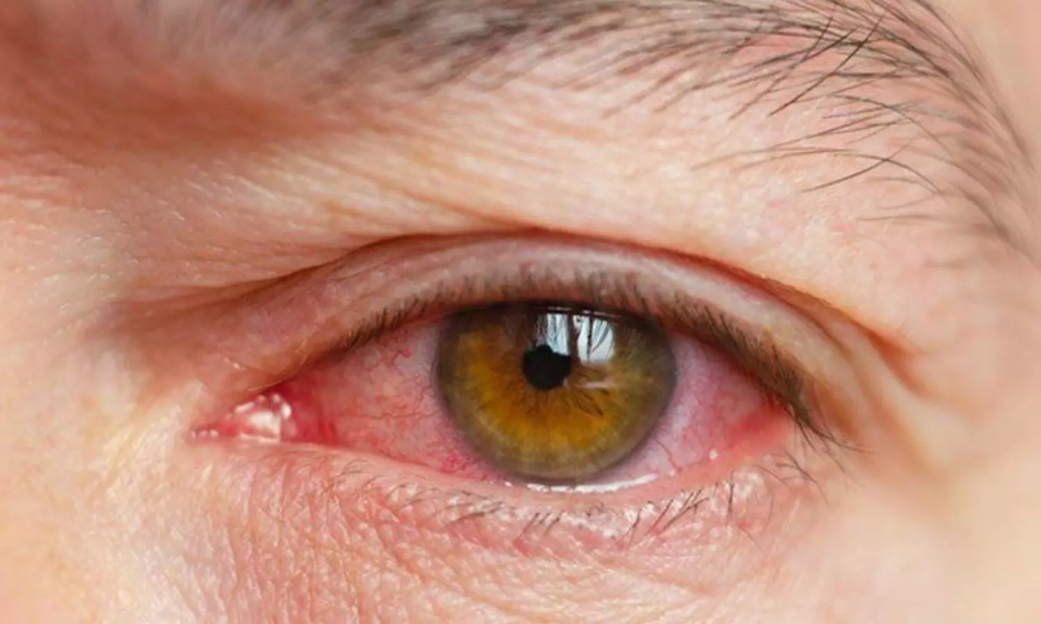
China: A recent meta-analysis published in BMC Ophthalmology has revealed worse sleep quality in individuals with dry eye than in the healthy population. They also reported a higher risk of unhealthy sleep duration such as insufficient sleep or excessive sleepiness, longer sleep latency, and poorer subjective sleep quality.

Dry eye disease (DED) is a common ocular condition affecting millions worldwide, characterized by discomfort, visual disturbances, and potential damage to the ocular surface. Recent research has begun to uncover a potential link between DED and sleep quality, suggesting that the health of our eyes may be intertwined with the quality of our sleep.

Against the above background, Lei Tian, Capital Medical University, Beijing, China, and colleagues aimed to review the association between dry eye and sleep quality systematically.

For this purpose, they searched the online databases for observational studies published before April 2023. Meta-analysis was performed using STAT15 software. The meta-analysis included a total of 21 studies with 419,218 participants.

Based on the review, the researchers reported the following findings:

  • Dry eye subjects had a worse sleep quality than the healthy population, with poorer subjective sleep quality, a higher risk of unhealthy sleep duration such as insufficient sleep or excessive sleep, and longer sleep latency.
  • The Pittsburgh Sleep Quality Index (PSQI) scores of subjects with dry eye were significantly higher than those of the control subjects (WMD = 1.78).
  • The dry eye subjects scored higher than the control subjects in sleep quality, sleep latency, and sleep disturbance in PSQI; there was no difference between the dry eye individuals and control subjects in sleep duration, sleep efficiency, daytime dysfunction, and sleep medication scores.
  • The risk of sleep disorders in the dry eye subjects was significantly higher than that in the non-dry eye subjects (RR = 2.20); the risk of insufficient sleep in the dry eye subjects was higher than that in the control subjects (RR = 3.76), and the prevalence of excessive sleepiness in dry eye subjects was higher than that in the control subjects (RR = 5.53).
  • The ESS scores of the dry eye subjects were significantly higher than those of the control subjects (WMD = 3.02).

The findings indicate that dry eye patients have a lower sleep quality than the healthy population, with longer sleep latency, poorer subjective sleep quality, and a higher risk of unhealthy sleep duration such as insufficient sleep or excessive sleep.

“However, so far, there is insufficient evidence to establish a causal relationship and related mechanisms between dry eye and sleep disorder,” the researchers wrote. “In the future, there will be a need for more large-scale prospective studies to provide more assistance in patient management and treatment.”

Reference:

Gu, Y., Cao, K., Li, A. et al. Association between sleep quality and dry eye disease: a literature review and meta-analysis. BMC Ophthalmol 24, 152 (2024). https://doi.org/10.1186/s12886-024-03416-7

Powered by WPeMatico

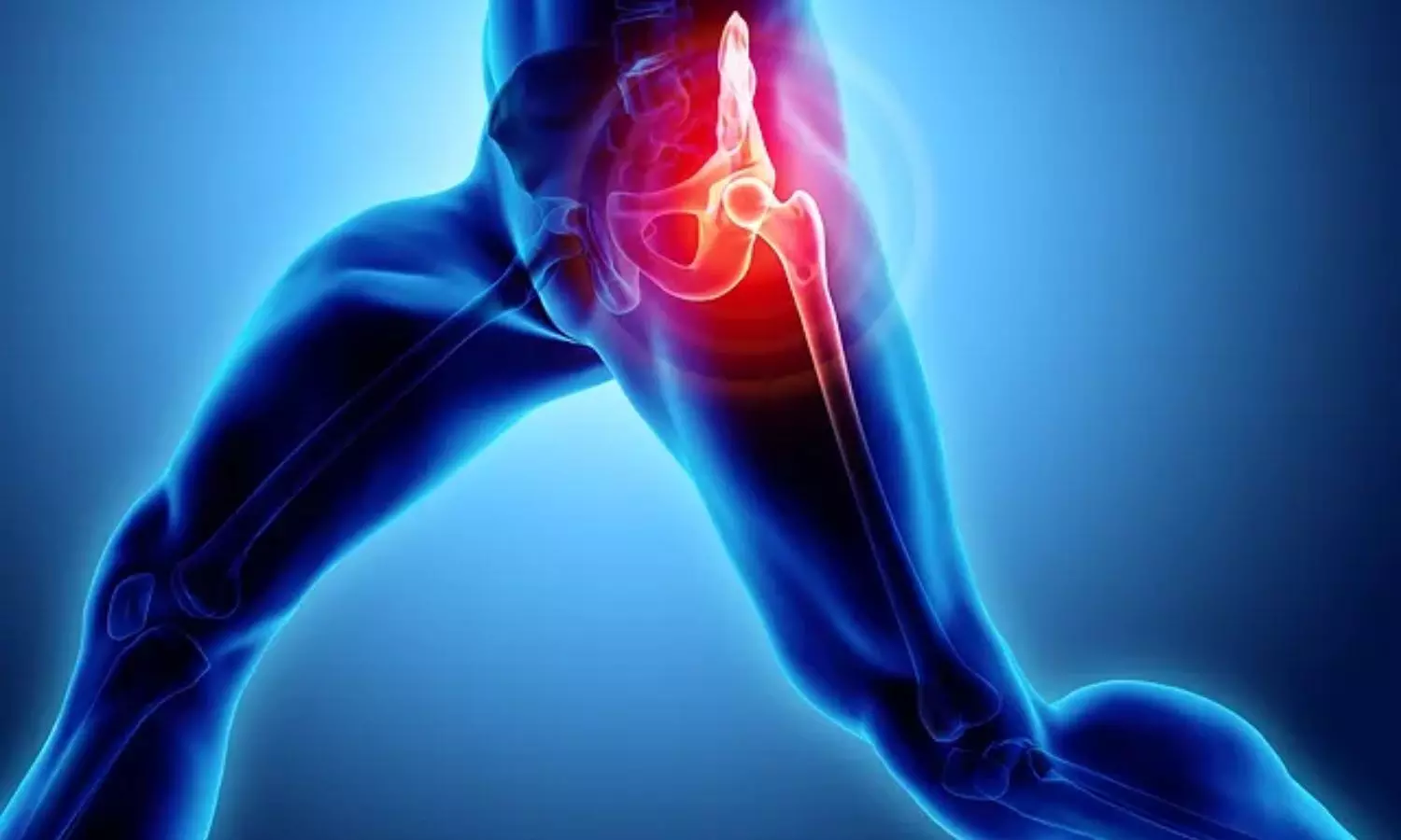
Progressive resistance training as good as neuromuscular exercise for relieving pain in hip OA patients: Study

Denmark: In patients with hip osteoarthritis (OA), progressive resistance training (PRT) is not superior to neuromuscular exercise (NEMEX) for improving hip pain, functional performance, or hip-related quality of life (QoL), a recent study published in Annals of Internal Medicine has revealed.

NEMEX emphasized sensorimotor control & stability, while PRT focused on high-intensity resistance training. The primary outcome, change in the 30-second chair stand test (30s-CST), showed no significant difference between the groups.

Hip osteoarthritis is a prevalent and debilitating condition characterized by stiffness, joint pain, and reduced mobility. Traditional management approaches often include medication and lifestyle modifications. However, emerging research suggests that targeted exercise interventions, such as progressive resistance training or neuromuscular exercise hold significant promise in managing hip OA symptoms and improving overall function. However, randomized controlled trials providing evidence for the optimal exercise type are lacking.

To fill this knowledge gap, Troels Kjeldsen, Næstved-Slagelse-Ringsted Hospitals, Slagelse, Denmark (T.K.), and colleagues aimed to investigate whether PRT is superior to NEMEX for improving functional performance in patients with hip OA.

For this purpose, the researchers conducted a multicenter, cluster-randomized, controlled, parallel-group, assessor-blinded, superiority trial enrolling 160 participants with clinically diagnosed hip OA from 2021 to 2023. They were randomly assigned to PRT (n = 82) or NEMEX (n = 78).

They received twelve weeks of PRT or NEMEX with 2 supervised 60-minute group sessions each week. The PRT intervention comprised of high-intensity resistance training exercises targeting muscles at the hip and knee joints. The NEMEX intervention comprised 10 exercises and emphasized functional stability and sensorimotor control.

The primary outcome was a change in the 30-second chair stand test. Key secondary outcomes included changes in scores on the pain and hip-related QoL subscales of the Hip Disability and Osteoarthritis Outcome Score (HOOS).

Following were the study’s key findings:

  • The mean changes from baseline to 12-week follow-up in the 30s-CST were 1.5 chair stands with PRT and 1.5 chair stands with NEMEX (difference, 0.0 chair stands).
  • For the HOOS pain subscale, mean changes were 8.6 points with PRT and 9.3 points with NEMEX (difference, −0.7 points).
  • For the HOOS QoL subscale, mean changes were 8.0 points with PRT and 5.7 points with NEMEX (difference, 2.3 points).

The main limitation of the study was that the participants and physiotherapists were not blinded.

In conclusion, both progressive resistance training and neuromuscular exercise showed comparable efficacy in improving functional performance, hip pain, and hip-related quality of life over 12 weeks in hip osteoarthritis patients.

Reference:

Kjeldsen T, Skou ST, Dalgas U, Tønning LU, Ingwersen KG, Birch S, Holm PM, Frydendal T, Garval M, Varnum C, Bibby BM, Mechlenburg I. Progressive Resistance Training or Neuromuscular Exercise for Hip Osteoarthritis : A Multicenter Cluster Randomized Controlled Trial. Ann Intern Med. 2024 Apr 9. doi: 10.7326/M23-3225. Epub ahead of print. PMID: 38588540.

Powered by WPeMatico

Physical activity reduces stress-related brain activity and lower CVD risk, suggests study

New research indicates that physical activity lowers cardiovascular disease risk in part by reducing stress-related signaling in the brain.

In the study, which was led by investigators at Massachusetts General Hospital (MGH), a founding member of the Mass General Brigham healthcare system and published in the Journal of the American College of Cardiology, people with stress-related conditions such as depression experienced the most cardiovascular benefits from physical activity.

To assess the mechanisms underlying the psychological and cardiovascular disease benefits of physical activity, Ahmed Tawakol, MD, an investigator and cardiologist in the Cardiovascular Imaging Research Center at Massachusetts General Hospital, and his colleagues analyzed medical records and other information of 50,359 participants from the Mass General Brigham Biobank who completed a physical activity survey.

A subset of 774 participants also underwent brain imaging tests and measurements of stress-related brain activity.

Over a median follow-up of 10 years, 12.9% of participants developed cardiovascular disease. Participants who met physical activity recommendations had a 23% lower risk of developing cardiovascular disease compared with those not meeting these recommendations.

Individuals with higher levels of physical activity also tended to have lower stress-related brain activity. Notably, reductions in stress-associated brain activity were driven by gains in function in the prefrontal cortex, a part of the brain involved in executive function (i.e., decision making, impulse control) and is known to restrain stress centers of the brain. Analyses accounted for other lifestyle variables and risk factors for coronary disease.

Moreover, reductions in stress-related brain signaling partially accounted for physical activity’s cardiovascular benefit.

As an extension of this finding, the researchers found in a cohort of 50,359 participants that the cardiovascular benefit of exercise was substantially greater among participants who would be expected to have higher stress-related brain activity, such as those with pre-existing depression.

“Physical activity was roughly twice as effective in lowering cardiovascular disease risk among those with depression. Effects on the brain’s stress-related activity may explain this novel observation,” says Tawakol, who is the senior author of the study.

“Prospective studies are needed to identify potential mediators and to prove causality. In the meantime, clinicians could convey to patients that physical activity may have important brain effects, which may impart greater cardiovascular benefits among individuals with stress-related syndromes such as depression.”

Effect of Stress-Related Neural Pathways on the Cardiovascular Benefit of Physical Activity, Journal of the American College of Cardiology (2024). DOI: 10.1016/j.jacc.2024.02.02

Powered by WPeMatico

New evidence links passive smoking with dangerous heart rhythm disorder

Exposure to secondhand smoke-even at small amounts-is linked with greater risk of a serious heart rhythm disorder, according to research presented at EHRA 2024, a scientific congress of the European Society of Cardiology (ESC).  The likelihood of atrial fibrillation increased as the duration of passive smoking lengthened.

“The dangers of secondhand smoke were significant regardless of whether individuals were at home, outdoors, or at work, indicating that exposure universally elevates the risk of atrial fibrillation,” said study author Dr. Kyung-Yeon Lee of Seoul National University Hospital, Seoul, Republic of Korea. “We should all make every effort to avoid spending time in smoky environments. The findings should also drive policymakers to further curb smoking in public areas and support smoking cessation programmes to improve public health.”

Atrial fibrillation is the most common heart rhythm disorder worldwide. Symptoms include palpitations, shortness of breath, fatigue, and difficulty sleeping. It is estimated that one in three Europeans will develop the condition during their lifetime. People with atrial fibrillation are five times more likely to have a stroke than their healthy peers. 

It is well established that passive smoking is linked to coronary artery disease and premature death.3-8 However, the links between secondhand smoke and atrial fibrillation have been unclear. This study examined the association between secondhand smoke exposure and the long-term risk of incident atrial fibrillation. The study included adults aged 40 to 69 years who had used the UK National Health Service (NHS) for any reason and were enrolled in the UK Biobank. Current smokers and those with atrial fibrillation at baseline were excluded from the study.

A total of 400,493 adults were included in the analysis. The average age of participants was 56.5 years and 55.2% were women. A touchscreen questionnaire was used to ask participants the number of hours they had been exposed to other people’s smoke in a typical week over the past year at home and in other environments. Participants were categorised into the ‘exposed group’ if they had any contact with secondhand smoke and the ‘non-exposed group’ if they had no contact with secondhand smoke. Some 85,984 (21%) participants had been exposed to secondhand smoke in the previous year, with an average exposure of 2.2 hours per week. During a median follow-up of 12.5 years, atrial fibrillation developed in 23,471 (6%) participants.

The researchers analysed the association between exposure to secondhand smoke and incident atrial fibrillation after adjusting for factors that could potentially affect the relationship, including age, sex, ethnicity, body mass index, daily alcohol consumption, moderate-to-vigorous physical activity, diabetes, high blood pressure, high blood lipids, socioeconomic status, and enrolment centre. The group exposed to secondhand smoke had a 6% higher risk of incident atrial fibrillation during follow-up compared with the non-exposed group after adjusting for the previously mentioned factors (hazard ratio 1.06, 95% confidence interval 1.03–1.10, p <0.001).

A dose-dependent relationship was observed, whereby each increase in the duration of weekly passive smoking was linked with an even greater risk of atrial fibrillation. For example, 7.8 hours of passive smoking per week was associated with an 11% higher likelihood of the heart rhythm disorder compared with no passive smoking. The risk of atrial fibrillation for passive smokers was raised in homes, workplaces, and outside spaces.

Dr. Lee said: “According to our study, once exposed to secondhand smoke, the likelihood of developing atrial fibrillation begins to increase, with the risk escalating significantly as the exposure time lengthens. The finding that passive smoking is harmful not only in enclosed indoor spaces but also outdoor environments underscores the importance of smoking bans to protect public health.” 

Powered by WPeMatico

PET/MRI accurately classifies prostate cancer patients, offers potential to avoid unnecessary biopsies: Study

China: Reston, VA-PET/MRI can improve diagnostic accuracy for prostate cancer patients and help avoid unnecessary biopsies, according to new research published in the April issue of The Journal of Nuclear Medicine.

By applying the PRIMARY scoring system to PET/MRI results, researchers found that more than 80 percent of unnecessary biopsies could be avoided at the expense of missing one in eight clinically significant prostate cancer cases.

The Prostate Imaging Reporting and Data System (PI-RADS) is a five-point scale used to evaluate suspected prostate cancer on MR images. PI-RADS category 3, which presents an unclear suggestion of clinically significant prostate cancer, remains a diagnostic challenge. Although biopsy is recommended under the current guidelines, less than 20 percent of PI-RADS 3 lesions contain clinically significant prostate cancer.

“PI-RADS 3 lesions present a dilemma to both urologists and patients because immediate biopsy could be unnecessary; however, a monitoring strategy could lead to some missed diagnoses of clinically significant prostate cancer,” stated Hongqian Guo, MD, a urologist at Nanjing Drum Tower Hospital at the Affiliated Hospital of Nanjing University Medical School in Nanjing, China. “Hence, specifically ruling out clinically significant prostate cancer among PI-RADS 3 lesions has significant clinical implications.”

In this study, 56 men with PI-RADS 3 lesions underwent 68Ga-PSMA PET/MRI. The five-level PRIMARY system, which is based on a combination of 68Ga-PSMA pattern, localization, and intensity information, was used to report prostate 68Ga-PSMA PET/MRI findings. After imaging, all patients underwent prostate systematic biopsy in combination with targeted biopsy to determine clinically significant prostate cancer.

Among the 56 patients, clinically significant prostate cancer was detected in eight patients (14.3 percent) by biopsy. When a PRIMARY score of at least four was used to make a biopsy decision in men with PI-RADS 3 lesions, 40 of 48 (83.3 percent) participants could have avoided unnecessary biopsies, at the expense of missing 1 in 8 (12.5 percent) of clinically significant prostate cancer cases.

“By demonstrating the additive value of 68Ga-PSMA PET/MRI in classifying PI-RADS 3 lesions, this study provides new insight into the clinical indication for 68Ga-PSMA PET/MRI,” noted Guo. “In the future, PI-RADS 3 patients could be referred for 68Ga-PSMA PET/MRI before prostate biopsy.”

References: Jingyan Shi, Danyan Li, Mengxia Chen, Yao Fu, Shan Peng, Qing Zhang, Jing Liang, Qun Lu, Jiaming Lu, Shuyue Ai, Feng Wang, Xuefeng Qiu and Hongqian Guo Journal of Nuclear Medicine April 2024, 65 (4) 555-559; DOI: https://doi.org/10.2967/jnumed.123.266742

Powered by WPeMatico