Erenumab: A Potential Breakthrough in Rosacea Management

Denmark: A nonrandomized controlled trial showed the importance of the calcitonin gene-related peptide (CGRP) pathway in the pathophysiology of erythema and flushing in rosacea. The findings suggested that calcitonin gene-related peptide receptor inhibition holds potential in treating rosacea-associated erythema and flushing.

“The study comprising 30 individuals with rosacea-associated flushing and erythema found that subcutaneous injections of monoclonal antibodies against the calcitonin gene-related peptide receptor administered every four weeks for 12 weeks significantly decreased days with erythema and flushing by weeks 9 through 12 compared with baseline,” the researchers reported. The findings were published online in JAMA Dermatology on April 17, 2024. 

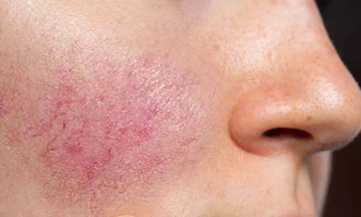
Rosacea is characterized by facial redness, visible blood vessels, and in some cases, papules and pustules. Persistent erythema and flushing are hallmark features, often resistant to conventional therapies. This affects patients’ physical appearance and psychological well-being, highlighting the need for effective treatment options.

While several treatment modalities exist, managing these symptoms remains a challenge. However, recent research suggests a promising avenue with erenumab use, a monoclonal antibody targeting CGRP. Nita K. F. Wienholtz, Copenhagen University Hospital-Rigshospitalet, Copenhagen, Denmark, and colleagues aimed to examine the tolerability, effectiveness, and safety of erenumab, an anti–CGRP-receptor monoclonal antibody, for the treatment of rosacea-associated erythema and flushing.

For this purpose, the researchers conducted a single-center, open-label, single-group, nonrandomized controlled trial between 2020 and 2021. Eligible participants included patients with rosacea with at least 15 days of either moderate to severe erythema and/or moderate to extreme flushing. Throughout the study period, no concomitant rosacea treatment was allowed.

Participants received 140 mg of erenumab subcutaneously every four weeks for 12 weeks. At week 20, a safety follow-up was performed. A total of 30 participants (mean age, 38.8 years; 77% females were included, of whom 27 completed the 12-week study.

The primary outcome was a mean change in the number of days with moderate to extreme flushing during weeks 9 through 12, compared with the 4-week run-in period (baseline). The secondary outcome was a mean change in the number of days with moderate to severe erythema. Adverse events were recorded for patients who received at least one dose of erenumab.

Following were the study’s key findings:

  • There was a reduction in the mean number of days with moderate to extreme flushing by −6.9 days from 23.6 days at baseline.
  • There was a reduction in the mean number of days with moderate to severe erythema by −8.1 days from 15.2 days at baseline.
  • Adverse events included transient mild to moderate constipation (33% of participants), transient worsening of flushing (13% of participants), bloating (10% of participants), and upper respiratory tract infections (10% of participants), consistent with previous data.
  • One participant discontinued the study due to a severe adverse event (hospital admission due to gallstones deemed unrelated to the study), and 2 participants withdrew consent due to lack of time.

In conclusion, the findings suggest that erenumab might be effective in reducing rosacea-associated flushing and chronic erythema (participants generally tolerated the treatment well, which was consistent with previous data). The study also showed that CGRP-receptor inhibition holds potential in treating flushing and erythema associated with rosacea.

“Larger randomized clinical trials are needed to confirm this finding,” the researchers wrote.

Reference:

Wienholtz NKF, Christensen CE, Do TP, et al. Erenumab for Treatment of Persistent Erythema and Flushing in Rosacea: A Nonrandomized Controlled Trial. JAMA Dermatol. Published online April 17, 2024. doi:10.1001/jamadermatol.2024.0408

Powered by WPeMatico

Advancements in detecting interstitial lung disease in rheumatoid arthritis: Thoracic ultrasound takes the lead

Denmark: Systematic screening for respiratory symptoms combined with thoracic ultrasound (TUS) can reduce the diagnostic delay of interstitial lung disease (ILD) in rheumatoid arthritis (RA), a recent study published in Arthritis Care & Research has shown.

“To our knowledge, this prospective study is the first to use respiratory symptoms in rheumatoid arthritis as inclusion criteria,” the researchers wrote. TUS offers several advantages, including its non-invasive nature, absence of radiation exposure, and cost-effectiveness.

Rheumatoid arthritis is a chronic autoimmune disorder primarily affecting the joints, but it can also have systemic manifestations, including ILD. Early detection of ILD in RA is crucial for timely intervention and management. Traditional diagnostic methods, such as chest X-rays and high-resolution computed tomography (HRCT), have limitations, including radiation exposure and cost. However, a recent study suggests that thoracic ultrasound could revolutionize the detection of ILD in RA patients.

Against the above background, Bjørk K. Sofíudóttir, PUlmo-RhEuma Frontline Center (PURE), Department of Rheumatology Odense University Hospital, Odense, Denmark, and colleagues aimed to determine the diagnostic accuracy of thoracic ultrasound for detecting interstitial lung disease in rheumatoid arthritis with respiratory symptoms.

For this purpose, the researchers systematically screened individuals with RA visiting Rheumatological outpatient clinics in the Region of Southern Denmark for cough, dyspnoea, recurrent pneumonia, prior severe pneumonia, or a chest X-ray indicating interstitial abnormalities. Eighty participants with a positive screening were consecutively included.

Individuals were not eligible if they had a chest HRCT <12 months or were already diagnosed with ILD. A blinded TUS expert evaluated thoracic ultrasound, and TUS was registered as positive for ILD if ≥10 B-lines or bilateral thickened and fragmented pleura were present.

The study’s primary outcomes were TUS’s specificity, sensitivity, negative predictive value (NPV), and positive predictive value (PPV). HRCT was assessed by an ILD-specialised thoracic radiologist, followed by a multidisciplinary team discussion. The accepted window of HRCT was <30 days after TUS was performed.

The key findings of the study were as follows:

  • 77 participants received HRCT <30 days after TUS, and 30% were diagnosed with ILD.
  • TUS had a sensitivity of 82.6% and a specificity of 51.9%, corresponding to a PPV of 42.2% and an NPV of 87.5%.

In conclusion, this prospective study is the first to use respiratory symptoms in rheumatoid arthritis as inclusion criteria. Systematic screening for respiratory symptoms combined with thoracic ultrasound can reduce the diagnostic delay of interstitial lung disease in rheumatoid arthritis.

Reference:

Sofíudóttir, B. K., Harders, S., Laursen, C. B., Lage-Hansen, P. R., Nielsen, S. M., Just, S. A., Christensen, R., Davidsen, J. R., & Ellingsen, T. Detection of Interstitial Lung Disease in Rheumatoid Arthritis by Thoracic Ultrasound. A Diagnostic Test Accuracy study. Arthritis Care & Research. https://doi.org/10.1002/acr.25351

Powered by WPeMatico

Admission blood sugar independent predictor of coagulopathy in multiple Trauma Patients, finds study

Admission blood sugar independent predictor of coagulopathy finds study published in the European Journal of Trauma and Emergency Surgery

Coagulopathy is prevalent in multiple trauma patients and worsens bleeding complications, leading to higher morbidity and mortality rates. Hyperglycemia upon admission predicts hemorrhagic shock and mortality in severely injured patients. This study aimed to assess admission glucose levels as an independent prognostic factor for coagulopathy in multiple injured patients. This retrospective cohort study observed multiple trauma patients treated at a level I trauma center between January 1, 2005, and December 31, 2020. Coagulopathy was defined as an international normalized ratio (INR) > 1.4 and/or activated thromboplastin time (APTT) > 40 s. Analysis of variance compared clinical and laboratory parameters of patients with and without coagulopathy. Receiver-operating-characteristic (ROC) and multivariate logistic regression analyses identified risk factors associated with coagulopathy. RESULTS: The study included 913 patients, of whom 188 (20%) had coagulopathy at admission. Coagulopathy patients had higher mortality than those without (26% vs. 5.0%, p < 0.001). The mean glucose level in coagulopathy patients was 10.09 mmol/L, significantly higher than 7.97 mmol/L in non-coagulopathy patients (p < 0.001). Admission glucose showed an area under the curve (AUC) of 0.64 (95% CI [0.59-0.69], p < 0.001) with an optimal cut-off point of 12.35 mmol/L. After adjusting for other factors, patients with high admission glucose had a 1.99-fold risk of developing coagulopathy (95% CI 1.07-3.60). Other laboratory parameters associated with coagulopathy included haemoglobin, bicarbonate (HCO3), and lactate levels. This study emphasizes the significance of admission blood glucose as an independent predictor of coagulopathy. Monitoring hyperglycemia can aid in identifying high-risk patients.

Reference:

Mayor, Jorge, et al. “Do Admission Glucose Levels Independently Predict Coagulopathy in Multiple Trauma Patients? A Retrospective Cohort Analysis.” European Journal of Trauma and Emergency Surgery : Official Publication of the European Trauma Society, 2024.

Keywords:

Admission blood sugar, independent predictor, coagulopathy, Mayor, Jorge, European Journal of Trauma and Emergency Surgery

Powered by WPeMatico

Subconjunctival injection of triamcinolone acetonide may prevent complications after phacoemulsification: Study

In recent research conducted by Kaiser Permanente Northern California, a single injection of triamcinolone acetonide (TA) was identified to be more effective and safer than traditional topical prednisolone acetate (PA) in preventing complications following cataract surgery. The findings were published in the Ophthalmology journal.

This retrospective study spanned from 2018 to 2021 and included 69,832 patient-eyes which underwent phacoemulsification to remove cataract. The patients were categorized into groups receiving either topical PA with or without a nonsteroidal anti-inflammatory drug (NSAID) or a subconjunctival injection of TA, at varying doses and concentrations. The effectiveness of these treatments was evaluated based on the incidence of postoperative macular edema (ME) and iritis and the safety was assessed via the occurrence of glaucoma-related events within a year following the surgery.

The results highlighted that the high-dose TA (10 mg/mL) group showed a statistically significant reduction in the risk of developing postoperative ME when compared to the group treated with topical PA. The odds ratio (OR) was 0.64 which suggests a 36% lower risk and provides promising insight into the potential benefits of opting for an injection over topical solutions. Further, the combination of PA and NSAID did not show a statistically significant benefit in reducing ME.

This study observed trends in the reduction of postoperative iritis in patients who were treated with high concentration TA (40 mg/mL), although these findings did not reach statistical significance. The low dose TA (10 mg/mL) group experienced markedly lower odds of encountering glaucoma-related events when compared to the topical PA group by indicating a safer profile for this particular injection dosage.

Higher doses of TA at 40 mg/mL were associated with an elevated risk of glaucoma-related events which suggesting that while effective, the higher concentrations of the drug might increase the risk of adverse effects. This underlines the importance of dose management in the use of injectable steroids to balance efficacy and safety.

This  findings are significant as they could influence future protocols in the management of inflammation and prevention of complications post-cataract surgery. The subconjunctival injections of TA with optimized dosages lower risks of ME and iritis and might become a preferred strategy over the conventional topical treatments. This offers a simpler and potentially more effective prophylactic option for the patients who undergo cataract surgery every year.

Reference:

Shorstein, N. H., McCabe, S. E., Alavi, M., Kwan, M. L., & Chandra, N. S. (2024). Triamcinolone Acetonide Subconjunctival Injection as Stand-alone Inflammation Prophylaxis after Phacoemulsification Cataract Surgery. In Ophthalmology. Elsevier BV. https://doi.org/10.1016/j.ophtha.2024.03.025

Powered by WPeMatico

FDA fast tracks enibarcimab for the treatment of septic shock

 It is believed that modulation of ADM pathway may improve outcomes in patients with septic shock.

The Food and Drug Administration has granted Fast Track designation to enibarcimab for management of septic shock. Enibarcimab is a first-in-class non-neutralizing monoclonal antibody that binds to adrenomedullin (ADM), a vasoprotective hormone.

AdrenoMed is now preparing a confirmatory Phase IIb/III clinical trial to confirm the reduced septic shock mortality under enibarcimab treatment employing a precision medicine approach.

AdrenoMed’s CEO Dr. Richard Jones commented: “We are very pleased that enibarcimab has received Fast Track designation from the FDA, recognizing its potential as an innovative biomarker-guided treatment against septic shock to fill the unmet medical need in this very serious condition with a high death toll. This is a great confirmation of AdrenoMed’s efforts to reduce patient heterogeneity and develop effective treatments by bringing precision medicine also to intensive care units.”

Enibarcimab is a non-blocking antibody binding to the vasoprotective hormone adrenomedullin. Applying AdrenoMed’s precision medicine approach, enibarcimab treatment of patients with septic shock resulted in improved organ dysfunction and a relevant reduction of day 28 all-cause mortality from 24% to 8% in the patient population defined by the two biomarkers adrenomedullin and DPP3 in the AdrenOSS-2 trial.

With a mortality rate of 20-30% for sepsis and 30%-50% for septic shock in developed countries, sepsis represents an enormous public health burden and is responsible for almost 20% of all deaths worldwide.

Fast Track is a process designed by the FDA to facilitate the development of promising drugs for the treatment of serious conditions that fill an unmet medical need and to accelerate review by the regulatory authority, aimed at getting important new drugs to the patient earlier. A drug that receives Fast Track designation is eligible for more frequent meetings with FDA to discuss development plans for the drug regarding collection of data needed to support its approval; more frequent communication about such things as the design of the proposed clinical trials and use of biomarkers; eligibility for Accelerated Approval and Priority Review, if relevant criteria are met; and Rolling Review.

Dr. Stephan Witte, CMO of AdrenoMed, said: “We are very confident that the use of enibarcimab in combination with two biomarkers, Adrenomedullin (bio-ADM) and circulating dipeptidyl peptidase 3 (cDPP3), holds the promise to become the first effective targeted treatment against septic shock. With AdrenoMed’s biomarker-guided approach it is possible to clearly define the patient population benefitting most from enibarcimab, resulting in a more pronounced treatment effect and leading to improved mortality in septic shock.”

This was the conclusion also drawn by Prof. Peter Pickkers from the Department of Intensive Care Medicine and Radboud Center for Infectious Diseases (RCI), Nijmegen, Netherlands, in a poster session during the recent ISICEM 2024, which took place in Brussels, Belgium, from March 19 – 22, 2024, where he presented previously unpublished data from a prespecified subgroup of the AdrenOSS-2 trial.

About AdrenOSS-2

The double-blind, randomized, placebo-controlled, biomarker-guided Phase II trial AdrenOSS-2 (n = 301) had included a prespecified analysis of the role of cDPP3 as a second biomarker (next to ADM) to exclude patients who are unlikely to respond to enibarcimab treatment.

DPP3 is a cytosolic enzyme involved in the degradation of various cardiovascular and endorphin mediators. High levels of cDPP3 indicate a high risk of organ dysfunction and mortality. This pathway is mechanistically independent from the loss of vascular integrity, which is known to be the main driver of mortality in septic shock and is indicated by elevated plasma levels of ADM (>70 pg/mL). Consequently, the aim of additional analyses was to investigate the impact of different cDPP3 levels on the treatment effect (28-day all-cause mortality) and to identify a suitable cut-off level.

It was shown that the effect of enibarcimab on mortality improves with lower cDPP3 values and that the patients with elevated ADM, but non-elevated baseline cDPP3 (below the upper normal range, ≤ 30-50 ng/mL), benefitted the most from enibarcimab treatment. At 28 days, this patient subgroup had a statistically significant >60% relative reduction in mortality compared to placebo.

Powered by WPeMatico

New Care Approach Lowers Opioid Use and Hospital Stay in Infants with opioid Withdrawal Syndrome: Study

A recent study published in the Journal of American Medical Association demonstrated that a novel care method, the eat, sleep, console (ESC) approach, substantially lowers opioid exposure and the length of hospital stays for infants with neonatal opioid withdrawal syndrome (NOWS) when compared to the traditional care practices.

The study was conducted from September 2020 to March 2022 and included a total of 463 infants across 26 U.S. hospitals as part of the ESC-NOW clinical trial. These infants were born at 36 weeks of gestation or later with evidence of antenatal opioid exposure and were pharmacologically treated for NOWS under two different care protocols, which is the ESC care approach and the usual care using the Finnegan scoring tool.

The results revealed that infants managed with the ESC care approach had significantly lower total opioid exposure, with a mean difference of 4.1 morphine milligram equivalents per kilogram (MME/kg) when compared to the individuals who receive usual care. Also, infants in the ESC group received an average of 4.8 MME/kg, whereas the infants in the usual care group received 8.9 MME/kg.

This study found that initiation of pharmacologic treatment was delayed by an average of 22.4 hours in the ESC group that allowed more time for non-pharmacological interventions and assessments. Also, there was no significant increase in the peak opioid dose required by suggesting that the ESC approach does not compromise the efficacy of the treatment when pharmacological intervention is necessary.

One of the most significant findings was the reduction in both the length of opioid treatment and total length of hospital stay. Infants in the ESC care group completed their treatment 6.3 days earlier than the infants in the usual care group and were discharged from the hospital an average of 6.2 days earlier. This not only suggests better outcomes for the infants but also indicates potential cost savings and more efficient use of hospital resources. Overall, the ESC method offers a more compassionate and sustainable option for treating vulnerable newborns exposed to opioids in utero by focusing on supportive care measures that allow for natural parental involvement and less reliance on medications.

Source:

Devlin, L. A., Hu, Z., Merhar, S. L., Ounpraseuth, S. T., Simon, A. E., Lee, J. Y., Das, A., Crawford, M. M., Greenberg, R. G., Smith, P. B., Higgins, R. D., Walsh, M. C., Rice, W., Paul, D. A., Maxwell, J. R., Fung, C. M., Wright, T., Ross, J., … McAllister, J. M. (2024). Influence of Eat, Sleep, and Console on Infants Pharmacologically Treated for Opioid Withdrawal. In JAMA Pediatrics. American Medical Association (AMA). https://doi.org/10.1001/jamapediatrics.2024.0544

Powered by WPeMatico

Full pulpotomy bests partial pulpotomy for managing of spontaneous symptomatic pulpitis: Study

Full pulpotomy bests partial pulpotomy for managing spontaneous symptomatic pulpitis suggests a study published in the Journal of Endodontics.

The aim of this study was to compare the outcome and prognostic factors for partial and full pulpotomy in the management of mature teeth with spontaneous symptomatic pulpitis. The study was a parallel double blind randomized clinical trial, 200 carious mature permanent teeth with spontaneous symptomatic pulpitis were randomized using a block randomization technique to either partial pulpotomy (n=99) or full pulpotomy (n=101). Intraoperative assessment of the pulp under magnification was done, hemostasis was achieved with 2.5% NaOCl moist pellet, and NeoPUTTY was the pulpotomy material. Preoperative pain levels were recorded and revaluated after 1 week. Clinical and radiographic evaluation was done after 6 and 12 months. Data were analyzed using Chi-square, Wilcoxon rank tests and regression analysis. Results: At 1week immediate failure occurred in 4 cases in partial pulpotomy, and 196/200 subjects reported pain relief and were satisfied with the treatment with no significant difference. At 6 months, 6 teeth failed in partial pulpotomy and 1 tooth in full pulpotomy, with a higher success rate for full pulpotomy (98.96 vs 89.69, p= 0.003). At 12 months the recall rate was 98% (96/200). Full pulpotomy was more successful than partial pulpotomy (98.98% (98/99), vs 84.53% (82/97), p< 0.001). Multivariate analysis revealed that the odds of success for full pulpotomy were 13.6 times higher than partial pulpotomy. Increased age and higher time to hemostasis were significantly associated with decreased odds of success. Full pulpotomy has higher success rate than partial pulpotomy in the management of spontaneous symptomatic pulpitis. Hemostasis within 4 minutes in partial pulpotomy can be set as cutoff point beyond which further tissue removal is indicated.

Reference:

Taha NA, Albakri SW. Outcome and prognostic factors for partial and full pulpotomy in the management of spontaneous symptomatic pulpitis in carious mature permanent teeth: A randomized clinical trial. J Endod. 2024 Apr 5:S0099-2399(24)00224-3. doi: 10.1016/j.joen.2024.03.012. Epub ahead of print. PMID: 38583758.

Keywords:

Full pulpotomy, bests, partial pulpotomy, managing, spontaneous, symptomatic pulpitis, study, Taha NA, Albakri SW, age; bleeding time; full pulpotomy; outcome; partial pulpotomy; spontaneous symptomatic pulpitis.

Powered by WPeMatico

Time-restricted fasting may effect weight loss and improve cardiometabolic function: Study

China: A recent meta-analysis published in BMC Cardiovascular Disorders has shown the potential of time-restricted fasting (TRF) as a treatment strategy for total body weight reduction by targeting adipose tissue with potential improvements in cardiometabolic function.

The study suggests that time-restricted fasting interventions can effectively reduce fat mass and body weight while retaining lean body mass; indicating that subjects lost fat rather than muscle and water.

According to the study, the results showed slight differences when the calculation method was modified, for example, by introducing the Hartung-Knapp adjustment. Although the reduction in body fat percentage remained statistically significant, the researchers noted no significant difference in weight loss. However, a significant decrease in systolic blood pressure was observed in this context.

Time-restricted fasting, an eating pattern that limits food intake to a specific time window each day, has garnered attention for its potential health benefits. In recent years, researchers have conducted numerous studies to investigate its effects on various aspects of health, including cardiovascular disease (CVD) risk factors.

A meta-analysis pooling the results of these studies aimed to investigate the impact of TRF on key risk factors, including glucose metabolism, body weight, blood pressure, and lipid profile.

Juntao Kan, Nutrilite Health Institute, Shanghai, China, and colleagues conducted a systematic search in five databases for relevant studies up to January 2023. Twelve studies were eligible for analysis after applying inclusion criteria. Quality assessment was conducted using the ROBINS-I and ROB-2.0 tools. The risk of bias was mapped using Revman 5.3, and data analysis included Hartung-Knapp adjustment using R 4.2.2.

“The group that underwent the TRF intervention exhibited a significant decrease in body weight (SMD: -0.22) and fat mass (SMD: -0.19), while maintaining lean mass (SMD: -0.09),” the researchers reported.

In conclusion, the research showed that time-restricted fasting effectively reduces body weight and fat mass without affecting lean body mass, indicating a preference for fat loss.

This finding highlights the potential of TRF as a strategy for improving body composition. However, there is a need for further clinical trials to explore the underlying mechanisms and broader impacts of this intervention, particularly with cardiovascular health.

“Future research should aim to clarify gender-specific responses to TRF, considering differences in adipose tissue characteristics and metabolism,” the researchers wrote. “This will enhance our understanding of the role of time-restricted fasting in promoting metabolic health.”

Reference:

Qiu, Z., Huang, E.Y.Z., Li, Y. et al. Beneficial effects of time-restricted fasting on cardiovascular disease risk factors: a meta-analysis. BMC Cardiovasc Disord 24, 210 (2024). https://doi.org/10.1186/s12872-024-03863-6

Powered by WPeMatico

Photon-counting CT improves coronary artery disease assessment, reveals study

Ultrahigh-spatial-resolution photon-counting detector CT improved assessment of coronary artery disease (CAD), allowing for reclassification to a lower disease category in 54% of patients, according to a new study published  in Radiology, a journal of the Radiological Society of North America (RSNA). The technology has the potential to improve patient management and reduce unnecessary interventions.

Coronary CT angiography is a first-line test in the assessment of coronary artery disease. However, its diagnostic value is limited in patients with severe calcifications, or calcium buildup in the plaque of the coronary arteries.

Ultrahigh-spatial-resolution photon-counting detector CT (PCD-CT) improves image quality compared to conventional CT. Additionally, it provides better spatial resolution, or the ability to differentiate two adjacent structures as being distinct from one another.

“Our study provides a glimpse into the potential impact of performing coronary CT angiography using ultrahigh spatial resolution technology on risk reclassification and recommended downstream testing,” said study co-author Tilman Emrich, M.D., attending radiologist at the University Medical Center Mainz in Germany, and assistant professor of radiology at the Medical University of South Carolina in Charleston.

For the study, researchers evaluated coronary stenoses, or narrowing in the coronary arteries, in a vessel phantom (in-vitro) containing two different stenosis grades (25%, 50%), and retrospectively in 114 patients (in-vivo) who underwent ultrahigh-spatial-resolution cardiac PCD-CT for the evaluation of coronary artery disease. In-vitro values were compared to the phantom’s manufacturer specifications, and patient results were assessed regarding effects on coronary artery disease reporting and data system reclassification (CAD-RADS).

“The study used a combination of artificial vessel models and real-world patient data,” Dr. Emrich said. “It simulated three types of reconstructions from a single PCD-CT scan, resembling conventional CT, high-resolution, and ultrahigh-spatial-resolution scans. Observers evaluated the severity of stenosis and generated CAD-RADS classifications, guiding further patient management decisions.”

In-vitro results demonstrated a reduced overestimation of the stenosis by ultrahigh-spatial-resolution scans by reducing the adverse effects of the calcifications on the image.

Results from the patients with suspected or diagnosed coronary artery disease confirmed a lower median degree of stenosis for calcified plaques (29% vs. 42%) with ultrahigh-spatial-resolution PCD-CT compared to standard CT. Ultrahigh-spatial-resolution often led to patients being reclassified to a lower CAD-RADS category. Of the 114 patients, 54% were given a lower CAD-RADS classification than they were originally assigned. The researchers found in-vitro quantification of the 193 coronary CT angiography-based stenoses was also more accurate using ultrahigh-spatial-resolution than standard resolution.

“We found that ultrahigh-spatial-resolution reconstructions resulted in significant changes in recommendations for over 50% of patients,” Dr. Emrich said. “The impact was particularly notable in cases with calcified plaques, where ultrahigh-spatial-resolution reduced the overestimation of stenosis.”

Dr. Emrich explained that ultrahigh-spatial-resolution may address the current limitations of conventional cardiac CT angiography by reducing the overestimation of stenosis due to calcium blooming, an effect which can cause small, high-density structures-such as calcifications-to appear larger than their true size.

“This could significantly alter recommendations for downstream testing, potentially leading to a reduction of unnecessary procedures (and their potential complications) and reduced healthcare costs,” he said.

No substantial benefits of ultrahigh-spatial-resolution were observed for mixed and non-calcified plaques.

“It is important to note that these findings are from a simulation study, and further validation is needed in real-world comparisons,” Dr. Emrich said.

Reference:

Moritz C. Halfmann, Stefanie Bockius, Tilman Emrich , Michaela Hell, U. Joseph Schoepf, Gerald S. Laux, Larissa Kavermann, Dirk Graafen, Tomasso Gori, Yang Yang, Roman Klöckner, Ultrahigh-Spatial-Resolution Photon-counting Detector CT Angiography of Coronary Artery Disease for Stenosis Assessment, Radiology, https://doi.org/10.1148/radiol.231956

Powered by WPeMatico

Healthy diet lowers heart disease risk in breast cancer survivors

A new paper in JNCI Cancer Spectrum, published by Oxford University Press, finds that following a healthy diet lowers the risk of cardiovascular disease in breast cancer survivors.

Cardiovascular disease is the top non-breast cancer related cause of death in women with breast cancer. There are more than 3.8 million female breast cancer survivors in the United States. These women are at higher risk for cardiovascular disease than women who have not had breast cancer. This is likely due to the cardiotoxic effects of breast cancer treatment, as well as common risk factors for both breast cancer and cardiovascular disease, such as aging, lack of exercise, and smoking. Dietary guidance for breast cancer survivors is limited and until recently has been based primarily on research related to cancer prevention.

Researchers used data from the Pathways Study, a prospective cohort study of women diagnosed with invasive breast cancer, to examine associations between diet quality and cardiovascular-related events. The analysis included 3,415 women diagnosed with invasive breast cancer at Kaiser Permanente Northern California between 2005 and 2013 and monitored through 2021.

To assess diet quality, researchers used a scoring system based on the Dietary Approaches to Stop Hypertension (DASH) diet which was developed in the 1990s to manage and treat hypertension. The diet emphasizes fruits, vegetables, whole grains, lean protein, and low-fat dairy. It also limits sodium, red and processed meats, and sugar sweetened beverages. The diet is similar to that recommended by the American Cancer Society, but also encourages consumption of low-fat dairy and nuts, and discourages sodium. The study evaluated heart health tied to these two diets as well as a plant-based diet, the 2020 Healthy Eating Index, and the alternate Mediterranean diet.

The researchers found that women whose diets were most similar to DASH at the time of their breast cancer diagnosis had a 47% lower risk of heart failure, a 23% lower risk of arrhythmia, a 23% lower risk of cardiac arrest, a 21% lower risk of valvular heart disease, and a 25% lower risk of venous thromboembolic disease than the women whose diets were least aligned with DASH.

In a closer examination the researchers found that higher consumption of low-fat dairy reduced the risk for cardiovascular disease-related death, after adjusting for all other food groups. They also found that the relationship between DASH and cardiovascular disease appeared to be modified by the type of chemotherapy treatment a woman received. For example, women whose treatment included an anthracycline and had diets closely aligned with the diet had a lower risk of cardiovascular disease than women least aligned with DASH, a relationship that was not apparent among women on other types of chemotherapy regimens.

“Our findings suggest that we need to begin talking to breast cancer survivors about the potential heart benefits of the DASH diet,” said the paper’s lead author, Isaac J. Ergas, PhD, a staff scientist at the Kaiser Permanente Division of Research. “We know that breast cancer survivors have an elevated risk for cardiovascular disease, and the diet might be able to help improve the overall health of this population.”

Reference:

Isaac J Ergas, Richard K Cheng, Janise M Roh, Lawrence H Kushi, Jacob K Kresovich, Carlos Iribarren, Mai Nguyen-Huynh, Jamal S Rana, Eileen Rillamas-Sun, Cecile A Laurent, Valerie S Lee, Charles P Quesenberry, Heather Greenlee, Marilyn L Kwan, Diet quality and cardiovascular disease risk among breast cancer survivors in the Pathways Study, JNCI Cancer Spectrum, Volume 8, Issue 2, April 2024, pkae013, https://doi.org/10.1093/jncics/pkae013.

Powered by WPeMatico