IV iron therapy effectively improves anemia in pediatric patients with IBD: Study

A recent study published in the World Journal of Clinical Pediatrics unveiled the effectiveness and safety of intravenous (IV) iron therapy for treating iron deficiency anemia (IDA) in children with inflammatory bowel disease (IBD). Iron deficiency anemia is a common complication in pediatric patients with IBD, primarily due to chronic inflammation that disrupts iron absorption and metabolism. Despite the recognized need for aggressive treatment, there has been hesitance to prescribe IV iron, largely due to concerns over potential adverse reactions. The study by Krishanth Manokaran and team addressed these concerns by evaluating the safety and efficacy of IV iron therapy in this group.

This study spanned from September 2017 to December 2019 and included a cohort of 236 pediatric patients by underlining a major step forward in pediatric IBD management. Out of the total 236 patients admitted during the study period, 92 met the criteria for IDA. The treatment modalities were split where 57 patients received IV iron, 17 patients were given oral iron supplements and 18 patients were discharged before any iron therapy could be administered. The findings observed that patients receiving IV iron showed a significant improvement in their health outcomes.

These patients experienced an increase in mean hemoglobin (Hb) levels by 1.9 g/dL at their first outpatient follow-up which is markedly higher than the increases noticed in the individuals who received oral iron or no iron treatment, which were both only 0.8 g/dL. This study also highlighted the safety of IV iron therapy with only one reported adverse reaction among the 57 patients treated through this method which translated to an incidence rate of just 1.8%. This low rate of adverse events suggests that IV iron is both a safe and more effective alternative to oral iron supplements for managing IDA in pediatric IBD patients.

These results provide empirical support for clinicians who consider IV iron therapy for young patients. Improved hemoglobin and iron levels significantly improve the quality of life and overall health outcomes for children suffering from this chronic condition. Overall, the findings from this study pave the way for more standardized use of IV iron in pediatric IBD treatment to reduce the reluctance among healthcare providers to utilize this treatment.

Source:

Manokaran, K., Spaan, J., Cataldo, G., Lyons, C., Mitchell, P. D., Sare, T., Zimmerman, L. A., & Rufo, P. A. (2024). Inpatient management of iron deficiency anemia in pediatric patients with inflammatory bowel disease: A single center experience. In World Journal of Clinical Pediatrics (Vol. 13, Issue 1). Baishideng Publishing Group Inc. https://doi.org/10.5409/wjcp.v13.i1.89318

Powered by WPeMatico

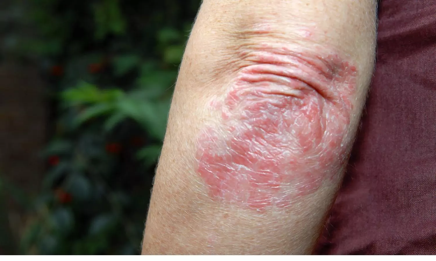
Unlocking Insights: The real-world BrIDGE study on brodalumab effectiveness in psoriasis patients

Greece: A large-scale study evaluating brodalumab for moderate-to-severe plaque psoriasis revealed significant improvements in patient outcomes.

The study of 638 patients showed that brodalumab led to substantial reductions in the Psoriasis Area and Severity Index (PASI) scores, with 74.0% achieving PASI ≤3 by week 12. In particular, 54.1% of patients achieved complete clearance (PGA0). The findings were published online in the Journal of the European Academy of Dermatology and Venereology on February 03, 2024.

In the psoriasis treatment landscape, it is crucial to understand the real-world effectiveness of medications for healthcare professionals to make informed decisions. The BrIDGE study in Greece offers valuable insights into brodalumab effectiveness in patients with moderate-to-severe plaque psoriasis.

Brodalumab, a human monoclonal antibody targeting the interleukin-17 receptor, has shown promising results in clinical trials for treating psoriasis. However, real-world data is essential to confirm its efficacy and safety in diverse patient populations. Therefore, Dimitrios Rigopoulos, 1st Department of Dermatology-Venereology, University of Athens, Athens, Greece, and colleagues conducted an observational, prospective, single-cohort, multicentre study (BrIDGE) that recruited patients with moderate-to-severe plaque psoriasis in Greece.

The primary objective was to assess the proportion of patients who achieved PASI 100 after 24 weeks. Other endpoints included: the short-term response [PASI75/90/100 and static Physician’s Global Assessment (sPGA) 0/1] to brodalumab at 12–16 weeks, the maintenance of PASI90/100 through to 104 weeks, and the time to complete clearance. Moreover, the researchers explored the change in the quality of life [Dermatology Life Quality Index (DLQI) 0/1] and adherence to brodalumab.

The researchers recruited two hundred patients initiating treatment with or switching to brodalumab. Analyses were conducted using the observed data and three imputation approaches were applied for the missing data.

The study led to the following findings:

  • Based on the ‘as observed data’, 42.0% of patients achieved PASI100 at Week 24 after 25.9 ± 3.5 weeks and 65% of patients attained PASI100 at Week 104.
  • In total, 70.2%, 47.5%, and 32.0% achieved PASI75/90/100, respectively, whereas 72.6% of patients achieved sPGA 0/1, at Weeks 12–16.
  • For sPGA status 82.8%, 89.2%, and 92.5% of patients achieved sPGA 0/1 at Weeks 24, 52, and 104, respectively.
  • The time to achieve PASI100 at Weeks 12–16 was 13.7 ± 1.3, 52.1 ± 3.4 weeks at Week 52 and 105.5 ± 4.8 weeks at Week 104.
  • Mean DLQI and Psoriasis Symptom Inventory (PSI) scores decreased by 11.4 ± 7.0 and 15.4 ± 6.5 points from baseline to Week 104, respectively. Treatment adherence was equal to 98.9%.

In conclusion, the study found that brodalumab confers rapid and durable responses and improvements in the quality of life of patients with moderate-to-severe psoriasis.

Reference:

Rigopoulos, D., Tampouratzi, E., Angelakopoulos, C., Apalla, Z., Barkis, I., Georgiou, S., Delli, F., Drosos, A., Zafiriou, E., Katsantonis, J., Lazaridou, E., Panagakis, P., Papadavid, E., Papakonstantis, M., Roussaki-Schulze, V., Sotiriou, E., Anastasiadis, G., Chasapi, V., Sfaelos, K., . . . Ioannides, D. Real-world data on the effectiveness of brodalumab in patients with moderate-to-severe plaque psoriasis in the Greek clinical setting (the BrIDGE study). Journal of the European Academy of Dermatology and Venereology. https://doi.org/10.1111/jdv.19816

Powered by WPeMatico
Endobronchial valve therapy for emphysema improves lung function and quality of life, finds research

A new study published in the Annals of the American Thoracic Society revealed the long-term efficacy of the Spiration Valve System (SVS) in treating severe heterogeneous emphysema. The EMPROVE study was conducted over a 24-month period that marks a significant advancement in understanding and managing this debilitating respiratory condition. EMPROVE trial is one of the first rigorous comparison between treatment and control groups over an extended follow-up period.

The findings from EMPROVE paint a promising picture for emphysema patients. The patients receiving SVS treatment demonstrated a remarkable and sustained improvement in lung function measured by the forced expiratory volume in 1 second (FEV1), even after 24 months. Also, significant enhancements in quality-of-life (QOL) measures were observed, including the marked improvements in respiratory symptoms and reduced dyspnea when compared to the control group.

The absence of significant adverse events associated with SVS treatment was a remarkable finding of this study. Despite the prevailing concerns about potential complications, the incidence of acute chronic obstructive pulmonary disease exacerbations and pneumothorax remained low and comparable between the treatment and control groups.

The durability of SVS treatment demonstrated in this study offers hope for the patients with emphysema who seek long-lasting relief from their symptoms. These results validate the effectiveness of endobronchial valve therapy, and also they highlight its safety and potential to transform the management of severe emphysema.

Research:

Criner, G. J., Mallea, J. M., Abu-Hijleh, M., Sachdeva, A., Kalhan, R., Hergott, C. A., Lazarus, D. R., Mularski, R. A., Calero, K., Reed, M. F., Nsiah-Dosu, S., Himes, D., Kubo, H., Kinsey, C. M., Majid, A., Hogarth, D. K., Kaplan, P. V., Case, A. H., Makani, S. S., … Shepherd, R. W. (2024). Sustained Clinical Benefits of Spiration Valve System in Patients with Severe Emphysema: 24-Month Follow-Up of EMPROVE. In Annals of the American Thoracic Society (Vol. 21, Issue 2, pp. 251–260). American Thoracic Society. https://doi.org/10.1513/annalsats.202306-520oc

Powered by WPeMatico
Baked Milk Immunotherapy Offers Relief for Cow’s Milk Allergy Patients: Study

According to a recent study being presented at the 2024 American Academy of Allergy, Asthma & Immunology (AAAAI) Annual Meeting, Baked Milk Oral Immunotherapy (BMOIT) is well-tolerated and results in significant desensitization to both baked and unheated milk.

Cow’s milk allergy is the most common food allergy among young children. This condition persists into adolescence and adulthood for about 20% of cases. Due to the ubiquity of milk, avoidance is challenging, leading to frequent severe reactions as well as social and dietary restrictions. This significantly impacts the quality of life. To address this, an effective treatment is essential. This study was conducted to test the safety and efficacy of a baked milk oral immunotherapy for children with severe milk allergies, and the results were promising, according to primary author Jennifer Dantzer, MD, MHS.
In a phase II double-blind, placebo-controlled study, researchers compared BMOIT to a placebo over 12 months. After one year, the study was unblinded, and the placebo group switched to active therapy while the active baked milk group continued to receive 2000mg of BMOIT for another 12 months. Participants underwent oral food challenges two years later for baked and unheated milk.
Key findings from the study are:
· 24/ 30 randomized patients completed the year two oral food challenges.
· 92% of participants tolerated approximately 2g doses of baked milk (BM)
· 79% of participants tolerated the maximum cumulative dose of 4g, with no difference between those on BMOIT for 12 or 24 months.
· The group receiving BM for 24 months showed a greater tolerance for unheated milk compared to those receiving 12 months of BMOIT or the placebo group. Longer treatment duration also demonstrated increased efficacy.
· Symptoms occurred in 12% of BM doses, primarily mild and without severe reactions. Common symptoms were oropharyngeal and gastrointestinal.
Cow’s milk allergy affects many young children, and currently, no known treatments exist. This study provides promising evidence for potential therapies in the future. After 12 and 24 months of treatment, participants showed significantly reduced sensitivity to milk products.
Reference:
Baked Milk Oral Immunotherapy Found to be Effective Treatment for Cow’s Milk Allergy. American Academy of Allergy, Asthma & Immunology. February 5, 2024. https://www.aaaai.org/about/news/news/2024/baked. Date accessed: February 21, 2024.

Powered by WPeMatico

Addition of Metronidazole to Cefazolin Reduces Surgical Site Infections risk in Gynecologic Surgery, finds study

Surgical site infections (SSI) pose a considerable risk in gynecologic cancer surgery due to their potential for complications such as wound dehiscence, abscess formation, and prolonged hospitalization. These infections not only impact patient morbidity and mortality but also contribute to increased healthcare costs. To mitigate this risk, pre-operative antibiotic prophylaxis is routinely administered. Cefazolin is commonly used for this purpose in clean-contaminated surgeries like hysterectomy.

However, recent evidence suggests that adding metronidazole to cefazolin may further reduce the incidence of SSIs by targeting anaerobic bacteria, which are prevalent in the pelvic region. This study was published in the American Journal Of Obstetrics and Gynecology. The study was conducted by Anne K. and colleagues.

Gynecologic cancer surgeries, including hysterectomy and oophorectomy, are associated with an increased risk of SSIs due to factors such as the proximity to the perineum, the potential for contamination from the genital tract, and the complexity of the procedures. SSIs can lead to adverse outcomes such as delayed wound healing, prolonged hospital stays, and the need for additional interventions.

This retrospective cohort study was conducted at a comprehensive cancer center and included patients undergoing gynecologic surgery between May 2017 and June 2023. The study compared two groups of patients: those who received cefazolin alone as antibiotic prophylaxis (pre-intervention group) and those who received a combination of cefazolin and metronidazole (post-intervention group). The primary outcome assessed was the incidence of SSI within 30 days post-surgery. Multivariable logistic regression analysis was performed to identify factors independently associated with SSI while adjusting for potential confounding variables.

The key findings of the study were:

  • Among the 3343 patients included in the study, the majority (76.9%) belonged to the pre-intervention group, while the remaining 23.1% were in the post-intervention group.

  • The overall SSI rate prior to the intervention was 4.7%, compared to 2.6% after the intervention, indicating a significant reduction in SSIs following the addition of metronidazole to cefazolin.

  • The addition of metronidazole to cefazolin resulted in a notable decrease in the odds of developing an SSI.

  • Patients receiving the combination regimen had a 49% lower odds of experiencing an SSI compared to those receiving cefazolin alone.

  • This reduction in SSI risk remained significant even after adjusting for potential confounders.

The findings of this study underscore the significant impact of adding metronidazole to cefazolin in reducing the incidence of SSIs in gynecologic cancer surgery. Providers should consider implementing this combination regimen as part of their antibiotic prophylaxis protocols, especially for patients undergoing procedures such as hysterectomy. By optimizing antibiotic prophylaxis, healthcare providers can improve patient outcomes and enhance the quality of care in gynecologic oncology.

Reference:

Knisely, A., Iniesta, M. D., Marten, C. A., Chisholm, G., Schmeler, K. M., & Taylor, J. S. (2024). Metronidazole and cefazolin versus cefazolin alone for surgical site infection prophylaxis in gynecologic surgery at a comprehensive cancer center. American Journal of Obstetrics and Gynecology. https://doi.org/10.1016/j.ajog.2024.03.043

Powered by WPeMatico

Novel CT exam reduces need for invasive artery treatment, study finds

A new study showed that a non-invasive imaging test can help identify patients with coronary artery blockage or narrowing who need a revascularization procedure.

The findings were published as a Special Report in Radiology: Cardiothoracic Imaging, a journal of the Radiological Society of North America (RSNA).

Doctors use coronary CT angiography (CTA) to diagnose narrowed or blocked arteries in the heart. A CTA exam receives a score from mild (0-1) to moderate (2-3) to severe (4-5). Patients with scores above 3 typically require medical treatments and can potentially benefit from stents or surgeries (revascularization) to restore blood flow to the heart.

“CTA tells you the degree to which a vessel is blocked,” said Mangun Kaur Randhawa, M.D., a post-doctoral research fellow in the Department of Radiology at Massachusetts General Hospital (MGH) in Boston. “But the degree of blockage doesn’t always reliably predict the amount of blood flow in the vessel.”

Doctors have traditionally relied on an invasive procedure known as invasive coronary angiography to image vessels and more recently have added other invasive tests like fractional flow reserve (FFR) to identify and assess significant blockages in the vessels. CT-FFR is a relatively new alternative that non-invasively models a patient’s coronary blood flow using CTA images of the heart, AI algorithms, and/or computational fluid dynamics.

To assess the impact of the selective use of CT-FFR on clinical outcomes, Dr. Randhawa’s research team conducted a retrospective study of patients who underwent coronary CTA at MGH between August 2020 and August 2021.

During the study period, 3,098 patients underwent coronary CTA. Of these, 113 coronary bypass grafting patients were excluded. Of the remaining 2,985 patients, 292 (9.7%) were referred for CT-FFR analysis, and eight of these exams were excluded, leaving a final study group of 284.

As expected, most referrals to CT-FFR were patients with scores of 3 or above. CT-FFR was requested in the majority (73.5 %) of patients with a score of 3 (moderate narrowing/blockage).

“In patients with moderate narrowing or blockage of the arteries, there can be ambiguity about who would benefit from invasive testing and revascularization procedures,” Dr. Randhawa said. “CT-FFR helps us identify and select those patients who are most likely to benefit.”

Out of the 284 patients, 160 (56.3%) had a negative CT-FFR result of > 0.80, 88 patients (30.9%) had a positive (abnormal) result of ≤ 0.75, and the remaining 36 patients (12.6%) had a borderline result between 0.76-0.80.

Patients with significant narrowing/blockages on coronary CTA who underwent CT-FFR had lower rates of invasive coronary angiography (25.5% vs. 74.5%) and subsequent percutaneous coronary intervention (21.1% vs. 78.9%) than patients who were not referred for a CT-FFR.

“CT-FFR helps us identify patients who would most benefit from undergoing invasive procedures and to defer stenting or surgical treatment in patients who likely won’t,” said senior author Brian B. Ghoshhajra, M.D., M.B.A., associate chair for operations and academic chief of cardiovascular imaging at MGH. “CT-FFR makes the CT ‘better’, but we found that the benefits were highest when used selectively.”

Dr. Ghoshhajra added that their CT-FFR analysis was successful in the large majority of patients, regardless of challenging factors such as elevated or irregular heart rates and obesity.

“When you objectively measure coronary artery flow with CT-FFR, you induce fewer patients to be further investigated and treated, because you tend to treat not just what the eyeball sees, but what the physiology supports,” he said.

The researchers said the study results demonstrate the utility of CT-FFR in clinical practice, when used selectively, highlighting its potential to reduce the frequency of invasive procedures in patients with significant coronary artery narrowing or blockages without compromising safety.

References: Mangun K. Randhawa* , Angelo K. Takigami*, Vikas Thondapu, Praveen G. Ranganath, Eric Zhang, Anushri Parakh, Reece J. Goiffon, Vinit Baliyan, Borek Foldyna, Michael T. Lu, Albree Tower-Rader, Nandini M. Meyersohn, Sandeep Hedgire, Brian B. Ghoshhajra * M.K.R. and A.K.T. contributed equally to this work. Published Online:Apr 4 2024https://doi.org/10.1148/ryct.230073

Powered by WPeMatico

Study reports high success rate for spontaneous space closure in patients treated with early extraction of first permanent molar

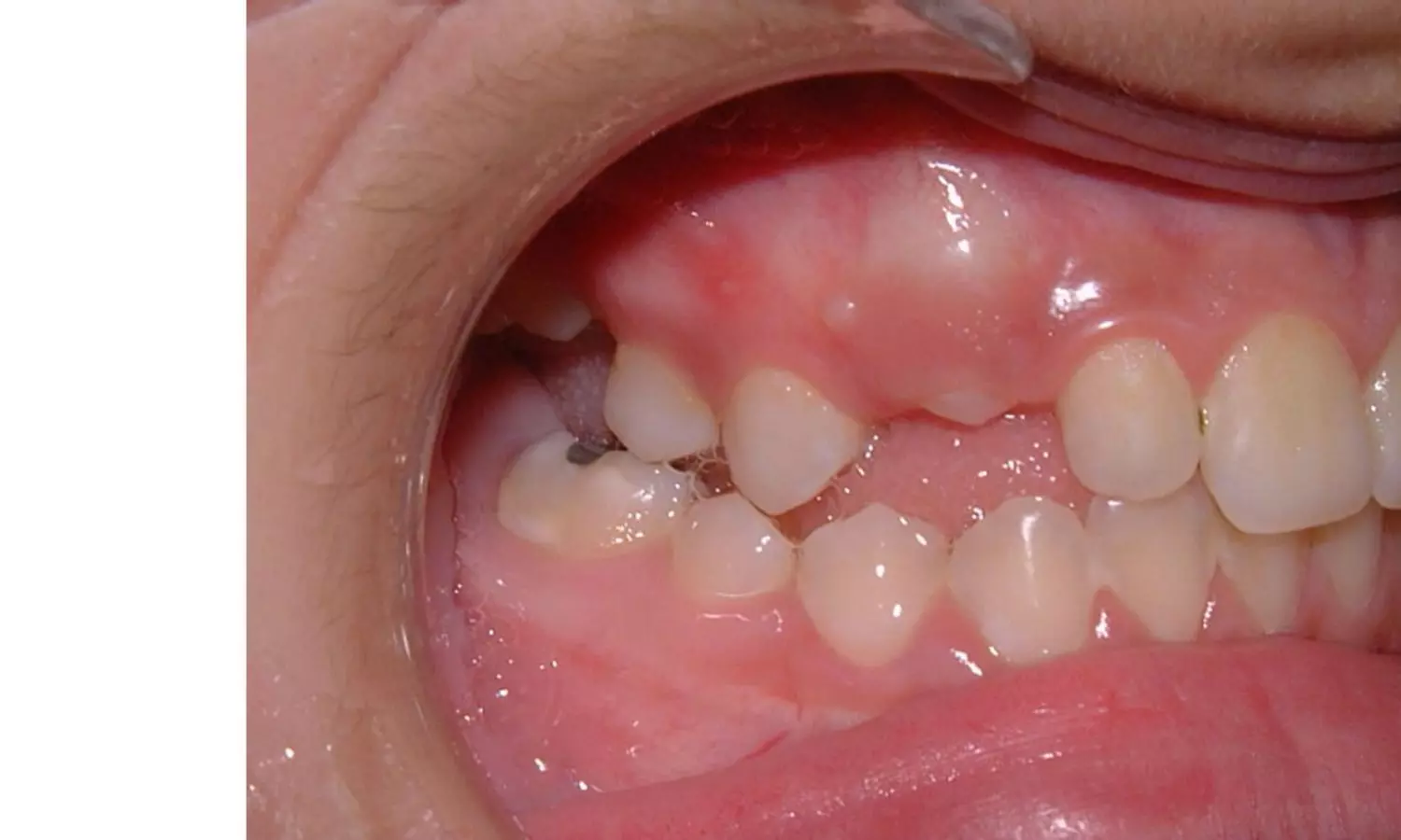
Sweden: The early extraction of the first permanent molar (FPM) in orthodontic treatment has long been a debatable topic among dental professionals. A recent retrospective records-based cohort study published in The Angle Orthodontist sheds new light on this subject, focusing specifically on spontaneous space closure following first permanent molar (FPM) extraction.

“The success rate of spontaneous space closure was high (84.3%) and was greater in the maxilla (94.1%) than the mandible (74.1%),” the researchers reported.

Age at the time of extraction and the dental developmental stage of the second permanent molar (SPM) were significant factors for successful spontaneous space closure in the maxilla and mandible, respectively.

FPM extraction is often considered a strategic orthodontic intervention, particularly in cases of crowding or malocclusion. Early extraction provides space for the eruption of permanent teeth, facilitating orthodontic treatment and potentially reducing the need for more invasive interventions later in life.

Against the above background, Yasser Aldahool, Folktandvården Stockholm, Spånga, Stockholm, Sweden, and colleagues aimed to assess the success rate of spontaneous space closure after extraction of the first permanent molar in the maxilla and the mandible. They also identified the factors that make spontaneous space closure most favorable.

For this purpose, the researchers searched the database of the Public Dental Service, Stockholm County Council, Stockholm, for young adults born between 2000 and 2001 who underwent extraction of one or more FPM between 2006 and 2016. They identified 995 extracted teeth, of which 203 teeth in 155 patients met the inclusion criteria.

The researchers reported the following findings:

  • Of the 203 extracted teeth, 81.8% received no orthodontic treatment.
  • The success rate for space closure in orthodontically treated patients was 91.9%.
  • The success rate for spontaneous space closure was 84.3%.
  • All unsuccessful spontaneous space closures in the maxilla occurred in patients older than 12 years.
  • The dental developmental stage of the second permanent molar had a statistically significant association with spontaneous space closure in the mandible.

In conclusion, the following variables were significant for successful spontaneous space closure:

  • Maxillary extractions before age 12 were more favorable than extractions done at or after 12.
  • Extraction site, with more favorable results for extractions in the maxilla compared to the mandible.
  • Dental development of the SPM in stages E and F at the time of the extraction had the most favorable results in the mandible.
  • This study could not verify that engagement of the second premolar under the roots of the primary second molar, mesial angulation of the SPM, or the presence of the third molar was significant for successful spontaneous space closure.

“There has been a lack of agreement on the most important factors for spontaneous space closure following the early extraction of FPMs,” the researchers wrote.

“Spontaneous space closure after FPM extraction is significantly associated with age and the developmental stage of the SPM in the maxilla and mandible, respectively.”

Powered by WPeMatico

Breakthrough Ultrasound Technology Offers Noninvasive Pain Relief through Brain Modulation, reveals study

In a groundbreaking study, researchers have explored the
potential of low-intensity focused ultrasound (LIFU) to noninvasively target
and modulate the dorsal anterior cingulate cortex (dACC), a key brain region
associated with pain and autonomic processing. The findings present a promising
avenue for noninvasive therapeutic interventions in pain management.


The study results were published in The Journal of Neuroscience.

The dACC, a critical area in the brain linked to pain and
autonomic responses, has long been a target for researchers seeking innovative
ways to alleviate discomfort. The study, conducted with 16 healthy volunteers
(6 males and 10 females), employed LIFU to modulate the dACC while participants
experienced transient heat pain stimuli.


Findings:


  • The results were significant, with LIFU demonstrating a
    remarkable reduction in pain ratings by an average of 1.09 points compared to
    Sham stimulation.

  • This achievement opens doors to noninvasive methods of pain
    relief, potentially revolutionizing how we approach discomfort and its
    management.

  • Crucially, the study also delved into the impact of LIFU on
    cardiac autonomic responses during acute heat pain stimuli.
  • The volunteers’ electrocardiograms (ECG) revealed intriguing
    changes, as LIFU increased heart rate variability, as indicated by higher
    standard deviation of normal sinus beats (SDNN), low-frequency (LF) power, and
    an altered low-frequency/high-frequency (LF/HF) ratio.

  • Despite the substantial effects on pain and cardiac
    responses, LIFU did not show significant alterations in blood pressure or
    electrodermal response (EDR), indicating a targeted and specific impact on
    pain-related brain regions.


  • One particularly noteworthy finding was the 38.1% reduction
    in the P2 amplitude of the contact heat-evoked potential (CHEP) observed with
    LIFU.
  • This result further supports the effectiveness of LIFU in modulating
    neural responses associated with pain, providing a tangible marker of the
    intervention’s impact.


These breakthrough results mark a significant stride in
understanding the brain’s role in pain processing and offer a potential
noninvasive solution for pain relief. The ability to modulate the dACC using
LIFU introduces novel possibilities for therapeutic interventions that go
beyond traditional approaches.

Researchers believe that these findings hold implications
not only for pain management but also for advancing our understanding of human
pain perception and autonomic processing. This research sets the stage for
future investigations into the therapeutic applications of LIFU, with potential
benefits for individuals suffering from chronic pain or seeking alternative
pain relief methods.


In conclusion, the study signifies a major leap forward in
the field of pain research, showcasing the potential of LIFU as a noninvasive
tool for influencing pain perception and autonomic responses. As scientists
continue to unlock the mysteries of the brain, this innovative approach could
pave the way for transformative advancements in pain management and
neurological interventions.

Further reading: Low-Intensity Focused Ultrasound to the Human Dorsal Anterior Cingulate Attenuates Acute Pain Perception and Autonomic Responses. DOI: https://doi.org/10.1523/JNEUROSCI.1011-23.2023

Powered by WPeMatico

Netarsudil and latanoprost combo useful fixed dose option for open-angle glaucoma and IOP: Study

A recent study published in the Graefe’s Archive for Clinical and Experimental Ophthalmology compared the effectiveness and safety of two fixed-dose combination (FDC) ophthalmic solutions for treating open-angle glaucoma (OAG) and ocular hypertension (OHT). This study evaluated the efficacy of the FDC of netarsudil 0.02%/latanoprost 0.005% (NET/LAT; Roclanda®) against bimatoprost 0.03%/timolol maleate 0.5% (BIM/TIM; Ganfort®).

Over a 6-month period, the MERCURY-3 study included patients over 18 years with OAG or OHT that was insufficiently controlled by topical medication. The efficacy and safety of NET/LAT and BIM/TIM were assessed at various intervals throughout the study.

The results indicated that NET/LAT was non-inferior to BIM/TIM in reducing intraocular pressure (IOP), with no statistically significant difference between the two treatments. Both treatments demonstrated a mean diurnal IOP ranging from 15.2 to 15.6 mmHg, where NET/LAT showed comparable efficacy to BIM/TIM. Also, no treatment-related adverse events were observed, with most adverse events being mild to moderate in severity.

The most common treatment-related adverse events were conjunctival hyperemia and cornea verticillata, where NET/LAT showed slightly higher rates of conjunctival hyperemia when compared to BIM/TIM. Further, these adverse events were consistent with previous findings and did not pose significant safety concerns.

This research emphasized the significance of the findings by stating that once-daily NET/LAT provides a compelling alternative to the fixed-dose combination treatment option for patients with OAG and OHT. This study offers valuable insights into the efficacy and safety profile of NET/LAT that expands the treatment options for individuals with these sight-threatening conditions.

Reference:

Stalmans, I., Lim, K. S., Oddone, F., Fichtl, M., Belda, J. I., Hommer, A., Laganovska, G., Schweitzer, C., Voykov, B., Zarnowski, T., & Holló, G. (2023). MERCURY-3: a randomized comparison of netarsudil/latanoprost and bimatoprost/timolol in open-angle glaucoma and ocular hypertension. In Graefe’s Archive for Clinical and Experimental Ophthalmology (Vol. 262, Issue 1, pp. 179–190). Springer Science and Business Media LLC. https://doi.org/10.1007/s00417-023-06192-0

Powered by WPeMatico

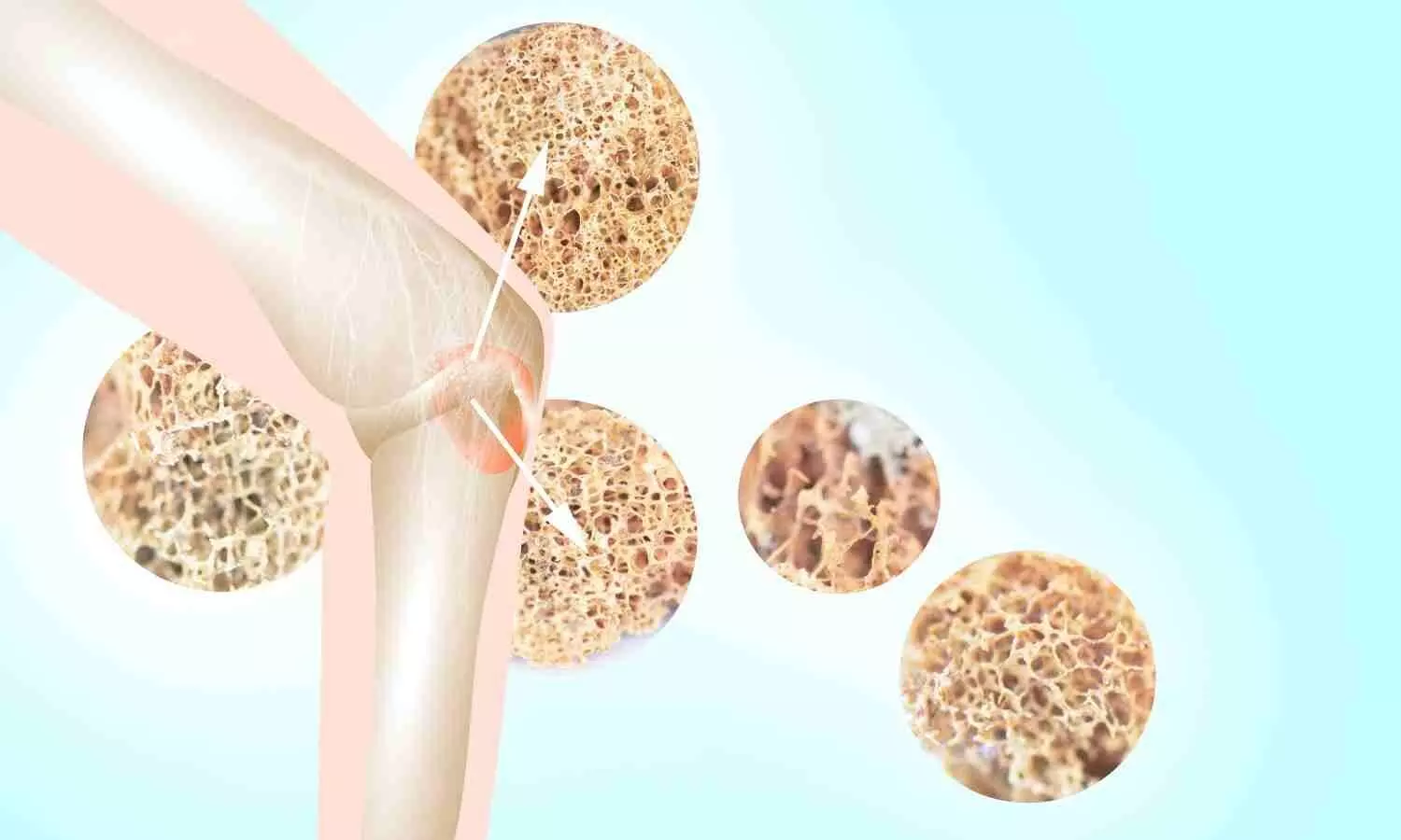
Compounds in female ginseng could lead to new osteoporosis treatments, claims study

With ever-increasing life expectancy comes the challenge of treating age-related disorders such as osteoporosis. Although there are effective drugs for treating this metabolic bone disease, they can be expensive and have side effects, limiting their availability to some people. In the search for alternative drug candidates, researchers reporting in ACS Central Science have discovered and fully replicated a compound from a botanical source, female ginseng, that had potent anti-osteoporotic activity in cellular tests.

Osteoporosis and low bone mass impact 54 million American adults over the age of 50, according to the International Osteoporosis Foundation. The disease can progress to significant disability, such as hip and spine fractures, and financial burdens, such as lost wages and hospitalization. Several drugs have proven effective in either preventing bone loss or promoting bone formation, but each comes with potential side effects, including injury to jaw and leg bones. Searching for alternative treatments, Hao Gao, Xin-Luan Wang and colleagues turned to female ginseng (Angelica sinensis), which has long been used in traditional Chinese medicine to treat osteoporosis.

The researchers performed chemical extraction on the medicinal plant and identified two new compounds, calling them falcarinphthalide A and B, that were structurally unlike anything previously discovered in female ginseng. They also determined potential biosynthetic precursors and metabolic pathways that the plants use to form these compounds. Then, with these mechanisms as starting points, the team devised lab synthesis methods and produced the compounds at quantities sufficient for biological testing. Inspired by the traditional efficacy of female ginseng, the team tested the compounds for their impact on the formation of cells called osteoclasts, which facilitate bone loss. They observed that only falcarinphthalide A and its precursors showed osteoclast inhibitory activity and an anti-osteoporotic effect. Further analysis showed that falcarinphthalide A blocked key molecular pathways involved in osteoclast generation. The researchers say that this study opens up the possibilities for new osteoporosis treatments based on the female ginseng compound, whether in its current form or as a structural template for further drug development.

Reference:

Jian Zou, Zuo-Cheng Qiu, Qiang-Qiang Yu, Jia-Ming Wu, Yong-Heng Wang, Ke-Da Shi, Yi-Fang Li, Rong-Rong He, Ling Qin, Xin-Sheng Yao, Xin-Luan Wang*, and Hao Gao, Discovery of a Potent Antiosteoporotic Drug Molecular Scaffold Derived from Angelica sinensis and Its Bioinspired Total Synthesis, https://doi.org/10.1021/acscentsci.3c01414.

Powered by WPeMatico