Doctors at Surya Mother and Child Hospital perform surgery to treat 4-year-old boy with Midgut Volvulus

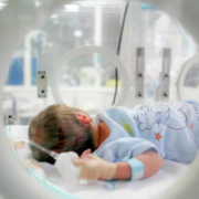
New Delhi: The doctors at Surya Mother and Child Hospital, Pune successfully performed surgery to save the life of a four-year-old boy suffering from Midgut Volvulus, a condition where the intestine becomes twisted.

Midgut volvulus is a severe condition that is common among children and infants and often occurs within the first few weeks of life, caused by a congenital anomaly of the intestines — leaving the child susceptible to sudden twisting of most of the intestines.

Symptoms include upper abdominal distension, bilious vomiting, and abdominal tenderness, signalling the need for immediate medical attention. While the condition is treatable, late detection of the condition could rapidly deteriorate the patient’s health and can be fatal.

Also Read:Medical experts concerned over lack of quality neonatal care

According to an IANS report, The boy, Sankalp, was admitted to Surya Mother and Child Hospital, Pune in an extremely deteriorated condition with wasted muscles, an immensely bloated abdomen, and dehydration.

“Delayed detection of midgut volvulus increases treatment challenges and risks of a high fatality rate,” said Sachin Shah, Director – Neonatal and Pediatric Intensive Care Services, Surya Mother and Child Super Speciality Hospital, Pune.

Sankalp was initially treated at a tertiary care hospital in Varanasi, where he underwent surgery. However, his recovery was fraught with challenges, leading to multiple corrective surgeries that failed to improve his condition.

Despite the efforts, Sankalp’s health continued to deteriorate, and his family was informed that further medical intervention seemed futile.

At the Pune hospital, the team of doctors assessed Sankalp’s condition and opted for a re-surgery to address the condition.

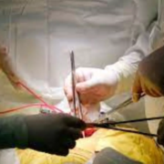
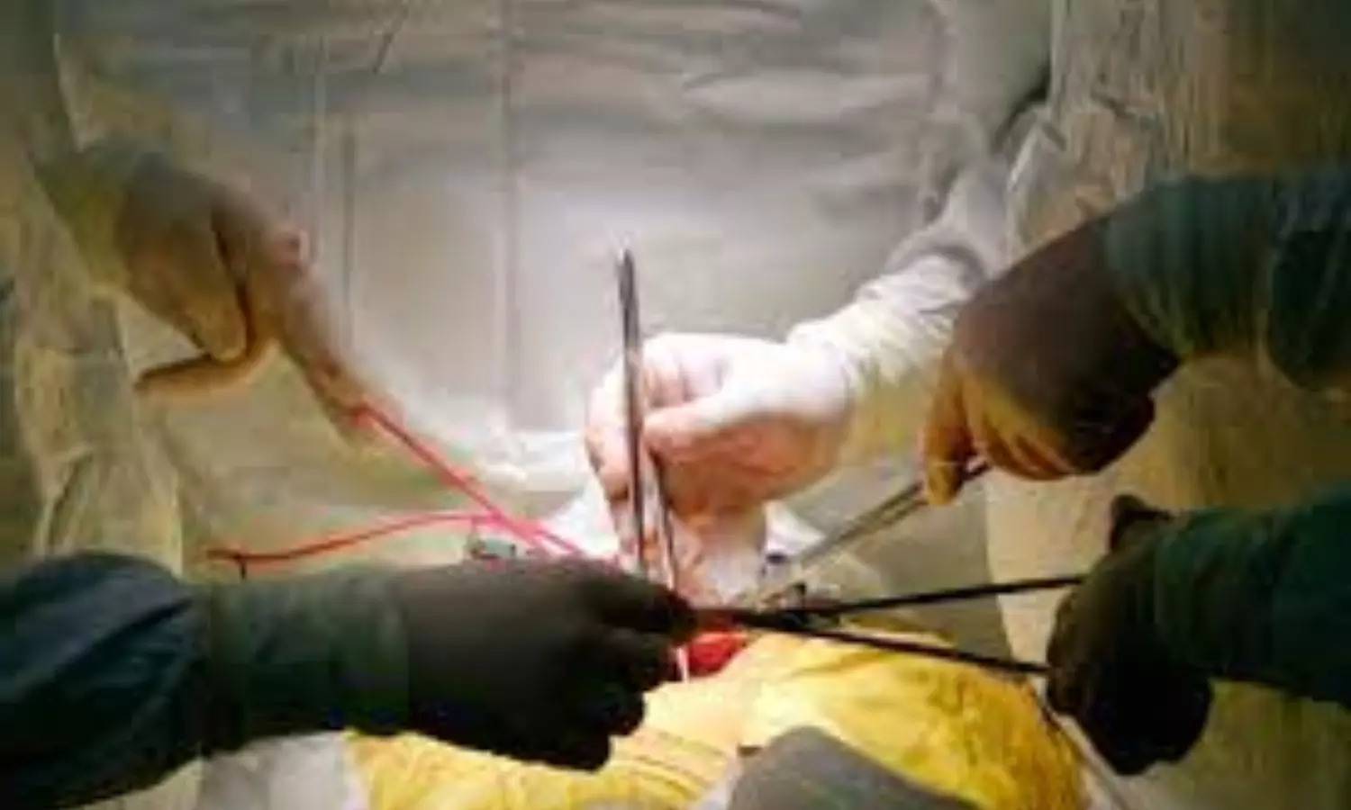
During the extensive four-hour procedure, they encountered adhesions in Sankalp’s abdomen, which had caused the intestines to adhere together, severely impairing their function.

“Despite these odds, the surgical team meticulously separated the intestines, repaired the damage, and restored their function during the extensive surgery,” Sachin said.

With their effort, Sankalp made a “remarkable recovery within 48 hours post-surgery”, reports news agency

After six days of abstaining from solid food, Sankalp was finally able to consume real food again, marking a significant milestone in his journey to health and recovery.

“Following a successful recovery, Sankalp was discharged after 10 days and closely monitored for the next three months. Once the doctors were satisfied with the healthy weight gain after a three-month follow-up period, Sankalp was discharged,” the doctor said.

Powered by WPeMatico

Contracting RSV Before Age 2 Can Cause Long-term Lung Changes and functional Impairment, finds study

Infants and children who have severe cases of respiratory syncytial virus (RSV) before age 2 are likely to have changes to their lung structure and function that could affect respiratory health later in life. The study is published ahead of print in the American Journal of Physiology-Lung Cellular and Molecular Physiology and has been chosen as an APSselect article for April.

Most children contract RSV, a lower respiratory tract disease, before they are 2 years old. Mild forms of the disease mimic the common cold with symptoms such as coughing, runny nose, congestion and sneezing. Severe forms of RSV can include wheezing, trouble breathing and, when oxygen levels are compromised, a blue tinge to the skin. A severe infection can affect a child’s future health as well. “[S]evere early-life RSV infection is associated with childhood wheezing related to respiratory viral infection exacerbation, allergies and asthma, that is accompanied by compromised lung function,” the researchers wrote.

To study severe RSV, the researchers measured lung function and alveolarization in infant mice. Alveolarization is the development of alveoli, the sacs in the lungs responsible for exchanging oxygen and carbon dioxide. “Alveolarization continues into adulthood with maximum alveolarization occurring between [2 and 3 years old] in humans,” the team wrote. Production of large numbers of immune cells occurs around the same time.

The researchers measured these markers at five weeks and three months after the initial RSV infection and again after a reinfection with the virus. They found “significant defects” in the ability of the lungs to stretch and expand during breathing. Structural changes to the mice’s lungs included an increase in alveoli size but fewer individual alveoli after RSV infection.

“These data indicate that the lungs of mice following an early-life RSV infection have a decreased lung function even at [three months] postinfection,” the research team wrote. “Importantly, the structural defects of the early-life infected mice largely mimic the clinical setting where severe exacerbations are observed in children for several years following a severe early-life respiratory infection, especially RSV.”

Reference:

Carrie-Anne Malinczak et al, Early-life pulmonary viral infection leads to long-term functional and lower airway structural changes in the lungs, American Journal of Physiology-Lung Cellular and Molecular Physiology (2024). DOI: 10.1152/ajplung.00300.2023.

Powered by WPeMatico

FFR-guided complete revascularization fails to improve outcomes in patients with MI: NEJM

Sweden: Fractional flow reserve (FFR)-guided complete revascularization does not result in better outcomes compared with primary percutaneous coronary intervention (PCI) of the culprit lesion among patients with STEMI or very-high-risk NSTEMI and multivessel coronary artery disease, randomized FULL REVASC trial has shown. 

“At a median follow-up of 4.8 years, a primary outcome event — myocardial infarction, death from any cause, or unplanned revascularization — occurred in 19% of patients in the complete-revascularization group versus 20.4% of those in the culprit-lesion-only group (HR 0.93),” the researchers reported during the American College of Cardiology annual meeting. The findings were subsequently published in The New England Journal of Medicine. 

Also, there were no differences in the composite of death from any cause or myocardial infarction between the complete-revascularization group and the culprit-lesion-only group (16.5% vs 15.3%; HR 1.12) or unplanned revascularization (9.2% vs 11.7%; HR 0.76).

When a patient presents with myocardial infarction, the urgency to restore blood flow to the affected area is paramount. PCI has long been the standard of care, but the debate continues between complete revascularization and culprit-only intervention. FFR has emerged as a valuable tool in guiding this decision-making process, offering insights into the hemodynamic significance of coronary lesions.

Against the above background, Felix Böhm, the Karolinska Institutet and Danderyd Hospital in Sweden, and colleagues explored the role of FFR guidance in determining whether complete or culprit-only PCI is more appropriate for patients with MI.

For this purpose, the researchers conducted a multinational, registry-based, randomized trial, including patients with STEMI or very-high-risk non-STEMI (NSTEMI) and multivessel disease undergoing primary PCI of the culprit lesion. A total of 1542 patients underwent randomization, 764 were assigned to receive FFR-guided complete revascularization, and 778 were assigned to receive culprit-lesion-only PCI.

The two key secondary outcomes were a composite of death from any cause or myocardial infarction and unplanned revascularization.

The following were the key findings of the study:

  • At a median follow-up of 4.8 years, a primary-outcome event had occurred in 19.0% of patients in the complete-revascularization group and in 20.4% in the culprit-lesion-only group (hazard ratio, 0.93).
  • For the secondary outcomes, the researchers observed no apparent between-group differences in the composite of death from any cause or myocardial infarction (hazard ratio, 1.12) or unplanned revascularization (hazard ratio, 0.76).
  • There were no apparent between-group differences in safety outcomes.

In conclusion, among patients with STEMI or very-high-risk NSTEMI and multivessel coronary artery disease, FFR-guided complete revascularization did not result in a lower risk of a composite of death from any cause, myocardial infarction or unplanned revascularization than culprit-lesion-only PCI at 4.8 years.

The study’s main limitation was that it had to be terminated early for feasibility and ethical reasons, which resulted in a long duration of follow-up for enough events to accrue.

Reference:

Böhm F, Mogensen B, Engstrøm T, Stankovic G, Srdanovic I, Lønborg J, Zwackman S, Hamid M, Kellerth T, Lauermann J, Kajander OA, Andersson J, Linder R, Angerås O, Renlund H, Ērglis A, Menon M, Schultz C, Laine M, Held C, Rück A, Östlund O, James S; FULL REVASC Trial Investigators.. FFR-Guided Complete or Culprit-Only PCI in Patients with Myocardial Infarction. N Engl J Med. 2024 Apr 8. doi: 10.1056/NEJMoa2314149. Epub ahead of print. PMID: 38587995.

Powered by WPeMatico

High Lipoprotein(a) Levels Increase Risk of Heart Disease in Diverse Population, finds study

A recent study published in the Journal of the American College of Cardiology highlighted the increased risk of heart disease associated with higher Lipoprotein(a) or Lp(a) levels in a large, multi-ethnic U.S. cohort. The study utilized data from five major U.S. prospective studies that provided significant insights into the genetic risk factors which contributes to atherosclerotic cardiovascular disease (ASCVD).

The research analyzed data from the Multi-Ethnic Study of Atherosclerosis (MESA), the Coronary Artery Risk Development in Young Adults (CARDIA), the Jackson Heart Study (JHS), the Framingham Heart Study-Offspring (FHS-OS) and the Atherosclerosis Risk In Communities (ARIC) study. These studies included a diverse group of 27,756 participants aged between 20 to 79 years without any previous cardiovascular incidents of which 55% were women and 35.6% were identified as Black. The participants were tracked for an average of 21.1 years, during which their Lp(a) levels were closely monitored. Lp(a) levels were categorized based on cohort-specific percentiles, the association between these levels and the incidence of ASCVD events was analyzed using multivariable Cox regression.

The results from this study indicated a clear correlation between higher Lp(a) levels and an increased risk of ASCVD. The participants with Lp(a) levels in the highest percentile (≥90th) experienced a 46% higher risk of cardiovascular events when compared to them with levels below the 50th percentile. This risk appeared consistent across various demographics which included sex and ethnicity. However, the impact of high Lp(a) levels was particularly more pronounced in individuals with diabetes with the risk increased to 92% for the individuals in the highest Lp(a) percentile.

The elevated Lp(a) levels were linked to specific outcomes such as myocardial infarction, revascularization, stroke and coronary heart disease death. Also, the study found no significant association between high Lp(a) levels and overall mortality rates. These findings underline the importance of Lp(a) as a potent genetic risk factor for ASCVD in patients with diabetes which highlights the need for targeted strategies to manage and potentially reduce this risk in the vulnerable populations. The comprehensive analysis of this study reinforces the pivotal role of genetic factors in cardiovascular health and paves the way for future research and treatment approaches that are tailored to individual genetic profiles.

Reference:

Wong, N. D., Fan, W., Hu, X., Ballantyne, C., Hoodgeveen, R. C., Tsai, M. Y., Browne, A., & Budoff, M. J. (2024). Lipoprotein(a) and Long-Term Cardiovascular Risk in a Multi-Ethnic Pooled Prospective Cohort. In Journal of the American College of Cardiology (Vol. 83, Issue 16, pp. 1511–1525). Elsevier BV. https://doi.org/10.1016/j.jacc.2024.02.031

Powered by WPeMatico

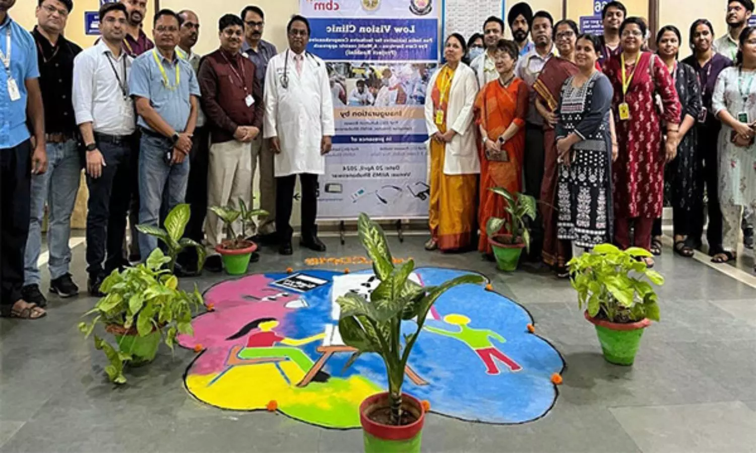
AIIMS Bhubaneswar inaugurates Low Vision Clinic for visually impaired patients

Bhubaneswar: In an aim to illuminate the lives of patients who have almost lost hope due to severe visual impairments, AIIMS Bhubaneswar has started a ”Low Vision Clinic” in the Department of Ophthalmology.

The one of its kind vision clinics will help a special group of patients like the socially blind and other patients having very low vision due to glaucoma, age-related macular degeneration, diabetes, retinal diseases, and other blinding diseases, who do not benefit from surgery or spectacle correction.

These types of patients usually lose hope and get frustrated with life due to being a burden to others in their family. The low vision aids will help these patients to do their routine work and lead an independent life. This is the first of its kind vision clinic in any government institute in Odisha.

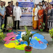
AIIMS Bhubaneswar Executive Director Dr Ashutosh Biswas inaugurated the clinic in the presence of Dr Praveen Vasisth from Dr R.P.Centre, AIIMS Delhi, Medical Superintendent Dr. Dillip Kumar Parida, DEAN Dr. Prashant Raghab Mohapatra.

Also Read:First in Odisha: AIIMS Bhubaneswar launches Advanced Artificial Urinary Sphincter Implantation Service

In a first-of-its-kind endeavour within any government institution in Odisha, AIIMS Bhubaneswar has collaborated with Dr. R.P.Centre, AIIMS Delhi, and Cristoffel Blinden Mission (CBM), Germany, to establish this visionary clinic. Operational daily, the clinic pledges to offer visual rehabilitation services to those in need, thereby reshaping lives and fostering a sense of empowerment.

Inaugurating the pioneering initiative Executive Director Dr Biswas praised the initiatives of the Ophthalmology department and acknowledged the transformative impact of the clinic. Emphasizing the importance of regular patient follow-up and expressed gratitude to AIIMS Delhi for their support under the “ROSHINI” project. He also unveiled training manuals aimed at empowering Asha workers and volunteers with essential eye care knowledge.  

Dr. Praveen Vasisth from Dr. R.P.Centre, AIIMS Delhi, shed light on the collaborative efforts between the Government of India and NGOs like CBM, culminating in the establishment of similar clinics nationwide under the “ROSHINI” project.

Dr Sucheta Parija, Head of the Department, Ophthalmology, AIIMS Bhubaneswar, provided insights into the clinic’s operational data, highlighting its impact on patient welfare. With over 75 patients already benefiting from the Low Vision Clinic at AIIMS Bhubaneswar, Dr Parija envisioned future collaborations with blind schools for enhanced rehabilitation efforts and the training of Asha workers for primary eye care at the community level.

The inauguration ceremony witnessed the presence of CMFM HoD Dr Sonu Subba, Physiology HoD Dr Prananti Nanda, Dr. Prabhas Tripahty, Dr (Maj). Mukund Sahu, Dr Bhagabat Nayak, Dr Priyadarshini Mishra, Dr Sandip Sahu, Dr Bruttendu Moharana, Dr Bijnya Panda, resident doctors, nursing officers, staffs and low vision patients, marking a collective commitment towards enhancing the quality of life for visually impaired individuals.

Powered by WPeMatico

Specific probiotic strain in yogurt may improve glycemic control in individuals with prediabetes: Study

A recent study published in the Diabetes, Obesity and Metabolism journal found promising outcomes of the probiotic strain Lactiplantibacillus plantarum OLL2712 in improving glycemic control among adults with prediabetes and prevents the onset of type 2 diabetes. Prediabetes is a condition characterized by slightly increased blood glucose levels which is a significant global health concern due to its high risk of progressing to type 2 diabetes. Addressing this condition early on is crucial in the fight against the diabetes epidemic.

This study was conducted over a period of 12 weeks, with an additional 8-week follow-up to illuminate the path for integrating dietary interventions in prediabetes management. The study included a total of 148 adults who were diagnosed with prediabetes and looked into the impacts of consuming yogurt enriched with the heat-treated OLL2712 cells when compared to a placebo.

The participants were meticulously selected by ensuring a diverse group with glycated hemoglobin (HbA1c) levels that ranged from 5.6% to 6.4% and aged between 20 and 64 years. They were randomly assigned to two groups where one received conventional yogurt and the other group received yogurt containing over 5 × 10^9 OLL2712 cells daily. The primary metric for assessing the efficacy of intervention through the covariance analyzed the change in HbA1c levels at the 12th and 16th weeks.

The study unveiled that while both groups expressed a significant reduction in HbA1c and glycoalbumin levels at the 12-week mark, this improvement persisted only in the OLL2712 group by the 16th week. At weeks 12 and 16, the decrease in HbA1c levels was significantly more pronounced in the OLL2712 group when compared to the placebo that showed the potential of this strain in enhancing the glycemic control. However, by the 20th week following the discontinuation of the yogurt consumption, no significant differences were noticed between the two groups which underscore the need for continuous intake of the probiotic to maintain its beneficial effects.

These findings suggests that daily ingestion of OLL2712-enriched yogurt could play a vital role in preventing the deterioration of glycemic control in the individuals with prediabetes. Overall, the outcomes of this study highlights the potential of yogurt in contributing to glycemic regulation and could pave the way for future dietary recommendations and interventions for the early stages of diabetes

Reference:

Toshimitsu, T., Gotou, A., Sashihara, T., Hojo, K., Hachimura, S., Shioya, N., Iwama, Y., Irie, J., & Ichihara, Y. (2024). Ingesting probiotic yogurt containing Lactiplantibacillus plantarum OLL2712 improves glycaemic control in adults with prediabetes in a randomized, double‐blind, placebo‐controlled trial. In Diabetes, Obesity and Metabolism. Wiley. https://doi.org/10.1111/dom.15534

Powered by WPeMatico

IP University Introduces Nurse Practitioner Critical Care Programme for Nursing Students, admissions open

New Delhi- Guru Gobind Singh Indraprastha University (GGSIU) has launched a new programme called ‘Nurse Practitioner Critical Care’ for nursing students for the academic session 2024-25.

As per the official statement as reported by PTI, admission for the said program has been opened therefore candidates who are registered nurses or midwives are eligible to apply for this program. Along with this, the applicant must have passed Nursing, Post-Basic Nursing or MSc Nursing with 55 percent marks as well as one year work experience required to apply for the program.

Applicants must submit the duly filled application form along with a demand draft of Rs 1,500 in favour of the Registrar at the University Facilitation Centre, IPU, Dwarka by May 15.

The programme will be available at the Max Institute of Nursing Education and Training, Max Super Speciality Hospital, Saket. It is a two-year program where the university offers 10 seats. However, at the time of admission, 75 per cent weightage will be given to the final qualifying examination and the remaining 25 per cent will be given to the personal interview.

Guru Gobind Singh Indraprastha University, formerly Indraprastha University (IP or IPU), is a state university located in Dwarka, Delhi, India. There are more than 8,000 students enrolled in the university. The university has been ranked 74th in the National Institutional Ranking Framework 2023. The university is organised around fourteen university schools and three university centres that focus on programs in law, medicine, technology, education, entrepreneurship, science and business. The university has more than 120 associate institutions, which run as per the rules and regulations laid down by the university. GGSIPU University is home to 14 university schools operating from its Dwarka and East Delhi campuses.

Powered by WPeMatico

Pragmatic Online Program tied to Long-Term Success in Primary Care Weight Loss: JAMA

Obesity is a prevalent health concern, and effective weight loss interventions are crucial. However, sustaining weight loss over the long term poses significant challenges. Pragmatic and scalable interventions are needed to address this issue, especially in primary care settings where patients often seek guidance for weight management. A recent clinical trial aimed to evaluate the long-term effectiveness of an automated, online, behavioral obesity treatment program in primary care and to compare different weight loss maintenance approaches.

This study was published in the journal JAMA Internal Medicine by J. Graham and colleagues. To assess the effectiveness of an automated, online, behavioral obesity treatment program over 12 and 24 months in primary care patients with overweight or obesity and to compare various weight loss maintenance approaches.

A randomized clinical trial recruited participants from a Rhode Island primary care network and offered a 3-month weight loss program followed by randomization to one of three 9-month maintenance programs. Data were collected from electronic medical records and analyzed for weight change outcomes.

The key findings of the study were:

  • Among 540 participants, the mean 3-month weight loss was 3.60 kg.

  • At 12 months, participants in the monthly and refresher maintenance groups regained significantly less weight compared to the control group (0.37 kg and 0.45 kg vs. 1.28 kg, respectively).

  • This trend persisted for 24 months, indicating sustained benefits of maintenance interventions.

  • In the per-protocol analysis of 253 participants, mean weight loss at 12 months was 6.19 kg, with less weight regained in the monthly and refresher maintenance groups compared to the control group.

The study demonstrates that a 12-month automated, online, behavioral obesity treatment program, coupled with active maintenance interventions, produces clinically significant weight loss over 2 years in primary care patients with overweight or obesity. These findings emphasize the importance of ongoing maintenance interventions to prevent weight regain and highlight the potential of scalable, technology-based approaches in addressing obesity in primary care settings.

Reference:

Thomas JG, Panza E, Goldstein CM, et al. Pragmatic Implementation of Online Obesity Treatment and Maintenance Interventions in Primary Care: A Randomized Clinical Trial. JAMA Intern Med. Published online March 11, 2024. doi:10.1001/jamainternmed.2023.8438

Powered by WPeMatico

Blood Urea Nitrogen, Creatinine and Proteinuria as Novel Biomarkers in Parkinson’s Disease: Study

Parkinson’s disease (PD) is a progressive neurodegenerative disorder affecting movement and quality of life in the elderly. The causes of PD remain unclear, though mitochondrial dysfunction, oxidative stress and neuroinflammation are implicated. Recent evidence suggests chronic conditions like chronic kidney disease (CKD) and related markers of renal dysfunction may be associated with higher PD risk.

A recent paper published in Neurology India by researchers in China analysed the correlation of PD with blood urea nitrogen, creatinine and proteinuria levels to assess their potential as predictive biomarkers.This retrospective study included 200 hospitalized PD patients diagnosed per 2015 MDS criteria along with 110 healthy controls. Medical history, demographics, and clinical rating scales were recorded. Blood samples were assessed for renal function markers – urea nitrogen, creatinine, and estimated glomerular filtration rate (eGFR). Urine protein level was measured. Comparative statistics were computed and multivariate logistic regression conducted to determine risk factors. Correlation analysis was performed between biomarker levels and PD severity scores.

The authors found that the PD group showed significantly higher creatinine, urea and proteinuria versus controls, while total cholesterol and LDL-cholesterol were lower. Adjusted multivariate regression revealed elevated urea nitrogen, creatinine and proteinuria levels were independent risk factors for PD. ROC analysis indicated urea nitrogen predicted PD onset with AUC 0.680, 75% sensitivity and 53% specificity at 4.97μmol/L threshold. Creatinine predicted at 63.5μmol/L with 53% sensitivity, 79% specificity and AUC 0.673. Positive correlations were observed between blood urea nitrogen and PD duration, Hoehn-Yahr stage, and UPDRS score (r=0.309-0.540). Similarly, creatinine levels correlated positively with clinical PD progression scores (r=0.139-0.320). The findings suggest these renal markers track with advancing disease.

The identification of modifiable risk factors like proteinuria, elevated creatinine and urea for PD development allows possibilities for early intervention and prevention. The ability to predict onset using easily measurable blood-based biomarkers aids diagnosis and prognostics. Renal dysfunction may relate to PD pathophysiology through vascular damage, inflammation and toxin buildup. 

The study summarises that impaired renal function markers – proteinuria, high creatinine and urea nitrogen levels correlate with increased PD risk as well as disease progression. These markers may serve as predictive biomarkers in at-risk elderly. The results warrant validation through larger randomized controlled trials. Early treatment of renal dysfunction may impact the occurrence and advancement of PD.

ReYang, Guang1,2; Wang, Ling Zhi1; Zhang, Rong1; Zhang, Xiao Yu1; Yu, Yue1; Ma, Hai Rong1,; He, Xiao Gang1,. Study on the Correlation between Blood Urea Nitrogen, Creatinine Level, Proteinuria and Parkinson’s Disease. Neurology India 71(6):p 1217-1221, Nov–Dec 2023.

DOI: 10.4103/0028-3886.391388ference

Powered by WPeMatico

Signs of Multiple Sclerosis Show Up in Blood Years Before Symptoms Appear

In a discovery that could hasten treatment for patients with multiple sclerosis (MS), UC San Francisco scientists have discovered a harbinger in the blood of some people who later went on to develop the disease. 

In about 1 in 10 cases of MS, the body begins producing a distinctive set of antibodies against its own proteins years before symptoms emerge. These autoantibodies appear to bind to both human cells and common pathogens, possibly explaining the immune attacks on the brain and spinal cord that are the hallmark of MS.

The findings were published in Nature Medicine.

MS can lead to a devastating loss of motor control, although new treatments can slow the progress of the disease and, for example, preserve a patient’s ability to walk. The scientists hope the autoantibodies they have discovered will one day be detected with a simple blood test, giving patients a head start on receiving treatment.

“Over the last few decades, there’s been a move in the field to treat MS earlier and more aggressively with newer, more potent therapies,” said UCSF neurologist Michael Wilson, MD, a senior author of the paper. “A diagnostic result like this makes such early intervention more likely, giving patients hope for a better life.”

Linking infections with autoimmune disease

Autoimmune diseases like MS are believed to result, in part, from rare immune reactions to common infections.

In 2014, Wilson joined forces with Joe DeRisi, PhD, president of the Chan Zuckerberg Biohub SF and a senior author of the paper, to develop better tools for unmasking the culprits behind autoimmune disease. They took a technique in which viruses are engineered to display bits of proteins like flags on their surface, called phage display immunoprecipitation sequencing (PhIP-Seq), and further optimized it to screen human blood for autoantibodies.

PhIP-Seq detects autoantibodies against more than 10,000 human proteins, enough to investigate nearly any autoimmune disease. In 2019, they successfully used it to discover a rare autoimmune disease that seemed to arise from testicular cancer.

MS affects more than 900,000 people in the US. Its early symptoms, like dizziness, spasms, and fatigue, can resemble other conditions, and diagnosis requires careful analysis of brain MRI scans.

The phage display system, the scientists reasoned, could reveal the autoantibodies behind the immune attacks of MS and create new opportunities to understand and treat the disease.

The project was spearheaded by first co-authors Colin Zamecnik, PhD, a postdoctoral researcher in DeRisi’s and Wilson’s labs; and Gavin Sowa, MD, MS, former UCSF medical student and now internal medicine resident at Northwestern University.

They partnered with Mitch Wallin, MD, MPH, from the University of Maryland and a senior author of the paper, to search for autoantibodies in the blood of people with MS. These samples were obtained from the U.S. Department of Defense Serum Repository, which stores blood taken from armed service members when they apply to join the military.

The group analyzed blood from 250 MS patients collected after their diagnosis, plus samples taken five or more years earlier when they joined the military. The researchers also looked at comparable blood samples from 250 healthy veterans.

Between the large number of subjects and the before-and-after timing of the samples, it was “a phenomenal cohort of individuals to look at to see how this kind of autoimmunity develops over the course of clinical onset of this disease,” said Zamecnik.

A consistent signature of MS 

Using a mere one-thousandth of a milliliter of blood from each time point, the scientists thought they would see a jump in autoantibodies as the first symptoms of MS appeared.

Instead, they found that 10% of the MS patients had a striking abundance of autoantibodies years before their diagnosis. 

The dozen or so autoantibodies all stuck to a chemical pattern that resembled one found in common viruses, including Epstein-Barr Virus (EBV), which infects more than 85% of all people, yet has been flagged in previous studies as a contributing cause for MS.

Years before diagnosis, this subset of MS patients had other signs of an immune war in the brain. Ahmed Abdelhak, MD, co-author of the paper and a postdoctoral researcher in the UCSF laboratory of Ari Green, MD, found that patients with these autoantibodies had elevated levels of neurofilament light (Nfl), a protein that gets released as neurons break down.

Perhaps, the researchers speculated, the immune system was mistaking friendly human proteins for some viral foe, leading to a lifetime of MS.

“When we analyze healthy people using our technology, everybody looks unique, with their own fingerprint of immunological experience, like a snowflake,” DeRisi said. “It’s when the immunological signature of a person looks like someone else, and they stop looking like snowflakes that we begin to suspect something is wrong, and that’s what we found in these MS patients.”

A test to speed patients toward the right therapies

To confirm their findings, the team analyzed blood samples from patients in the UCSF ORIGINS study. These patients all had neurological symptoms and many, but not all, went on to be diagnosed with MS.

Once again, 10% of the patients in the ORIGINS study who were diagnosed with MS had the same autoantibody pattern. The pattern was 100% predictive of an MS diagnosis. Across both the Department of Defense group and the ORIGINS group, every patient with this autoantibody pattern had MS.

“Diagnosis is not always straightforward for MS, because we haven’t had disease specific biomarkers,” Wilson said. “We’re excited to have anything that can give more diagnostic certainty earlier on, to have a concrete discussion about whether to start treatment for each patient.”

Many questions remain about MS, ranging from what’s instigating the immune response in some MS patients to how the disease develops in the other 90% of patients. But the researchers believe they now have a definitive sign that MS is brewing.

“Imagine if we could diagnose MS before some patients reach the clinic,” said Stephen Hauser, MD, director of the UCSF Weill Institute for Neurosciences and a senior author of the paper. “It enhances our chances of moving from suppression to cure.” 

Reference:

Zamecnik, C.R., Sowa, G.M., Abdelhak, A. et al. An autoantibody signature predictive for multiple sclerosis. Nat Med (2024). https://doi.org/10.1038/s41591-024-02938-3.

Powered by WPeMatico