Prolonged use of desogestrel pill linked to small increased brain tumour risk: Study

Taking the progestogen-only contraceptive pill desogestrel continuously for more than five years is associated with a small increased risk of developing a type of brain tumour called an intracranial meningioma, finds a study from France published by The BMJ today.

However, the researchers stress that the risk is low compared with some other progestogens (for every 67,000 women taking desogestrel, one might need surgery for meningioma) and disappeared one year after stopping treatment.

Intracranial meningiomas are typically non-cancerous brain tumours that occasionally require surgery. They are more common in older women, but many studies lack information on the specific type of progestogen used, and risk has not been measured for continuous, current, and long term use.

To address this knowledge gap, researchers set out to assess the real-life risk of intracranial meningioma associated with short-term (less than 1 year) and prolonged (1-7 or more years) use of oral contraceptives containing desogestrel 75µg, levonorgestrel 30µg, or levonorgestrel 50-150 µg combined with oestrogen.

Their findings are based on data from the French national health data system (SNDS) for 8,391 women (average age 60; 75% older than 45) who had undergone surgery for intracranial meningioma in 2020-2023. Each case was matched to 10 control women without meningioma (total 83,910) of the same age and area of residence.

Potentially influential factors, including use of a known high-risk progestogen in the six years before the study, were taken into account.

The results show a small increased risk associated with use of desogestrel for more than five continuous years.

An increased risk was not found for shorter durations or when desogestrel had been discontinued for more than one year, but it was if other progestogens of known associated increased risk of meningioma had been used in the six years before desogestrel.

This excess risk was greater in women older than 45 years, those with a meningioma located in the front or middle of the skull, and after prolonged use of one of the known high risk progestogens before desogestrel.

The authors estimate that 67,000 women would need to use desogestrel for one woman to need surgery for intracranial meningioma, and 17,000 women if current use was for more than five years.

Reassuringly, no increased risk of meningioma was found for levonorgestrel, alone or combined with oestrogen, regardless of duration of use.

This is an observational study so can’t establish cause and effect, and the authors acknowledge that some information may be missing from the SNDS database. Nor were they able to account for genetic predisposition and exposure to high dose radiation – factors that may have affected the results.

However, by using comprehensive, real-life data, including history of using other known high risk progestogens, they were able to improve reliability and minimise bias.

As such, they suggest that desogestrel should be discontinued if an intracranial meningioma is identified and patients monitored rather than undergoing immediate surgery.

Although direct evidence is still lacking, stopping treatment when desogestrel related meningioma is diagnosed may preclude the need for surgery, says neurosurgeon Gilles Reuter in a linked editorial.

“It is already common knowledge that stopping cyproterone, nomegestrol, chlormadinone, promegestone, medroxyprogesterone, or medrogestone precludes the need for surgery,” he explains. “Now we know that stopping desogestrel may also avoid unnecessary potentially harmful treatments.”

Reference:

https://www.bmj.com/content/389/bmj-2024-083981

Powered by WPeMatico

New Study Links Triglyceride-Glucose Index to Increased Risk of Periodontitis

China: A recent study published in Frontiers in Endocrinology sheds light on a previously underexplored link between the triglyceride-glucose (TyG) index and periodontitis. Led by Jing Huang from the School of Nursing, Peking University, Beijing, China, the research utilized extensive datasets from two large-scale population surveys to explore this potential association.

“The observational analysis demonstrates a notable association between elevated TyG index levels and an increased risk of periodontitis,” the authors wrote.

The study aimed to clarify whether elevated TyG index, a surrogate marker of insulin resistance, correlates with a higher risk of developing periodontitis. To achieve this, the researchers examined data from the National Health and Nutrition Examination Survey (NHANES, 2009–2014) and the Korea National Health and Nutrition Examination Survey (KNHANES, 2007–2018, excluding 2011).

The analysis included a total of 2,511 individuals with periodontitis from NHANES and 16,239 from KNHANES. The researchers employed multivariate logistic regression models, stratified and subgroup analyses, as well as restricted cubic spline (RCS) models to assess the nature and strength of the relationship between TyG index levels and periodontitis. Furthermore, the predictive performance of the TyG index was evaluated using receiver operating characteristic (ROC) curve analysis, and mediation analyses were conducted to explore underlying metabolic and inflammatory contributors.

The key findings include the following:

  • Higher TyG index levels were significantly associated with an increased risk of periodontitis in both NHANES and KNHANES cohorts.
  • In the NHANES dataset, individuals in the second quartile had a 20% higher risk of periodontitis compared to those in the lowest quartile (OR 1.20).
  • In the same dataset, those in the highest quartile had a 23% increased risk (OR 1.23).
  • In the KNHANES cohort, individuals in the highest quartile had a 9% greater risk of periodontitis (OR 1.09).
  • These associations remained consistent even after adjusting for confounding variables, with a significant trend observed.
  • Restricted cubic spline (RCS) analysis showed a nonlinear relationship between TyG index levels and periodontitis risk.
  • Receiver operating characteristic (ROC) analysis showed the TyG index had moderate predictive capacity, with values of 8.24 in NHANES and 8.69 in KNHANES.
  • Mediation analysis indicated that inflammatory markers (alkaline phosphatase and white blood cells) and metabolic factors (vitamin D and HDL cholesterol) partially mediated the association between the TyG index and periodontitis.

The authors concluded that this observational evidence supports a significant connection between metabolic dysfunction, as captured by the TyG index, and oral health outcomes. They call for further research to unpack the biological mechanisms and to assess whether the TyG index could serve as an early indicator for periodontal disease prevention strategies.

Reference:

Huang J, Zhang D, Li H, Zhang Y, Long T, Guo X, Cui H, Wei Z, Zhao J, Li M and Wang P (2025) Triglyceride-glucose index and periodontitis: evidence from two population-based surveys. Front. Endocrinol. 16:1558692. doi: 10.3389/fendo.2025.1558692

Powered by WPeMatico

Antiviral treatment in HBV infection reduces risk of kidney disease development: Study

A new study published in the journal of BMC Nephrology revealed that kidney disease (KD) caused by an infection with the hepatitis B virus (HBV), can be reduced by anti-HBV therapy.

Chronic HBV infection is linked to extrahepatic comorbidities, such as renal symptoms, in addition to potential liver-related sequelae including cirrhosis and hepatocellular carcinoma (HCC). Kidney disease (KD) and HBV infection have been linked in a number of studies. According to research that looked at Medicare and Medicaid claims data from 2006 to 2015, patients with HBV had a greater frequency of KD than matched controls, and this prevalence rose with time. Moreover, an elevated risk of developing end-stage renal disease (ESRD) has also been linked to untreated HBV.

This research investigated whether KD development is linked to HBV-related liver illness, if anti-HBV medication reduces these risks, and whether patients with HBV have a greater chance of developing KD using a big US-based electronic medical record (EMR) database. From 2006 to 2020, information was retrieved from the IQVIA Ambulatory EMR database. To improve balance across analyses, propensity score matching was used. Hazard ratios (HRs) with 95% CIs for the start of KD between groups were estimated using a Cox proportional hazards model.

The majority of occurrences were beyond the age of 55. Among patients with and without HBV (n = 11,772 each), those with HBV had a more than twofold higher risk of developing KD compared to matched controls without HBV infection.

By the time they were 75 years old, patients with HBV and concurrent hypertension, diabetes, or obesity were more likely to develop KD (19% with HBV vs. 6% without HBV). In patients with HBV and concurrent comorbidities, the cumulative probability of developing KD exceeded the additive risk of developing KD for those with the comorbidities without HBV or just with HBV. Advanced liver disease was not substantially linked to KD in HBV patients.

Antiviral-treated individuals were less likely to develop KD than untreated HBV patients. Overall, the aging HBV population is significantly impacted by these discoveries. All of these results point to the necessity of prospective research to fully comprehend how antiviral medications affect the onset of KD. 

Source:

Ito, K. L., Zhang, Y., Li, B., King, A., Yee, L. J., Frenette, C., Abramov, F., Flaherty, J. F., & Malkov, V. A. (2025). Chronic hepatitis B virus infection increases the risk of kidney disease while antiviral therapy for hepatitis B virus can decrease kidney disease risk. BMC Nephrology, 26(1), 171. https://doi.org/10.1186/s12882-025-03991-x

Powered by WPeMatico

Vitamin D Deficiency linked to Pulsatile Tinnitus Due to Dehiscent Sigmoid Plates: Study

Researchers have found in a new study that Vitamin D deficiency may contribute to pulsatile tinnitus associated with dehiscent sigmoid plates. However further research is required to assess the effectiveness of vitamin D supplementation in managing symptoms in such cases. The study was conducted by Michael F. and colleagues published in The Egyptian Journal of Otolaryngology.

Pulsatile tinnitus, or a rhythmic noise heard within the ears, usually synchronized with the heartbeat, may be caused by vascular abnormalities of the brain, such as sigmoid sinus dehiscence. It is a condition whereby there is a loss of bony covering of the sigmoid sinus, leaving the vessel exposed and allowing sound to be conducted directly into the ear. It has come to be increasingly appreciated as a common venous etiology of tinnitus.

Although previous research has documented associations between tinnitus and various systemic conditions, no previous studies have addressed a possible relation between vitamin D deficiency and sigmoid sinus dehiscence, even though the firmly established function of vitamin D in bone health and cortical bone density has been documented.

The main objective of this study was to analyze the prevalence of vitamin D deficiency in patients diagnosed with pulsatile tinnitus caused by sigmoid sinus dehiscence, and to identify the association between the levels of vitamin D, the severity of bone dehiscence, and the intensity of tinnitus.

This cross-sectional study was carried out at Ain Shams University Hospitals between December 2023 and December 2024, a period of 12 months. The study recruited 36 patients who had pulsatile tinnitus and radiologically proven sigmoid sinus dehiscence. Tinnitus severity was clinically evaluated, and imaging grading of dehiscence was done. Blood samples were drawn to determine vitamin D levels, and statistical analysis of correlations between variables was done.

Results

There were three significant associations that were found to be present.

Severity of sigmoid sinus dehiscence was positively correlated with severity of tinnitus the greater the bone dehiscence, the louder or more chronic the tinnitus symptoms experienced by patients.

A negative significant correlation existed between dehiscence severity and vitamin D level as vitamin D level went down, the extent of bony dehiscence went up.

Comparable negative association was also found between tinnitus severity and vitamin D status patients with reduced vitamin D levels reported more severe tinnitus.

This study concludes that vitamin D deficiency could be a causative factor in the development and severity of pulsatile tinnitus caused by sigmoid sinus dehiscence.

Reference:

Fadel, M., Bedros, R.Y., Nassif, M.M. et al. Prevalence of vitamin D deficiency in patients presented by pulsatile tinnitus due to dehiscent sigmoid sinus. Egypt J Otolaryngol 41, 45 (2025). https://doi.org/10.1186/s43163-025-00787-6

Powered by WPeMatico

Combination therapy can prolong life in severe heart disease, reports research

Aortic valve narrowing (aortic stenosis) with concomitant cardiac amyloidosis is a severe heart disease of old age that is associated with a high risk of death. Until now, treatment has consisted of valve replacement, while the deposits in the heart muscle, known as amyloidosis, often remain untreated. An international research consortium led by MedUni Vienna and University College London has now been able to demonstrate for the first time that combined treatment consisting of heart valve replacement and specific drug therapy offers a significant survival advantage for patients. The study results have been published in the European Heart Journal.

As part of the study conducted by the research team led by Christian Nitsche (Department of Medicine II, Clinical Division of Cardiology, MedUni Vienna) and Thomas Treibel (Department of Cardiovascular Imaging, University College London), data from 226 patients with aortic stenosis and concomitant cardiac amyloidosis from ten countries were examined. Aortic stenosis is a narrowing of the heart valve that directs blood from the left ventricle into the bloodstream. In cardiac amyloidosis, misfolded proteins are deposited in the heart muscle. Both diseases occur in older people and often together. Until now, it was unclear whether treating amyloidosis in addition to valve surgery would benefit patients.

The analysis now published showed that both aortic valve replacement and treatment with the drug tafamidis for amyloidosis were associated with a significantly lower risk of death. The survival benefit was highest in patients who received both forms of treatment. “Our results even show that patients with both conditions who received valve replacement and specific amyloidosis therapy had similar long-term survival rates to people with aortic stenosis without amyloidosis,” emphasises study leader Christian Nitsche.

Targeted tests necessary

Both aortic stenosis and cardiac amyloidosis impair the heart’s pumping function and can lead to death if left untreated. Targeted therapy can slow the progression of amyloidosis, while valve replacement treats the mechanical stress caused by the narrowed heart valve. Around ten percent of patients with aortic stenosis also have amyloidosis, but this is often not diagnosed in everyday clinical practice. “Our findings also suggest that patients with severe aortic valve stenosis should be screened for amyloidosis so that we can offer them targeted life-prolonging treatment options,” emphasises Christian Nitsche.

Reference:

Christian Nitsche, Stephan Dobner, Hannah R Rosenblum, Kush P Patel, Simone Longhi, Ali Yilmaz, Marco Merlo, Maria Papathanasiou, Jan Griffin, Marish I F J Oerlemans, Francisco Gama, Ashraf Hamdan, Andrew D Kelion, Andreas Schuster, Sigita Glaveckaité, Nuriye Akyol, Aldostefano Porcari, Lara Schlender, Teresa Capovilla, Maximilian Autherith, Laurenz Hauptmann, Kseniya Halavina, João L Cavalcante, Marianna Fontana, Paul R Scully, James C Moon, Julia Mascherbauer, Robin Ristl, Elena Biagini, Stefan Stortecky, Matthew S Maurer, Thomas A Treibel, AS-Amyloidosis Consortium, Cardiac transthyretin amyloidosis treatment improves outcomes after aortic valve replacement for severe stenosis, European Heart Journal, 2025;, ehaf362, https://doi.org/10.1093/eurheartj/ehaf362.

Powered by WPeMatico

Psychiatric Medications Linked to Higher ALS Risk and Poorer Prognosis: JAMA

A new study published in the Journal of American Medical Association showed that prescription usage of sedatives, antidepressants, or anxiety medications was linked to a higher chance of receiving an ALS diagnosis in the future as well as a lower chance of surviving after receiving one.

While a number of studies have indicated that people with a history of mental illnesses are more likely to be diagnosed with amyotrophic lateral sclerosis (ALS), there is little and conflicting evidence linking the use of popular psychiatric drugs to ALS. Thus, this study investigated if the risk and course of ALS are related to the prescribed use of popular psychiatric drugs, such as antidepressants, hypnotics and sedatives, and anxiolytics.

According to the Swedish Motor Neuron Disease Quality Registry, this nationwide register-based case-control study was carried out in Sweden among all patients diagnosed with ALS between January 1, 2015, and July 1, 2023. These patients were matched for age and sex with up to 5 people who did not have ALS, as well as their spouses and full siblings. Following diagnosis, ALS patients were monitored for a median (IQR) of 1.33 (0.64-2.37) years. Prior to the diagnosis of ALS, at least two prescriptions for the psychiatric drugs under study were considered.

Prior use of psychiatric drugs was associated with an increased risk of developing ALS in a study of 1057 ALS cases and 5281 controls (mean age 67.5 years; 53.1% male). Notably, there was a high correlation between the use of hypnotics/sedatives 0–1 year before diagnosis (OR 6.10), anxiolytics 1–5 years prior (OR 1.60), and antidepressants over 5 years prior (OR 1.21).

The use of hypnotics/sedatives (OR 1.21), antidepressants (OR 1.26), and anxiolytics (OR 1.34), after excluding the year before diagnosis, continued to be linked to an elevated risk of ALS. Relative comparisons revealed similar results, allaying worries about family confounding (apart from hypnotics/sedatives). ALS patients who had previously used antidepressants or anxiolytics showed worse survival rates (HRs 1.72 and 1.52, respectively).

Overall, the use of antidepressants, hypnotics and sedatives, or anxiolytics was linked to an increased chance of receiving an ALS diagnosis in the future, according to this countrywide case-control research. Among ALS patients, prediagnostic usage of several of these drugs was likewise linked to a worse survival rate and a quicker functional deterioration.

Reference:

Chourpiliadis, C., Lovik, A., Ingre, C., Press, R., Samuelsson, K., Valdimarsdottir, U., & Fang, F. (2025). Use of common psychiatric medications and risk and prognosis of amyotrophic lateral sclerosis. JAMA Network Open, 8(6), e2514437. https://doi.org/10.1001/jamanetworkopen.2025.14437

Powered by WPeMatico

Intralesional Vitamin D3 Proven Effective and Affordable for Treating Cutaneous Warts: Study

A new study published in the Journal of the College of Physicians and Surgeons – Pakistan showed that for cutaneous warts, intralesional vitamin D3 is a cost-effective and efficient therapy alternative.

Common benign skin lesions called cutaneous warts are brought on by an infection with the human papillomavirus (HPV). In around 65–78% of instances, they resolve on their own and are often asymptomatic. The majority of wart patients seek treatment because they are unsightly and occasionally because of the pain or tenderness that specific wart types (such as plantar and periungual warts) cause.

Warts can be treated using a variety of modalities, such as topical medications (such as salicylic acid, trichloroacetic acid, etc.) and physical destructive techniques (such as electrocoagulation, cryotherapy, or laser therapy). Immunotherapeutic techniques are gaining popularity in the treatment of warts because of their shown effectiveness and simplicity of administration. Instead of merely removing the skin lesions, they work by promoting the host cell-mediated immunity to eradicate the virus.

Intralesional vitamin D has emerged as a promising therapy option for cutaneous warts in a number of recent trials. Thus, to ascertain the function of intralesional vitamin D3 in the management of cutaneous warts, Sabahat Shah and colleagues carried out this investigation.

This study from May to November 2023, was carried out from the dermatology department of P.N.S. Shifa Hospital in Karachi, Pakistan. 30 individuals with cutaneous warts of different sizes and lengths of time were chosen. Lignocaine (0.2 ml, 20 mg/ml) was used to anesthetize the warts before injecting vitamin D3 (0.2 ml, 15 mg/ml) into the base of the warts. The injections were administered again every 2 weeks for a maximum of four sessions, or until full recovery, whichever came first. The patients were monitored for 6 months following the final injection, and a maximum of two warts were treated each session.

The patients’ average length of sickness was 14.3 ± 6.5 months, and their average age was 32.20 ± 11.9 years. 10 (33.4%) of the 30 patients were female, and 20 (66.6%) were male. 3 instances (10%) had a modest reaction, 7 cases (23.3%) had a moderate response, and 20 cases (66.6%) had full clearance.

Mild injection site swelling and irritation were the only adverse effects seen in 17 (56.6%) instances, and they both resolved on their own in a matter of days. Overall, one possible therapy option for cutaneous warts is intralesional vitamin D3. The use of this therapy technique is increased by its affordability and absence of significant adverse effects.

Source:

Shah, S., Khan, S., & Sabah, N. U. (2025). Role of intralesional vitamin D3 in the treatment of cutaneous warts. Journal of the College of Physicians and Surgeons–Pakistan: JCPSP, 35(5), 642–645. https://doi.org/10.29271/jcpsp.2025.05.642

Powered by WPeMatico

Lead-Exposed Children More Likely to Have Hearing Loss, study finds

Researchers have discovered that children with high blood lead levels are much more likely to be diagnosed with sensorineural hearing loss (SNHL) even when the lead levels are only slightly increased. This correlation was found in a large American study which found that children who had blood lead levels of 3.5 µg/dL or greater had a 22% higher risk of developing hearing impairment compared to children with lower blood lead levels. The study was published in the International Journal of Pediatric Otorhinolaryngology by Neil R. and colleagues.

Even though lead-based paint was prohibited in the United States in 1978, lead exposure during childhood continues through environmental and in-home sources like older housing, soil, and pipes. Acknowledging that there is no safe level of lead, the Centers for Disease Control and Prevention (CDC) reduced the blood lead reference level to 3.5 µg/dL in 2021, which represents a change in the evaluation of pediatric lead exposure. Although lead’s harmful impact on the nascent nervous system is well understood, relatively few large-scale studies have looked at its particular association with hearing impairment. This new research provides compelling evidence that even quite low doses of lead can negatively affect a child’s hearing.

To assess the association between lead exposure and hearing loss, scientists employed data from the TriNetX Analytics Network, a federated research platform with more than 78 million electronic health records from all over the United States. The study involved children 18 years and below, grouping them into two categories according to their blood lead level:

  • Lead-exposed group: Children with blood lead levels ≥ 3.5 µg/dL

  • Control group: Young children with blood lead < 3.5 µg/dL

Young children with other causes of established hearing loss—such as congenital cytomegalovirus, inner ear malformations, congenital hearing loss, or noise-induced hearing loss—were eliminated from the analysis to provide reliable results.

Results

  • Total children examined: 136,280

  • 15,820 with blood lead level ≥ 3.5 µg/dL

  • 120,460 with blood lead level < 3.5 µg/dL

Prevalence of SNHL:

  • 2.83% in lead-exposed group (~448 cases)

  • 2.32% in control group (~2,796 cases)

Relative risk (RR): 1.22

  • Confidence interval (CI): 1.21–1.23

  • Statistical significance: p = 0.005

This population-based analysis confirms that children with a high level of blood lead (≥ 3.5 µg/dL) are significantly at increased risk for sensorineural hearing loss relative to their peers with low levels. As the largest study to date evaluating this association under the CDC’s new reference value, it presents strong evidence for improved screening and early audiologic treatment for lead-exposed children.

References

Rana, N., Cleveland, C., Karasik, D., & Otteson, T. (2025). Elevated lead levels as a risk factor for pediatric sensorineural hearing loss. International Journal of Pediatric Otorhinolaryngology, 112406, 112406. https://doi.org/10.1016/j.ijporl.2025.112406

Powered by WPeMatico

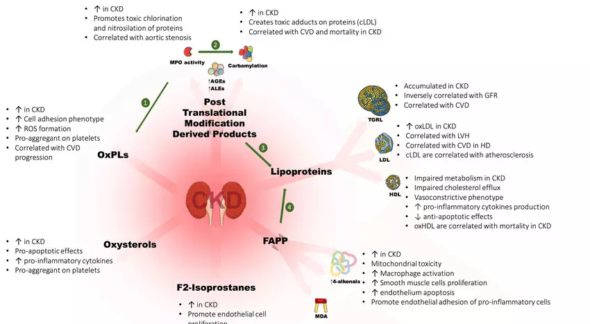
Lipid Profile Changes may predict Mortality Risk in hemodialysis Patients: Study

Researchers have found in a new research that in hemodialysis (HD) patients, baseline HDL-C levels and changes in LDL-C over three years were significant predictors of all-cause mortality. However in contrast, no significant risk factors were identified in the peritoneal dialysis (PD) group.

Changes in lipid levels over time and the associated all-cause mortality have not yet been studied in different populations of patients undergoing dialysis. This study aimed to investigate the differences in time-varying serum lipid levels and all-cause mortality among haemodialysis (HD) and peritoneal dialysis (PD) patients over a 5-year follow-up period. This observational study included Chinese patients with end-stage renal disease (ESRD) who started HD or peritoneal dialysis at Sun Yat-sen Memorial Hospital from January 2010 to February 2018. Changes in lipid profiles and trends of change in overall survival rates between the two groups were investigated. Risk factors for the outcome were identified, and the optimal cut-off values were determined using ROC analysis. Additionally, the relationship between the group variable and the outcome measure was assessed using linear regression with a generalized estimating equation (GEE) model. Results: A total of 141 patients (74 HD patients and 67 PD patients) were enrolled in the study. Forty-three (30.71%) patients died during the follow-up period. Compared with the HD group, the peritoneal dialysis group had significantly greater triglyceride (TG) (Year 1 and Year 2) and low-density lipoprotein cholesterol (LDL-C) (Year 2) levels and significantly lower high-density lipoprotein cholesterol (HDL-C) (Year 1 and Year 2) levels. There was no significant difference in total cholesterol (TC) levels. The GEE results revealed similar changes in lipid levels between HD patients and peritoneal dialysis patients over time. The Kaplan‒Meier survival curve revealed that there was no significant difference in overall survival between the two groups (log-rank test, P = 0.119). Furthermore, the multivariate Cox proportional hazard regression models demonstrated that baseline HDL-C levels (HR: 0, 95% CI: 0 to 0.11; P = 0.004) and changes in LDL-C levels from baseline to 3 years of follow-up(difference from 0 to 3 years of follow-up) (HR: 0.21, 95% CI: 0.09 to 0.53; P < 0.001) were associated with a greater risk of death in HD patients. An increase in LDL-C levels (difference from 0 to 3 years of follow-up) ≤ 0.24 mmol/L in HD patients and age ≥ 53 years in all patients initiating dialysis was associated with a significantly increased risk of mortality. The baseline levels of HDL-C and changes in LDL-C levels over a three-year period were significant predictors of all-cause mortality in HD patients, which differed from the lack of significant risk factors observed in the peritoneal dialysis group.

Reference:

Xu, Z., Zeng, Y., Liang, Y. et al. Association between time-varying serum lipid levels and all-cause mortality in haemodialysis and peritoneal dialysis patients. BMC Nephrol 26, 199 (2025). https://doi.org/10.1186/s12882-025-04119-x

Keywords:

Lipid, Profile, Changes, predict, Mortality Risk, hemodialysis Patients, Study , Xu, Z., Zeng, Y., Liang, Lipid, Haemodialysis, Peritoneal dialysis, Mortality, Time-varying

Powered by WPeMatico

Treating Sleep Problems Early May Reduce Pain in Rheumatoid Arthritis, suggests study

Sleep disturbances have been identified to significantly compromise pain interference in early rheumatoid arthritis (RA) patients, emphasizing the importance of early intervention aimed at sleep problems, as per a recent published study. The study discovers sleep disturbance in the early stages of RA to be potent predictors of future limitations of daily functioning due to pain. These findings indicate that early treatment of sleep disturbances can enhance long-term pain outcomes in RA patients. The study was conducted by Burcu A. and colleagues published in the journal of Arthritis Care & Research.

Rheumatoid arthritis is a long-standing inflammatory condition typically associated with fatigue, sleep disturbance, and chronic pain. Although medical management has improved considerably, pain continues to be a chief complaint among patients with RA. Poor sleep has been associated with increased pain perception in chronic diseases, but little research has addressed this relationship over a longitudinal period, especially in early RA. This investigation aimed to test whether sleep quality might predict pain interference with daily functioning in the long term, with the potential for informing more optimal pain management in clinical practice.

Data were gathered on 502 adults with early RA—diagnosed as having symptoms of 12 months or less in their joints—enrolled between 2016 and 2023 in the Canadian Early Arthritis Cohort. Patients received clinical assessment and filled out PROMIS-29 (Patient-Reported Outcomes Measurement Information System) questionnaires on five occasions: baseline (0 months), 6 months, 12 months, 18 months, and 24 months.

Sleep disturbance was targeted as the primary predictor and pain interference as the key outcome. Linear mixed effects regression models were employed to estimate the associations after controlling for age, sex, body mass index, education, income, smoking status, comorbidities, disease activity, treatment, and depression. A lagged analysis model was applied to analyze whether sleep disturbance at every time point predicted 6-month follow-up pain interference.

Results

  • Study population: 502 early RA patients (symptoms ≤12 months)

  • Demographics: 68% female, 81% White, mean age 56 years (SD = 14)

  • Disease duration at baseline: 5.4 months (SD = 2.9)

  • Measures used: PROMIS-29 measures of sleep disturbance and pain interference

  • Analysis model: Linear mixed effects, controlled for several confounders

  • Outcome: Low-quality sleep at a specific time point predicted higher pain interference scores 6 months later

  • Significance: The correlation held after controlling for demographics, clinical factors, and mental health

The research concludes that the early recognition and treatment of sleep disturbances can be an essential step in enhancing pain outcomes among patients with early rheumatoid arthritis. Providers should include assessment for sleep into standard care for patients with a new diagnosis of RA, as sleep treatments that enhance quality can become extremely significant in reducing long-term pain-associated functional impairment. This research highlights the often-overlooked role of sleep in the treatment of chronic inflammatory conditions like RA and supports an integrated model of care.

Reference:

Aydemir, B., Schieir, O., Valois, M.-F., Muhammad, L. N., Song, J., Dunlop, D., Chang, R. W., Bartlett, S. J., Bessette, L., Boire, G., Hazlewood, G., Hitchon, C., Pope, J., Thorne, C., Tin, D., Bykerk, V. P., & Lee, Y. C. (2025). Association between sleep disturbance and subsequent pain interference in patients with early rheumatoid arthritis. Arthritis Care & Research. https://doi.org/10.1002/acr.25568

Powered by WPeMatico