CCR2 Identified as Promising Biomarker for AAA-Associated Aortic Disease: Study

Researchers have found in a new study that CCR2 shows strong potential as a biomarker for diseased aortic tissue in abdominal aortic aneurysm (AAA), supporting its use in future molecular imaging and targeted therapies.

This study evaluated whether C-C Chemokine Receptor 2 (CCR2) is a tissue biomarker of aortic disease severity, particularly in patients with abdominal aortic aneurysm (AAA).

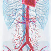
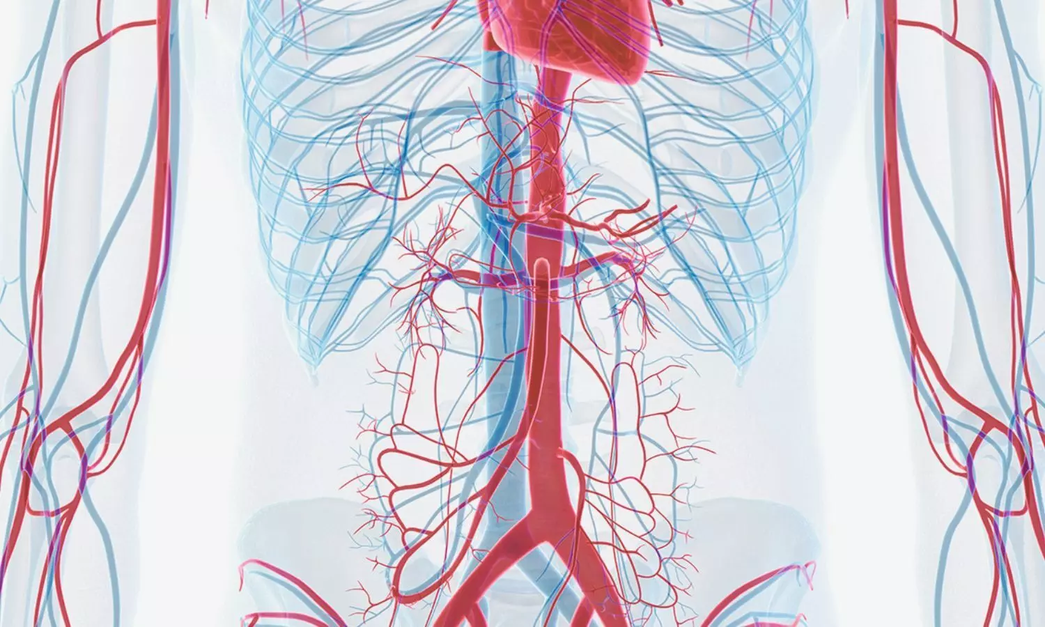
There is an unmet clinical need for tissue biomarkers that can monitor abdominal aortic aneurysm progression and predict rupture risk. Given the key role of inflammation in abdominal aortic aneurysm pathogenesis, we hypothesized that CCR2, involved in the recruitment and activation of immune cells, is upregulated in abdominal aortic aneurysms. They used our human vascular biobank to obtain aortic tissue from 42 individuals with non-ruptured and ruptured abdominal aortic aneurysm (rAAA), aortoiliac occlusive disease (AOD), or normal abdominal aorta (NAA).

We evaluated aortic wall CCR2-positive cellular content and aortic tissue levels of cytokines and CCR2 ligand, monocyte chemoattractant protein 1 (MCP1). Additionally, a single-cell RNA sequencing dataset was analyzed to assess Ccr2 expression in human abdominal aortic aneurysm tissues. Results: Compared with NAA, the aneurysmal aorta (AAA and rAAA) demonstrated significantly higher CCR2-positive cellular content (P<0.05). The number of aortic wall CCR2-positive macrophages was significantly elevated in individuals with abdominal aortic aneurysm (P<0.05), rAAA (P<0.01), and AOD (P<0.01). They also observed higher levels of MCP1 and inflammatory cytokines in diseased aortic tissue. Furthermore, single-cell transcriptomics revealed that in abdominal aortic aneurysm tissue, Ccr2 is predominantly expressed by macrophages (69%), followed by T-cells (22%), and B-cells (8%).

The findings indicate that CCR2 is a promising biomarker for diseased aortic tissue, particularly in the setting of abdominal aortic aneurysm. These findings suggest potential applications for novel molecular imaging and pharmacological targeting.

Reference:

Hafezi, Shahab MD*; Arif, Batool MS*; Ruhel, Rajrani PhD†; Zaghloul, Mohamed MD*; Elizondo-Benedetto, Santiago MD*; Pyeatte, Sophia R. MD*,‡; Joseph, Karan BS*; Zhang, Bo PhD†; Lin, Chieh-Yu MD PhD§; Gropler, Robert J. MD∥; Zayed, Mohamed A. MD PhD MBA*,‡,∥,¶,#,**. C-C Chemokine Receptor 2 is a Tissue Biomarker for Abdominal Aortic Aneurysmal and Occlusive Disease. Annals of Surgery ():10.1097/SLA.0000000000006778, June 11, 2025. | DOI: 10.1097/SLA.0000000000006778

Powered by WPeMatico

Combination of exercise and omega-3 reduces the severity of tooth root infections, suggests study

A study published in the journal Scientific Reports indicates that physical exercise combined with omega-3 supplementation considerably improves the immune response and reduces the severity of chronic apical periodontitis.

Inflammation at the apex of the tooth – the tip of the root – and in the surrounding area is primarily caused by caries. If left untreated, the bacteria can reach the root canal and pass through it to the apex, causing apical periodontitis. This condition leads to bone loss in the area.

The study is the first to demonstrate that a combination of moderate physical exercise and omega-3 supplementation significantly improves the inflammatory condition caused by apical periodontitis. This combination limited bacterial progression, reduced bone tissue loss, regulated the release of pro-inflammatory cytokines, and stimulated the activity of fibroblasts, the cells that create and maintain tissue.

If left untreated, the infection can lead to tooth loss. In addition, there is a two-way relationship between apical periodontitis and systemic changes in patients. Diabetes, metabolic syndrome, arteriosclerosis, and kidney disease, among others, can exacerbate apical periodontitis. At the same time, infection in the apex can exacerbate these diseases.

“It’s a condition that patients may not even know they have because of its chronic nature, but which can evolve and lead to bone destruction and tooth mobility. In addition, in specific situations, such as a drop in immunity, it can become acute, so the patient starts to feel pain, pus forms at the site, the face can become swollen,” explains Rogério de Castilho, a professor at the Araçatuba School of Dentistry at São Paulo State University (FOA-UNESP) in Brazil. Castilho supervised the study and is supported by FAPESP.

“In rats, physical exercise alone brought about a systemic improvement, regulating the local immune response. In addition, when combined with supplementation, it further reduced the destructive condition caused by endodontic pathology,” explains Ana Paula Fernandes Ribeiro, the first author of the study, carried out during her doctorate at FOA-UNESP.

Less inflammation

The researchers induced apical periodontitis in 30 rats and divided them into three groups. The first group received no intervention. The second and third groups underwent a 30-day swimming regimen.

The third group also received dietary supplementation of omega-3, a polyunsaturated fatty acid known for its therapeutic effects on chronic inflammatory diseases.

The group that only swam had better outcomes than the untreated control group. However, omega-3 supplementation combined with physical exercise regulated the immune response and infection control even better.

Immunohistochemical analyses, which assess how the immune system responds to infection, revealed varying levels of the cytokines interleukin 17 (IL-17) and tumor necrosis factor alpha (TNF-α), indicating the intensity of the inflammatory response.

While the rats that received no treatment had moderate levels of these cytokines, those that exercised had lower levels, and those that took supplementation had the lowest levels.

In addition to having lower levels of these cytokines, the group that exercised had fewer osteoclasts. These are cells that resorb bone tissue, indicating bone loss. The results were even better for the group that consumed omega-3, showing statistically significant differences compared to the animals that received no treatment.

Micro CT scans of the jaws showed that the animals that swam experienced less loss of volume of alveolar bone, which covers the teeth, than those in the control group. The loss was even less in the supplemented group.

For the authors, the study provides new evidence of the benefits of physical activity and omega-3 for the immune system, now with even more obvious repercussions for oral health.

“To know if the same would be true for humans, we’d need a clinical study with a significant number of patients. However, in addition to the many proven benefits of physical exercise and omega-3 consumption, this is yet another important piece of evidence,” Jacinto says.

Reference:

Ribeiro, A.P.F., de Lima Rodrigues, M., Loureiro, C. et al. Physical exercise alone or combined with omega-3 modulates apical periodontitis induced in rats. Sci Rep 15, 8760 (2025). https://doi.org/10.1038/s41598-025-90029-9

Powered by WPeMatico

Delayed First Childbirth Linked to Increased Risk of Pelvic Organ Prolapse, Review Finds

USA: As more women worldwide delay childbearing, emerging evidence suggests that having a first baby at an older age could increase the risk of developing pelvic organ prolapse (POP) later in life. A recent review conducted by researchers from the University of Utah, led by Hannah A. Zabriskie, has examined the link between maternal age at first delivery and the likelihood of POP, calling for greater clinical attention and further research on this growing concern.

The findings were published online in the American Journal of Obstetrics and Gynecology in the June 2025 issue.

Pelvic organ prolapse—a condition in which pelvic organs such as the bladder, uterus, or rectum descend from their normal position—has long been associated with vaginal delivery and advancing chronological age. However, the specific contribution of maternal age at the time of first childbirth has received limited investigation until now.

The review emphasizes that aging naturally leads to deterioration in muscle function, including a decline in strength, flexibility, and healing capacity—factors that can compromise the pelvic floor muscles during childbirth. As a result, older first-time mothers may be more vulnerable to pelvic floor injuries during delivery and less able to recover effectively afterward.

Zabriskie and her team analyzed available literature to assess how delayed childbirth influences known mechanisms involved in POP, such as pelvic muscle weakening, levator ani muscle defects, and widening of the genital hiatus. They found consistent evidence that advancing maternal age at first delivery is linked to an increased risk of pelvic floor trauma and subsequent development of prolapse symptoms.

Importantly, the authors highlighted that current human studies are limited in number and scope, especially regarding the underlying cellular and molecular processes. There is a need for more comprehensive research that examines these biological mechanisms, specifically concerning maternal age at first delivery.

The review outlines several key recommendations for future investigations. These include ensuring clearly defined study populations with consistent parity and delivery methods, using appropriate statistical models, and accounting for confounding variables such as the duration of labor. Additionally, the authors noted that older mothers are often more likely to attend postpartum follow-ups, potentially skewing data and underrepresenting younger women. Therefore, greater efforts are needed to engage and retain younger postpartum women in future cohort studies.

Ultimately, the authors advocate for age-specific analyses in future POP research and stress the need to stratify findings based on maternal age at first birth. By better understanding how age affects pelvic floor outcomes, clinicians may be able to more effectively identify women at higher risk and develop targeted therapies to prevent or treat POP.

Reference:

Zabriskie HA, Drummond MJ, Nygaard IE, Swenson CW. Older maternal age at first delivery as a risk factor for pelvic organ prolapse: what we know. Am J Obstet Gynecol. 2025 Jun;232(6):499-505. doi: 10.1016/j.ajog.2025.03.006. Epub 2025 Mar 8. PMID: 40064413.

Powered by WPeMatico

FDA Approves Prednisolone Acetate Ophthalmic Suspension for Ocular Inflammation

The FDA has approved preodnisolone acetate ophthalmic suspension, USP 1%, for treating steroid-responsive ocular inflammation. Amneal Pharmaceuticals plans to launch the product in Q3 2025.

Prednisolone acetate ophthalmic suspension, USP 1% is a sterile, topical anti-inflammatory 0agent for ophthalmic use and is indicated for treating steroid-responsive ocular inflammation.

“Our Affordable Medicines portfolio continues to grow with a strong and diverse pipeline that supports broader access to high-quality treatments across the U.S. healthcare system,” said Andy Boyer, Executive Vice President and Chief Commercial Officer, Affordable Medicines. “The approval of prednisolone acetate ophthalmic suspension-a complex product to develop and manufacture-highlights the depth of our R&D capabilities and the strength of our manufacturing and supply operations.”

The most commonly reported adverse reactions for prednisolone acetate ophthalmic suspension in clinical studies were elevation of intraocular pressure (IOP) with possible development of glaucoma and infrequent optic nerve damage, posterior subcapsular cataract formation, and delayed wound healing. For prescribing information, see package insert here.

According to IQVIA® U.S. annual sales for prednisolone acetate ophthalmic suspension for the 12 months ended April 2025 were approximately $201 million.

Powered by WPeMatico

COVID-19 Infection Linked to Higher Miscarriage Risk in Early Pregnancy: Study

A new study published in the journal of BMC Medicine showed women infected with COVID-19 before or during pregnancy faced a two to three times higher risk of miscarriage before 20 weeks of gestation. 

It is yet unclear how SARS-CoV-2 infection prior to or during pregnancy affects the course of the pregnancy. COVID-19 in late pregnancy or the peripartum period was associated with higher incidence of problems, including hypertensive disorders of pregnancy, preterm delivery, and maternal death, during the early waves of the pandemic (pre-Delta).

There is probably a complicated physiological link between maternal and newborn outcomes and SARS-CoV-2 infection at various periods of pregnancy or even before conception. Micaela Sandoval and colleagues therefore developed a tailored electronic health record (EHR)-based cohort of COVID-19 patients who experienced at least one pregnancy episode between 2019 and 2023 in order to investigate the association between COVID-19 and miscarriage.

Using a large, retrospective, electronic health record (EHR)-based cohort, this study investigated the association between COVID-19 and unfavorable pregnancy outcomes, like spontaneous abortion, ectopic pregnancy, and premature birth, between 2019 and 2023. Using generalized estimating equation modeling, risk variables for unfavorable pregnancy outcomes were determined. The following exposures were included in the study: age, race/ethnicity, comorbidity load, neighborhood-level social vulnerability, COVID-19 before pregnancy, and COVID-19 during pregnancy.

The risk of miscarriage was 6.3% among pregnancy episodes with a miscarriage, livebirth, or delivery result in the Southeast Texas Pregnancy and COVID Cohort (26,783 pregnancy episodes) (1514/ 24,119). A history of mild or moderate to severe COVID-19 before to pregnancy was linked to miscarriage in multivariable modeling (adjusted odds ratio (aOR) 2.48, CI 2.21–2.78 and aOR 2.81, CI 1.8–4.38, respectively).

Furthermore, miscarriage was linked to both mild and moderate to severe COVID-19 in the first trimester in the same model. Overall, COVID-19 was found to be a risk factor for spontaneous abortion both before and during pregnancy. These results emphasize how crucial COVID prevention is for expectant mothers and those preparing to become pregnant. 

Source:

Sandoval, M. N., Klawans, M. R., Bach, M. A., Mikhail, J., Graviss, E. A., Cao, T., Parchem, J. G., Husain, J., & Boerwinkle, E. (2025). COVID-19 infection history as a risk factor for early pregnancy loss: results from the electronic health record-based Southeast Texas COVID and Pregnancy Cohort Study. BMC Medicine, 23(1), 274. https://doi.org/10.1186/s12916-025-04094-y

Powered by WPeMatico

ASH and ISTH publish revised clinical practice guidelines for pediatric venous thromboembolism

The American Society of Hematology (ASH) and the International Society on Thrombosis and Haemostasis (ISTH) released new and updated clinical practice guidelines for the treatment of pediatric venous thromboembolism (VTE).

The guidelines, published in ASH’s peer-reviewed journal Blood Advances, were developed by an expert panel following a rigorous review process. They aim to improve health outcomes by providing evidence-based recommendations for managing VTE in children.

The new and updated pediatric VTE clinical guidelines incorporate the latest research in the field and mark the first update to the guidelines since their publication in 2018. In total, 16 recommendations were updated and four were added. One of the most notable changes is the new recommendation for direct oral anticoagulants over standard-of-care anticoagulants. Specifically, this recommendation advises clinicians to consider dabigatran or rivaroxaban over therapies such as low molecular weight heparin and vitamin k antagonist.

“These evidence-based guidelines are a key resource for clinicians to improve the quality of care for this vulnerable population,” said Belinda R. Avalos, MD, ASH president. “The recommendations are updated to incorporate the latest science, and we are glad to have collaborated with the ISTH on this project.”

“Collaborating with ASH on these updated pediatric VTE guidelines reflects the ISTH’s commitment to ensuring that children affected by VTE receive the most effective, evidence-based care,” said Pantep Angchaisuksiri, MD, ISTH president. “It’s critical that we continue advancing research and education in this area to support better outcomes for young patients worldwide.”

VTE sometimes manifests as deep vein thrombosis, when a blood clot forms in the deep veins, or pulmonary embolism, when a blood clot blocks an artery in the lung. Although the incidence of VTE in children at a population level is very low, it is higher in children who are hospitalized and can be life-threatening.

“Thrombosis in children is an increasingly common complication in children with complex illnesses and conditions and, if not managed well, can impact long-term outcomes for these patients,” said Paul Monagle, MD, MBBS, MSc, a pediatric hematologist and professor of pediatrics at the University of Melbourne and chair of the ASH ISTH Guidelines on Treatment of Pediatric VTE. “The care of children is important, and parents should know that there is a body of evidence that supports their child’s treatment options.”

Powered by WPeMatico

Sleep apnea more common than previously known in female athletes, suggests research

According to a new study, presented at the American College of Cardiology (ACC) Care of the Athletic Heart 2025 conference, sleep apnea may be more prevalent in younger female athletes than previously believed, especially among female athletes with higher levels of training. While obstructive sleep apnea has been observed in younger male athletes, the prevalence in female athletes and the association with cardiovascular risk is largely unknown.

Obstructive sleep apnea (OSA) is the most common sleep-related breathing disorder, impacting about 18 million Americans, and is prevalent in both men and women. It occurs when the throat muscles relax and block the airway, causing patients with OSA to repeatedly stop and start breathing when sleeping. Symptoms include excessive daytime sleepiness, loud snoring, waking during the night and gasping or choking. OSA increases the risk of high blood pressure and other cardiovascular issues, including arrhythmias, heart attack, heart failure and stroke.

“There is a lot of overlap in symptoms of sleep apnea and sleep deprivation, which is unsurprisingly quite common among these athletes. Some symptoms that might clue in an athletic trainer, coach or clinician that an athlete may be experiencing or at risk for sleep apnea include not feeling well-rested despite a full night of sleep, snoring very loudly, gasping or choking while sleeping,” said Austin Rim, MD, a cardiology fellow at Emory University in Atlanta and the study’s lead author. “There may be more subtle signs too, including difficulty concentrating or irritability. Accurately diagnosing sleep apnea is important, as a range of treatment options-such as CPAP, mandibular advancement devices, or even surgery-can significantly improve quality of life and, depending on disease severity, potentially reduce long-term cardiovascular risk.”

The study included 68 female collegiate athletes at two universities in the United States. Softball players made up the largest proportion of female athletes, followed by volleyball and basketball. According to the researchers, about 6 in 10 self-identified as White, a quarter as Black and the rest as Hispanic/Latino, Asian or another race/ethnicity. The average age of participants was 19 years old.

“We found in this cohort that sleep health, not even considering sleep apnea, was poor among athletes, with 36% reporting poor sleep quality and 22% reporting that they had trouble staying awake at least once in the past month while eating or engaging in social activities,” Rim said. “Ensuring good sleep hygiene among these athletes is low-hanging fruit for improving overall health in this population,”

The study recorded anthropometrics (the systematic measurements of the human body to define a person’s size and form), blood pressure and pulse wave velocity—a measurement that quantifies arterial stiffness, a precursor to hypertension. Participants also completed home sleep tests, which can detect abnormalities such as pauses in breathing during sleep. The primary outcomes were apnea-hypopnea index (AHI), systolic and diastolic blood pressure and pulse wave velocity.

AHI is a key metric to diagnose and assess the severity of sleep apnea by determining how many apneas and hypopneas occur per hour. An apnea is a complete pause of breathing lasting more than 10 seconds, and a hypopnea is a partial reduction in airflow lasting more than 10 seconds. OSA is diagnosed either by an AHI greater than five events per hour accompanied with associated symptoms like daytime sleepiness or unrefreshing sleep or an AHI score greater than 15 events per hour regardless of symptoms. According to Rim, between five events and 15 events per hour is “mild,” 15 to 30 is “moderate,” and anything over 30 is “severe.”

In this study participants ranged from 2.1 events to 7.7 events per hour and 25% were considered to have mild sleep apnea. No athletes had a measured AHI over 15 events per hour. Pulse wave velocity was higher in athletes with mild sleep apnea and was associated with higher AHI. The study found greater age and lean mass were independent predictors of sleep apnea, which the researchers said suggests sleep apnea may become more prevalent with higher levels of training in female athletes.

According to the researchers, while this is the largest study of sleep apnea among female athletes, it is limited in its generalizability given the small sample size. Future studies are needed to confirm the findings.

“Ideally, a future study would include multi-campus projects that track athletes over several seasons, compare men and women directly, and evaluate whether treating mild apnea reduces these early measures of cardiovascular risk. Treatment of OSA in older trials through CPAP haven’t always been shown to mitigate risk, so it would be helpful to see if treatment at a younger age could improve on this,” Rim said.

Reference:

Sleep apnea more common than previously known in female athletes, American College of Cardiology, Meeting: ACC Care of the Athletic Heart.

Powered by WPeMatico

Early Preemptive Kidney Transplants fail to Lower Mortality Risk in ESKD: Study

A new study published in the journal of Transplantation Proceedings showed that in individuals with end-stage kidney disease (ESKD), early preventive kidney transplants do not lower the chance of death.

Among other things, diabetes and high blood pressure can lead to end-stage kidney disease, sometimes referred to as renal or kidney failure. When starting dialysis, or even before, a patient with kidney failure needs a transplant. It is widely accepted that people with ESKD would fare better if they received a kidney transplant beforehand, before beginning dialysis, because dialysis strains the body and impairs immunity. With an emphasis on the mortality benefit of early preemptive transplants, Abhishek Kumar and colleagues therefore carried out this study to assess the present status of preemptive kidney transplants in the United States.

This research investigated patterns in preemptive kidney transplantation among first-time adult patients using the United Network of Organ Sharing database. Based on the estimated glomerular filtration rate [eGFR] at the time of transplant, they divided the population into 4 groups: ≥ 20 mL/min/1.73 m2, 10 to < 15 mL/min/1.73 m2, 15 to < 20 mL/min/1.73 m2, and < 10 mL/min/1.73 m2. The risk of ESKD was compared between the groups using the cumulative incidence competing risk (CICR) approach, and the difference in mortality was evaluated using multivariable Cox regression.

About 18% of all kidney transplants are still preventive (33% from dead donors and 67% from current donors). Preemptive kidney transplants were more likely to be given to white individuals with private insurance and greater educational attainment. The four eGFR groups did not vary in terms of mortality. Again, there was no difference in mortality between the four groups in a subgroup analysis that focused solely on recipients of preemptive kidney transplants from live donors.

Overall, the findings of study shows that even though the “Kidney First Initiative” was started more than ten years ago, the rates of preventive kidney transplants have been clinically unchanged over the past 20 years. 24% of recipients of preemptive LDKTs, where the transplant date can be somewhat controlled, have an eGFR ≥ 15 mL/min/1.73 m2, which is overly early given that there was no survival advantage and that patients were exposed to the dangers of immunosuppression and lifestyle changes.

Reference:

Kumar, A., Bonnell, L., & Kuppachi, S. (2025). Early pre-emptive kidney transplant does not offer any mortality benefits: A study of trends in pre-emptive kidney transplantation over the last two decades. Transplantation Proceedings, 57(4), 538–543. https://doi.org/10.1016/j.transproceed.2025.02.032

Powered by WPeMatico

Colchicine May Raise Stroke Risk in Elderly Patients With Intracranial Artery Stenosis: CHANCE-3 Trial

China: A new subgroup analysis from the CHANCE-3 randomized clinical trial suggests that the anti-inflammatory drug colchicine may have varying effects on the risk of recurrent stroke depending on the presence of symptomatic intracranial atherosclerotic stenosis (sICAS) and the patient’s age. The findings, published in eClinicalMedicine, raise important considerations for personalized stroke prevention strategies, especially in older adults.

The study, led by Dr. Jiejie Li and colleagues from Beijing Tiantan Hospital and Capital Medical University, evaluated data from 7,567 patients who had experienced an ischemic stroke or transient ischemic attack. All participants underwent intracranial artery assessment at baseline and were randomly assigned to receive colchicine or a placebo for 90 days. The primary goal was to assess the risk of recurrent stroke during this period.

The following were the key findings of the study:

  • Among patients with symptomatic intracranial atherosclerotic stenosis (sICAS), 10.5% of those on colchicine had a recurrent stroke compared to 8.5% in the placebo group, with an adjusted hazard ratio (HR) of 1.30, indicating a statistically significant increased risk.
  • In patients without sICAS, the recurrence rate was 4.2% in the colchicine group and 5.2% in the placebo group, with an adjusted HR of 0.80, suggesting no significant benefit.
  • These findings indicate that the effectiveness of colchicine may vary based on the presence or absence of intracranial artery stenosis.
  • In patients aged 65 and older with sICAS, colchicine was associated with a significantly higher risk of recurrent stroke (adjusted HR 1.58).
  • Serious adverse events did not differ notably between treatment groups, suggesting colchicine was generally well-tolerated.
  • Gastrointestinal side effects, particularly diarrhea, were more common in patients receiving colchicine, affecting 1.8% of them compared to 0.5%–0.8% in the placebo group, regardless of sICAS status.

The authors acknowledged certain limitations, including the exclusion of roughly 10% of participants due to unavailable imaging data and the concentration of the study within a Chinese population, which may limit generalizability. Additionally, because the analysis was a predefined subgroup study, the results should be interpreted with caution and confirmed by further research.

The authors note, “Despite these limitations, the findings highlight the importance of tailoring stroke prevention strategies. Colchicine may offer benefits to patients without intracranial stenosis, but its use in older adults with sICAS warrants careful consideration.”

They concluded, “Future international trials are needed to explore these interactions further and to inform clinical decision-making in diverse populations.”

Reference:

Li J, Jing J, Meng X, Shi FD, Gu HQ, Jin A, Jiang Y, Li H, Johnston SC, Hankey GJ, Easton JD, Xie X, Jin WN, Chang L, Shi P, Wang L, Zhuang X, Li H, Zang Y, Zhang J, Sun Z, Liu D, Li Y, Yang H, Zhao J, Yu W, Wang A, Pan Y, Lin J, Li S, Niu S, Wang Y, Zhao X, Li Z, Liu L, Zheng H, Wang Y; CHANCE-3 Investigators. Colchicine for preventing stroke in patients with and without intracranial atherosclerotic stenosis: a prespecified analysis of a randomized clinical trial. EClinicalMedicine. 2025 May 19;84:103226. Doi: 10.1016/j.eclinm.2025.103226. PMID: 40496882; PMCID: PMC12149651.

Powered by WPeMatico

AI tool enables real-world assessment of eczema severity via smartphone photos, reveals research

A team of researchers from Keio University School of Medicine, Kyoto Prefectural University of Medicine, and Teikyo University, in collaboration with Atopiyo LLC, has developed a novel artificial intelligence (AI) model that can objectively assess eczema severity using smartphone images uploaded by patients. Their findings were recently published in Allergy, the official journal of the European Academy of Allergy and Clinical Immunology.

Atopic dermatitis (AD) is a common chronic skin condition that flares repeatedly and often requires long-term monitoring and treatment adjustments. In recent years, smartphone apps and social media platforms have made it easier for patients to track their symptoms, learn about their condition, and document changes over time. However, patient-reported symptoms like itch or sleep loss don’t always align with visible disease severity. This gap underscores the need for more standardized, objective evaluation tools-and highlights the growing potential of digital biomarkers to fill that role.

To address this, the research team leveraged data from Atopiyo, Japan’s largest AD platform, where over 28,000 users have shared more than 57,000 symptom photos and personal comments since 2018. The AI model developed in this study integrates three key algorithms: body part detection, eczema lesion detection, and severity scoring using the Three Item Severity (TIS) scale, which evaluates redness, swelling, and excoriation.

Using a training dataset of 880 images with self-reported itch scores, the AI model demonstrated high diagnostic accuracy. In a validation study using 220 test images, the AI-based TIS (AI-TIS) showed a strong correlation with dermatologist-assessed TIS scores (R = 0.73, P < 0.001) and a meaningful correlation with objective SCORAD scores (R = 0.53, P = 0.04).

“Many patients with eczema struggle to evaluate their disease severity on their own,” said Dr. Takeya Adachi, the study’s corresponding author. “Our AI model allows for objective, real-time tracking using just a smartphone, empowering patients and potentially improving disease management.”

Interestingly, the study also found that AI-derived severity scores correlated only weakly with self-reported itch scores, highlighting the discrepancy between perceived symptoms and observable inflammation. This supports the growing need for digital biomarkers that can enhance precision in dermatological care.

The researchers aim to expand the model’s utility by incorporating broader skin types, age ranges, and integrating additional clinical features from scoring systems like SCORAD and EASI. This work paves the way for AI-driven teledermatology solutions that support both patients and clinicians in real-world settings.

Reference:

Utako Okata-Karigane, Masakazu Hirota, Chiaki Takahashi, Akihiro Miyagawa, Ryotaro Ako, Saeko Nakajima, Masaki Futamura, Satoru Yonekura, Yasushi Ogawa, Takenori Inomata, Tetsuo Ishikawa, Yoshihiro Ito, Katsunori Masaki, Sakura Sato, Norito Katoh, Hideaki Morita, Takeya Adachi, AI-Based Objective Severity Assessment of Atopic Dermatitis Using Patient Photos in a Real-World Setting: A Digital Biomarker Approach, Allergy, https://doi.org/10.1111/all.16586.

Powered by WPeMatico