USFDA issues one observation for Natco Pharma Mekaguda unit

Hyderabad: NATCO Pharma Limited has announced that the company has received one observation from the United States Food and Drugs Administration (USFDA) after the inspection at its Mekaguda, Hyderabad Active Pharmaceutical Ingredients (API) Unit.

The inspection was conducted from June 9th – June 13th, 2025.
“On conclusion of the inspection, the Company received 1 (One) observation in the Form-483,” the company stated. An FDA Form 483 is issued to firm management at the conclusion of an inspection when an investigator(s) has observed any conditions that in their judgment may constitute violations of the Food Drug and Cosmetic (FD&C) Act and related Acts. FDA investigators are trained to ensure that each observation noted on the FDA Form 483 is clear, specific and significant. 
“The Company believes that
the observation is procedural in nature. The Company is confident to address this observation comprehensively.
Company remains committed to being cGMP compliant and in supplying high-quality products to its customers and patients
globally,” Natco stated in a BSE filing.

NATCO Pharma Limited, headquartered at Hyderabad, India, develops, manufactures and distributes generic and branded pharmaceuticals, specialty pharmaceuticals, active pharmaceutical ingredients and crop protection products. The Company is a R&D oriented, and a science driven, leading Oncology player in the targeted therapies of domestic market and focuses on limited competition molecule in the US. The Company has 9 manufacturing sites and 2 R&D facilities in India. The Company’s manufacturing facilities are approved by several leading regulatory authorities like US FDA, Brazil ANVISA, Health Canada, WHO and others catering to 50+ global markets.

Read also: Natco Pharma Executive VP – Technology Transfer, Intellectual Property Rights & Regulatory Affairs (API), Dr Ramesh Dandala, superannuates

Powered by WPeMatico

Aundh District Hospital under scrutiny after inspection reveals lapses

Pune: Aundh District Hospital (ADH) has come under scrutiny following a surprise inspection that revealed serious lapses in hygiene, staff discipline, and attendance protocols. The Pune district civil surgeon has issued a strict warning to hospital staff, implementing disciplinary measures, pay cuts, and stricter attendance monitoring.

The inspection, conducted on May 30 by Dr. Radhakishan Pawar, Deputy Director of Health Services, revealed appalling conditions in the hospital’s wards and toilets. Despite the hospital having a significant number of sanitation and support staff, the premises were found to be in a filthy state. In addition, many staff members were absent during duty hours, and those present were not wearing the prescribed uniforms.

Also Read: Activists Call for Opening of 100-Bed Aundh District Hospital Amid HMPV Concerns

Dr. Nagnath Yempalay, Pune district civil surgeon, took immediate action and on June 10 issued a formal warning letter to ADH staff. The letter outlines the consequences for failing to adhere to basic standards of hygiene and discipline. These include strict disciplinary action, pay cuts, and withholding of salary for those not complying with the rules.

To ensure accountability, the hospital staff have been instructed to mark their attendance using the Aadhaar-based biometric facial recognition system. The official letter stated that failure to comply would result in non-payment of salaries.

Also Read: 100-bed Aundh District Hospital to be utilised for Ophthalmology department

Speaking to Hindustan Times, Dr Yempalay said, “The sanitation staff have been asked to make sure the toilets and wards are cleaned daily by the respective staff. If they are found unclean, the salary for that day will not be paid to the concerned employee. Additionally, class IV employees who do not wear the uniform while on duty daily will not be paid the washing allowance in their salary from May 2025 onwards.”

Dr Pawar said, during the inspection, several staff were not in uniform and were without ID cards, which confused if they were staff or visitors to the hospital. “Orders have been given to take strict action,” he said, reports Hindustan Times.

Powered by WPeMatico

Celiac and Thyroid Diseases Linked to Over Twofold Higher Risk of Type 1 Diabetes, Especially in Youth: Study Finds

USA: People diagnosed with celiac disease or thyroid disorders may be at significantly increased risk of developing type 1 diabetes (T1D), according to new findings published in Diabetes, Obesity and Metabolism. The study, led by Dr. Steve V. Edelman from the University of California, San Diego, highlights the importance of proactive screening for early-stage diabetes in these patient populations.

The retrospective, observational study analyzed real-world insurance claims data to assess the incidence of new-onset type 1 diabetes among individuals with celiac disease, hyperthyroidism (such as Graves’ disease), and hypothyroidism (such as Hashimoto’s thyroiditis), compared with matched individuals who had none of these autoimmune conditions. The research team used a 1:1 propensity score matching strategy to ensure similar baseline characteristics between groups, allowing for a more accurate comparison of diabetes risk.

The study led to the following findings:

  • During a median follow-up of two years, individuals with celiac disease, hyperthyroidism, or hypothyroidism were found to be significantly more likely to develop type 1 diabetes compared to those without these conditions.
  • In the celiac disease group, 0.14% (68 out of 47,099) developed T1D compared to 0.06% (27 out of 47,099) in the control group.
  • Among individuals with hyperthyroidism, 0.17% (281 out of 164,830) developed T1D, while only 0.06% (99 out of 164,830) of their matched counterparts did.
  • In the hypothyroidism cohort, 0.18% (1,756 out of 980,477) developed T1D, versus 0.08% (764 out of 980,477) in controls.
  • The risk of developing type 1 diabetes was more than double in all three disease groups compared to controls, with hazard ratios of 2.54 for celiac disease, 2.98 for hyperthyroidism, and 2.41 for hypothyroidism.
  • The risk of T1D was especially elevated among individuals younger than 18 years.
  • The time to T1D diagnosis was shorter in individuals with autoimmune diseases than in those without, suggesting a faster progression toward clinical diabetes in these groups.

The researchers suggest that individuals with celiac disease or thyroid disorders could benefit from regular screening for type 1 diabetes, particularly for markers like islet autoantibodies that can indicate the presence of stage 2 T1D before clinical symptoms appear. Early detection could allow timely interventions, including disease-modifying therapies, potentially preventing or delaying the onset of overt diabetes and its complications.

These findings reinforce the interconnectivity of autoimmune conditions and highlight the need for integrated care approaches. The authors advocate for increased vigilance and screening protocols to identify at-risk individuals early, offering a window of opportunity for preventive strategies.

Reference:

Edelman SV, Agardh D, Cui N, Hao L, Wieloch M, Meneghini L. Risk of new-onset type 1 diabetes in individuals with celiac disease and thyroid disease-An observational study. Diabetes Obes Metab. 2025 May 28. doi: 10.1111/dom.16454. Epub ahead of print. PMID: 40437819.

Powered by WPeMatico

MASLD Linked to Elevated ASCVD Risk in Type 1 Diabetes Patients: Study

MASLD (metabolic dysfunction-associated steatotic liver disease) is linked to elevated ASCVD Risk in Type 1 diabetes patients suggests a new study published in the Cardiology Diabetology.

This study aimed to investigate the correlation between metabolic dysfunction-associated steatotic liver disease (MASLD) and atherosclerotic cardiovascular disease (ASCVD) in individuals with type 1 diabetes (T1D). Adults with T1D (n = 659) were consecutively screened for liver steatosis via abdominal ultrasound.

The presence of macrovascular disease (including coronary artery disease [CAD], peripheral artery disease [PAD], or ischaemic stroke [CVA, cerebrovascular accident]) was identified via electronic medical records. The 5- and 10-year risks of fatal/nonfatal ASCVD were assessed via the Steno Type 1 Risk Engine. Insulin resistance was assessed via the estimated glucose disposal rate (eGDR). Results: The metabolic dysfunction-associated steatotic liver disease prevalence was 16.8%.

The prevalence of composite atherosclerotic cardiovascular disease, CAD, PAD and CVA was greater in people with metabolic dysfunction-associated steatotic liver disease. The 5-year and 10-year risks of atherosclerotic cardiovascular disease were greater in those with metabolic dysfunction-associated steatotic liver disease. Metabolic dysfunction-associated steatotic liver disease was associated with prevalent atherosclerotic cardiovascular disease, independent of age, sex, diabetes duration, smoking, statin use, LDL-cholesterol, the glomerular filtration rate, albuminuria, and metabolic syndrome.

Metabolic dysfunction-associated steatotic liver disease is associated with both an increased prevalence of atherosclerotic cardiovascular disease and an increased calculated risk of fatal/nonfatal atherosclerotic cardiovascular disease in people with T1D.

Reference:

Mertens J, Weyler J, Dirinck E, Vonghia L, Kwanten WJ, Van Gaal LF, De Winter BY, Francque S, De Block C. Increased prevalence and risk of atherosclerotic cardiovascular disease in individuals with Type 1 diabetes and metabolic dysfunction-associated steatotic liver disease. Cardiovasc Diabetol. 2025 May 29;24(1):230. doi: 10.1186/s12933-025-02764-y. PMID: 40442720; PMCID: PMC12124096.

Powered by WPeMatico

Elevated Atherogenic Index of Plasma Levels Predict Higher Death Risk in Cardiovascular-Kidney-Metabolic Syndrome: Study

Researchers have found in a new study that higher levels of the atherogenic index of plasma (AIP) is strongly associated with increased risks of all-cause and cardiovascular mortality among Cardiovascular-Kidney-Metabolic (CKM) syndrome patients. The study found AIP to be a good predictor of mortality among CKM patients, solidifying its position as a useful and easily available clinical biomarker. The study was conducted by Qianrong Z. and colleagues published in the journal of Cardiovascular Diabetology.

CKM syndrome is a newly developed clinical syndrome which recognizes the intertwined pathophysiology of metabolic disease, chronic kidney disease (CKD), and cardiovascular disease (CVD). As cardiovascular death remains the leading cause of death in the globe, it is necessary to identify early predictive indicators in patients with CKM. The AIP, as measured by the log ratio of triglycerides to high-density lipoprotein cholesterol (log10[TG/HDL-C]), has proven to be a sensitive marker of atherosclerosis and cardiovascular disease risk. Yet until recently, proof linking AIP directly to mortality in CKM patients was sparse.

This large scale observational study used 15,703 CKM syndrome cases diagnosed from the National Health and Nutrition Examination Survey (NHANES) data, conducted between 2005 and 2018. AIP was estimated for all participants and mortality was ascertained through linkage to the National Death Index (NDI), with follow-up through December 31, 2019.

Researchers used Kaplan-Meier survival curves, Cox proportional hazards models, restricted cubic spline (RCS) models, and subgroup analyses to examine the correlation of AIP levels with mortality outcomes. Participants were divided into tertiles according to their AIP values to be compared.

Key Findings

• A median follow-up duration of 7.67 years saw 1,570 deaths in the participants out of which 344 cardiovascular deaths were reported specifically.

• All-cause mortality: Participants in the uppermost AIP tertile had a 19% increased risk of all-cause death than those in the lowermost tertile (Hazard Ratio [HR] = 1.19; 95% Confidence Interval [CI]: 1.08–1.31; P < 0.001).

• Cardiovascular mortality: Risk of cardiovascular death was 38% higher in the highest AIP tertile (HR = 1.38; 95% CI: 1.22–1.57; P < 0.001).

• A positive linear dose-response association existed between both types of mortality and AIP levels when AIP was treated as a continuous variable.

• Kaplan-Meier curves illustrated considerably better survival outcomes among participants in the lowest tertile of AIP.

• Subgroup analyses revealed that the associations between higher AIP and higher mortality were sustained in all but most clinical and demographic variables, with the exception of gender, suggesting potential sex differences in risk associated with AIP.

This NHANES-derived study presents strong evidence that high AIP levels are independently and strongly linked to higher all-cause and cardiovascular mortality in patients with CKM syndrome. These findings highlight AIP’s utility as a simple, cost-effective, and non-invasive biomarker for mortality risk estimation. Routine clinical application of AIP could have a considerable advantage in managing and predicting patients with cardiometabolic and renal comorbidities.

Reference:

Zheng Q, Cao Z, Teng J, Lu Q, Huang P, Zhou J. Association between atherogenic index of plasma with all-cause and cardiovascular mortality in individuals with Cardiovascular-Kidney-Metabolic syndrome. Cardiovasc Diabetol. 2025 Apr 26;24(1):183. doi: 10.1186/s12933-025-02742-4. PMID: 40287685; PMCID: PMC12034140.

Powered by WPeMatico

Eating more fruits and veggies could help you sleep better: Study

From counting sheep to white noise and weighted blankets, people have tried innumerable ways to get a good night’s sleep. Sleep disruptions can have far-reaching negative consequences, impacting cardiovascular and metabolic health, memory, learning, productivity, mood regulation, interpersonal relationships and more.

It turns out that an important tool for improving sleep quality may have been hiding in plain sight…in the produce aisle. A new study led by researchers at the University of Chicago Medicine and Columbia University found that eating more fruits and vegetables during the day was associated with sleeping more soundly later that same night.

“Dietary modifications could be a new, natural and cost-effective approach to achieve better sleep,” said co-senior author Esra Tasali, MD, director of the UChicago Sleep Center. “The temporal associations and objectively-measured outcomes in this study represent crucial steps toward filling a gap in important public health knowledge.”

Uncovering the connection between diet and sleep

Studies have shown that inadequate sleep can cause people to gravitate towards unhealthier diets higher in fat and sugar. However, despite the far-reaching effects of sleep on public health and even economic productivity, doctors and scientists know far less about how diet can affect sleep patterns.

In previous observational studies, high fruit and vegetable intakes were associated with better self-reported overall sleep quality, but this new study was the first to draw a temporal connection between a given day’s dietary choices and objectively-measured sleep quality that same night.

Healthy young adults who participated in the study reported their food consumption each day with an app and wore a wrist monitor that allowed the researchers to objectively measure their sleep patterns. The researchers specifically looked at “sleep fragmentation,” an index that reflects how often someone awakens or shifts from deep to light sleep throughout the night.

Promising findings support dietary guidelines

The researchers found that each day’s diet was correlated with meaningful differences in the subsequent night’s sleep. Participants who ate more fruits and vegetables during the day tended to have deeper, more uninterrupted sleep that same night, as did those who consumed more healthy carbohydrates like whole grains.

Based on their findings and statistical modeling, the researchers estimate that people who eat the CDC-recommended five cups of fruits and veggies per day could experience a 16 percent improvement in sleep quality compared to people who consume no fruits or vegetables.

“16 percent is a highly significant difference,” Tasali said. “It’s remarkable that such a meaningful change could be observed within less than 24 hours.”

Future studies will help establish causation, broaden the findings across diverse populations, and examine the underlying mechanisms of digestion, neurology, and metabolism that could explain the positive impact of fruits and vegetables on sleep quality. But based on current data, the experts confidently advise that regularly eating a diet rich in complex carbohydrates, fruits, and vegetables is best for long-term sleep health.

“People are always asking me if there are things they can eat that will help them sleep better,” said co-senior author Marie-Pierre St-Onge, PhD, director of the Center of Excellence for Sleep & Circadian Research at Columbia. “Small changes can impact sleep. That is empowering-better rest is within your control.”

Reference:

Boege, Hedda L. et al., Higher daytime intake of fruits and vegetables predicts less disrupted nighttime sleep in younger adults, Sleep Health, DOI:10.1016/j.sleh.2025.05.003 

Powered by WPeMatico

Conservative Oxygen Therapy does not harm chances of survival in Ventilated ICU Patients: JAMA

Reducing supplementary oxygen given to intensive care patients does not deliver any clear and obvious health benefits or harm their chances of survival, according to new research.

Each year, around 184,000 patients are admitted to NHS intensive care units (ICUs) and over 30% need breathing support in the form of oxygen provided through mechanical ventilators.

However, there is insufficient evidence to guide the use of oxygen, with the possibility that administering too much, or too little, oxygen may not deliver the intended benefits and actually end up causing patients additional harm.

That prompted researchers to establish UK-ROX, the largest ever clinical trial of oxygen use in UK ICUs, and they recruited 16,500 patients across almost 100 intensive care units at UK hospitals.

They then sought to establish whether a strategy of conservative oxygen therapy – delivering less oxygen to maintain a person’s oxygen saturation at around 90% – would reduce mortality rates among ICU patients.

The results-published in the JAMA journal-showed there was no statistically significant difference in patient outcomes with 35.4% of patients receiving conservative oxygen therapy having died within 90 days of their admission compared with 34.9% of patients receiving usual oxygen therapy.

As a result, they believe it is safe to allow oxygen levels to be lower in ICU patients but that it may not necessarily be better for them in terms of survival, with the need to develop and evaluate more personalised therapies based on a person’s specific medical conditions.

The UK-ROX trial, funded by the National Institute for Health and Care Research (NIHR), was led by researchers at the University of Plymouth’s Peninsula Medical School and the Intensive Care National Audit & Research Centre (ICNARC).

Professor Daniel Martin OBE, Professor of Perioperative and Intensive Care Medicine at the University of Plymouth and a Consultant at University Hospitals Plymouth NHS Foundation Trust, is co-chief investigator of the UK-ROX trial. He said: “Administering supplemental oxygen through mechanical ventilation is a standard course of treatment for many of those admitted to intensive care. However, there is insufficient evidence to guide us on what oxygen levels deliver the greatest benefits or have the potential to cause harm. The results of this study show that reducing supplementary oxygen results in no overall benefit or harm to the patients, but that doesn’t mean it is not important for intensive care patients. On the contrary, it means that we will have to come up with ways of determining how much oxygen an individual patient needs and deliver it to them in a precise way if we are to improve patient outcomes.”

Paul Mouncey, Co-Director of the Intensive Care National Audit & Research Centre (ICNARC) and the other co-chief investigator of the UK-ROX trial, said: “UK-ROX is the largest individually randomised trial conducted in intensive care in the UK. The study has provided much needed robust evidence to inform clinicians in their day-to-day management of patients. A study of this size was only possible by using routinely collected data held within NHS England and within the Case Mix Programme, the national clinical audit of adult critical care in England, Wales and Northern Ireland. We would like to thank the 97 NHS hospitals and 16,500 patients and their families for taking part in the clinical trial.”

Co-investigator Professor Mike Grocott, Director of the NIHR Southampton Biomedical Research Centre, added: “This landmark study clarifies the safety of administering lower levels of oxygen to critically ill patients. Understanding how to individualise oxygen therapy will be the next step in improving patient care.”

In addition to being published in JAMA, Professor Martin and Mr Mouncey have today presented the results of the UK-ROX trial to delegates at the Critical Care Reviews Meeting 2025 in Belfast.

The study and presentation also include details of how they were able to deliver UK-ROX at a fraction of the usual cost (around £100 per patient) of a randomised control trial, showing that clinical trials can be conducted differently and cost-effectively in an increasingly challenging financial climate.

Reference:

Martin DS, Gould DW, Shahid T, et al. Conservative Oxygen Therapy in Mechanically Ventilated Critically Ill Adult Patients: The UK-ROX Randomized Clinical Trial. JAMA. Published online June 12, 2025. doi:10.1001/jama.2025.9663

Powered by WPeMatico

CCR2 Identified as Promising Biomarker for AAA-Associated Aortic Disease: Study

Researchers have found in a new study that CCR2 shows strong potential as a biomarker for diseased aortic tissue in abdominal aortic aneurysm (AAA), supporting its use in future molecular imaging and targeted therapies.

This study evaluated whether C-C Chemokine Receptor 2 (CCR2) is a tissue biomarker of aortic disease severity, particularly in patients with abdominal aortic aneurysm (AAA).

There is an unmet clinical need for tissue biomarkers that can monitor abdominal aortic aneurysm progression and predict rupture risk. Given the key role of inflammation in abdominal aortic aneurysm pathogenesis, we hypothesized that CCR2, involved in the recruitment and activation of immune cells, is upregulated in abdominal aortic aneurysms. They used our human vascular biobank to obtain aortic tissue from 42 individuals with non-ruptured and ruptured abdominal aortic aneurysm (rAAA), aortoiliac occlusive disease (AOD), or normal abdominal aorta (NAA).

We evaluated aortic wall CCR2-positive cellular content and aortic tissue levels of cytokines and CCR2 ligand, monocyte chemoattractant protein 1 (MCP1). Additionally, a single-cell RNA sequencing dataset was analyzed to assess Ccr2 expression in human abdominal aortic aneurysm tissues. Results: Compared with NAA, the aneurysmal aorta (AAA and rAAA) demonstrated significantly higher CCR2-positive cellular content (P<0.05). The number of aortic wall CCR2-positive macrophages was significantly elevated in individuals with abdominal aortic aneurysm (P<0.05), rAAA (P<0.01), and AOD (P<0.01). They also observed higher levels of MCP1 and inflammatory cytokines in diseased aortic tissue. Furthermore, single-cell transcriptomics revealed that in abdominal aortic aneurysm tissue, Ccr2 is predominantly expressed by macrophages (69%), followed by T-cells (22%), and B-cells (8%).

The findings indicate that CCR2 is a promising biomarker for diseased aortic tissue, particularly in the setting of abdominal aortic aneurysm. These findings suggest potential applications for novel molecular imaging and pharmacological targeting.

Reference:

Hafezi, Shahab MD*; Arif, Batool MS*; Ruhel, Rajrani PhD†; Zaghloul, Mohamed MD*; Elizondo-Benedetto, Santiago MD*; Pyeatte, Sophia R. MD*,‡; Joseph, Karan BS*; Zhang, Bo PhD†; Lin, Chieh-Yu MD PhD§; Gropler, Robert J. MD∥; Zayed, Mohamed A. MD PhD MBA*,‡,∥,¶,#,**. C-C Chemokine Receptor 2 is a Tissue Biomarker for Abdominal Aortic Aneurysmal and Occlusive Disease. Annals of Surgery ():10.1097/SLA.0000000000006778, June 11, 2025. | DOI: 10.1097/SLA.0000000000006778

Powered by WPeMatico

Combination of exercise and omega-3 reduces the severity of tooth root infections, suggests study

A study published in the journal Scientific Reports indicates that physical exercise combined with omega-3 supplementation considerably improves the immune response and reduces the severity of chronic apical periodontitis.

Inflammation at the apex of the tooth – the tip of the root – and in the surrounding area is primarily caused by caries. If left untreated, the bacteria can reach the root canal and pass through it to the apex, causing apical periodontitis. This condition leads to bone loss in the area.

The study is the first to demonstrate that a combination of moderate physical exercise and omega-3 supplementation significantly improves the inflammatory condition caused by apical periodontitis. This combination limited bacterial progression, reduced bone tissue loss, regulated the release of pro-inflammatory cytokines, and stimulated the activity of fibroblasts, the cells that create and maintain tissue.

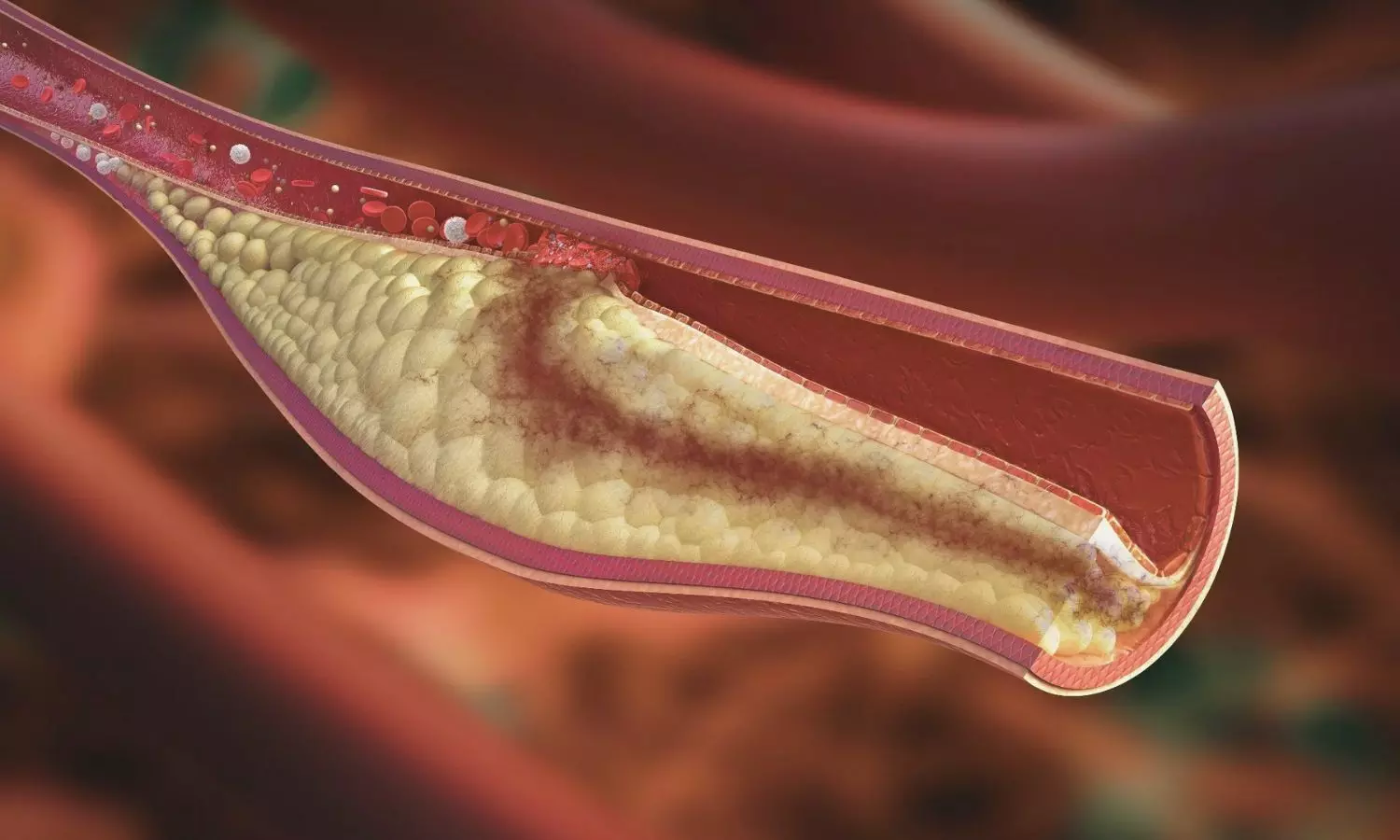
If left untreated, the infection can lead to tooth loss. In addition, there is a two-way relationship between apical periodontitis and systemic changes in patients. Diabetes, metabolic syndrome, arteriosclerosis, and kidney disease, among others, can exacerbate apical periodontitis. At the same time, infection in the apex can exacerbate these diseases.

“It’s a condition that patients may not even know they have because of its chronic nature, but which can evolve and lead to bone destruction and tooth mobility. In addition, in specific situations, such as a drop in immunity, it can become acute, so the patient starts to feel pain, pus forms at the site, the face can become swollen,” explains Rogério de Castilho, a professor at the Araçatuba School of Dentistry at São Paulo State University (FOA-UNESP) in Brazil. Castilho supervised the study and is supported by FAPESP.

“In rats, physical exercise alone brought about a systemic improvement, regulating the local immune response. In addition, when combined with supplementation, it further reduced the destructive condition caused by endodontic pathology,” explains Ana Paula Fernandes Ribeiro, the first author of the study, carried out during her doctorate at FOA-UNESP.

Less inflammation

The researchers induced apical periodontitis in 30 rats and divided them into three groups. The first group received no intervention. The second and third groups underwent a 30-day swimming regimen.

The third group also received dietary supplementation of omega-3, a polyunsaturated fatty acid known for its therapeutic effects on chronic inflammatory diseases.

The group that only swam had better outcomes than the untreated control group. However, omega-3 supplementation combined with physical exercise regulated the immune response and infection control even better.

Immunohistochemical analyses, which assess how the immune system responds to infection, revealed varying levels of the cytokines interleukin 17 (IL-17) and tumor necrosis factor alpha (TNF-α), indicating the intensity of the inflammatory response.

While the rats that received no treatment had moderate levels of these cytokines, those that exercised had lower levels, and those that took supplementation had the lowest levels.

In addition to having lower levels of these cytokines, the group that exercised had fewer osteoclasts. These are cells that resorb bone tissue, indicating bone loss. The results were even better for the group that consumed omega-3, showing statistically significant differences compared to the animals that received no treatment.

Micro CT scans of the jaws showed that the animals that swam experienced less loss of volume of alveolar bone, which covers the teeth, than those in the control group. The loss was even less in the supplemented group.

For the authors, the study provides new evidence of the benefits of physical activity and omega-3 for the immune system, now with even more obvious repercussions for oral health.

“To know if the same would be true for humans, we’d need a clinical study with a significant number of patients. However, in addition to the many proven benefits of physical exercise and omega-3 consumption, this is yet another important piece of evidence,” Jacinto says.

Reference:

Ribeiro, A.P.F., de Lima Rodrigues, M., Loureiro, C. et al. Physical exercise alone or combined with omega-3 modulates apical periodontitis induced in rats. Sci Rep 15, 8760 (2025). https://doi.org/10.1038/s41598-025-90029-9

Powered by WPeMatico

Delayed First Childbirth Linked to Increased Risk of Pelvic Organ Prolapse, Review Finds

USA: As more women worldwide delay childbearing, emerging evidence suggests that having a first baby at an older age could increase the risk of developing pelvic organ prolapse (POP) later in life. A recent review conducted by researchers from the University of Utah, led by Hannah A. Zabriskie, has examined the link between maternal age at first delivery and the likelihood of POP, calling for greater clinical attention and further research on this growing concern.

The findings were published online in the American Journal of Obstetrics and Gynecology in the June 2025 issue.

Pelvic organ prolapse—a condition in which pelvic organs such as the bladder, uterus, or rectum descend from their normal position—has long been associated with vaginal delivery and advancing chronological age. However, the specific contribution of maternal age at the time of first childbirth has received limited investigation until now.

The review emphasizes that aging naturally leads to deterioration in muscle function, including a decline in strength, flexibility, and healing capacity—factors that can compromise the pelvic floor muscles during childbirth. As a result, older first-time mothers may be more vulnerable to pelvic floor injuries during delivery and less able to recover effectively afterward.

Zabriskie and her team analyzed available literature to assess how delayed childbirth influences known mechanisms involved in POP, such as pelvic muscle weakening, levator ani muscle defects, and widening of the genital hiatus. They found consistent evidence that advancing maternal age at first delivery is linked to an increased risk of pelvic floor trauma and subsequent development of prolapse symptoms.

Importantly, the authors highlighted that current human studies are limited in number and scope, especially regarding the underlying cellular and molecular processes. There is a need for more comprehensive research that examines these biological mechanisms, specifically concerning maternal age at first delivery.

The review outlines several key recommendations for future investigations. These include ensuring clearly defined study populations with consistent parity and delivery methods, using appropriate statistical models, and accounting for confounding variables such as the duration of labor. Additionally, the authors noted that older mothers are often more likely to attend postpartum follow-ups, potentially skewing data and underrepresenting younger women. Therefore, greater efforts are needed to engage and retain younger postpartum women in future cohort studies.

Ultimately, the authors advocate for age-specific analyses in future POP research and stress the need to stratify findings based on maternal age at first birth. By better understanding how age affects pelvic floor outcomes, clinicians may be able to more effectively identify women at higher risk and develop targeted therapies to prevent or treat POP.

Reference:

Zabriskie HA, Drummond MJ, Nygaard IE, Swenson CW. Older maternal age at first delivery as a risk factor for pelvic organ prolapse: what we know. Am J Obstet Gynecol. 2025 Jun;232(6):499-505. doi: 10.1016/j.ajog.2025.03.006. Epub 2025 Mar 8. PMID: 40064413.

Powered by WPeMatico