Diet rich in omega-3 fatty acids may help ward off short sightedness in children: Research

A diet rich in omega-3 fatty acids, found predominantly in fish oils, may help ward off the development of short sightedness (myopia) in children, while a high intake of saturated fats, found in foods such as butter, palm oil, and red meat, may boost the risk of the condition, finds research published online in the British Journal of Ophthalmology.

The global prevalence of myopia is rising, especially in East Asia, and it’s predicted that around half of the world’s population will be affected by 2050, note the researchers.

Risk factors are thought to include excessive screen time and too little time spent outdoors, as well as inherited susceptibility, they explain.

Omega-3 polyunsaturated fatty acids (ω-3 PUFAs), which can only be obtained from the diet, are thought to improve/prevent several chronic eye conditions, including dry eye disease and age-related macular degeneration. But whether they can help ward off myopia isn’t clear as studies to date have been experimental and haven’t included people.

To explore this further, the researchers drew on 1005 Chinese 6-8 year olds, randomly recruited from the population based Hong Kong Children Eye Study, which is tracking the development of eye conditions and potential risk factors.

The children’s eyesight was assessed and their regular diet measured by a food frequency questionnaire, completed with the help of their parents. This included 280 food items categorised into 10 groups: bread/cereals/pasta/rice/noodles; vegetables and legumes; fruit; meat; fish; eggs; milk and dairy products; drinks; dim sum/snacks/fats/oils; and soups.

Intakes of energy, carbohydrate, proteins, total fat, saturated fats, monounsaturated fats, PUFAs, cholesterol, iron, calcium, vitamins A and C, fibre, starch, sugar and nutrients were then calculated, based on the questionnaire responses.

The amount of time the children spent outdoors in leisure and during sports activities, reading and writing, and on screens during weekdays and at the weekend was calculated from validated questionnaire responses.

In all, around a quarter of the children (276; 27.5%) had myopia. Higher dietary intake of omega-3 fatty acids was associated with a lower risk of the condition.

Axial length-measurement of the eye from the cornea at the front to the retina at the back, and an indicator of myopia progression—was longest in the 25% of children with the lowest dietary intake of omega-3 fatty acids, after accounting for influential factors, including age, sex, weight (BMI), the amount of time spent in close work and outdoors, and parental myopia.

It was shortest in the 25% of children with the highest dietary intake of omega-3 fatty acids.

Similarly, cycloplegic spherical equivalent (SE), which measures refractive error, such as the degree of shortsightedness, was highest in those with the lowest omega-3 fatty acid intake and lowest in those with the highest intake.

But these findings were reversed for the 25% of children with the highest saturated fat intake, compared with the 25% of those with the lowest. None of the other nutrients was associated with either measure or myopia.

This is an observational study, and as such, can’t establish causal and temporal factors. And the researchers acknowledge that food frequency questionnaires rely on recall and only provide a snapshot in time of diet. Nor was there objective evidence of nutritional intake from blood samples.

The prevalence of myopia in Hong Kong is also among the highest in the world. And whether the findings might apply to other ethnic groups with different lifestyles and less myopia remains to be verified, they add.

But omega-3 fatty acids may suppress myopia by increasing blood flow through the choroid, a vascular layer in the eye, responsible for delivering nutrients and oxygen, and so staving off scleral hypoxia-oxygen deficiency in the white of the eye and a key factor in the development of shortsightedness, they suggest.

And they conclude: “This study provides the human evidence that higher dietary ω-3 PUFA intake is associated with shorter axial length and less myopic refraction, highlighting ω-3 PUFAs as a potential protective dietary factor against myopia development.”

Reference:

Dietary omega-3 polyunsaturated fatty acids as a protective factor of myopia: the Hong Kong Children Eye Study Doi: 10.1136/bjo-2024-326872

Powered by WPeMatico

Er:YAG Laser Plus 60s Acid Etch Restores Bonding in Fluorotic Enamel: Study

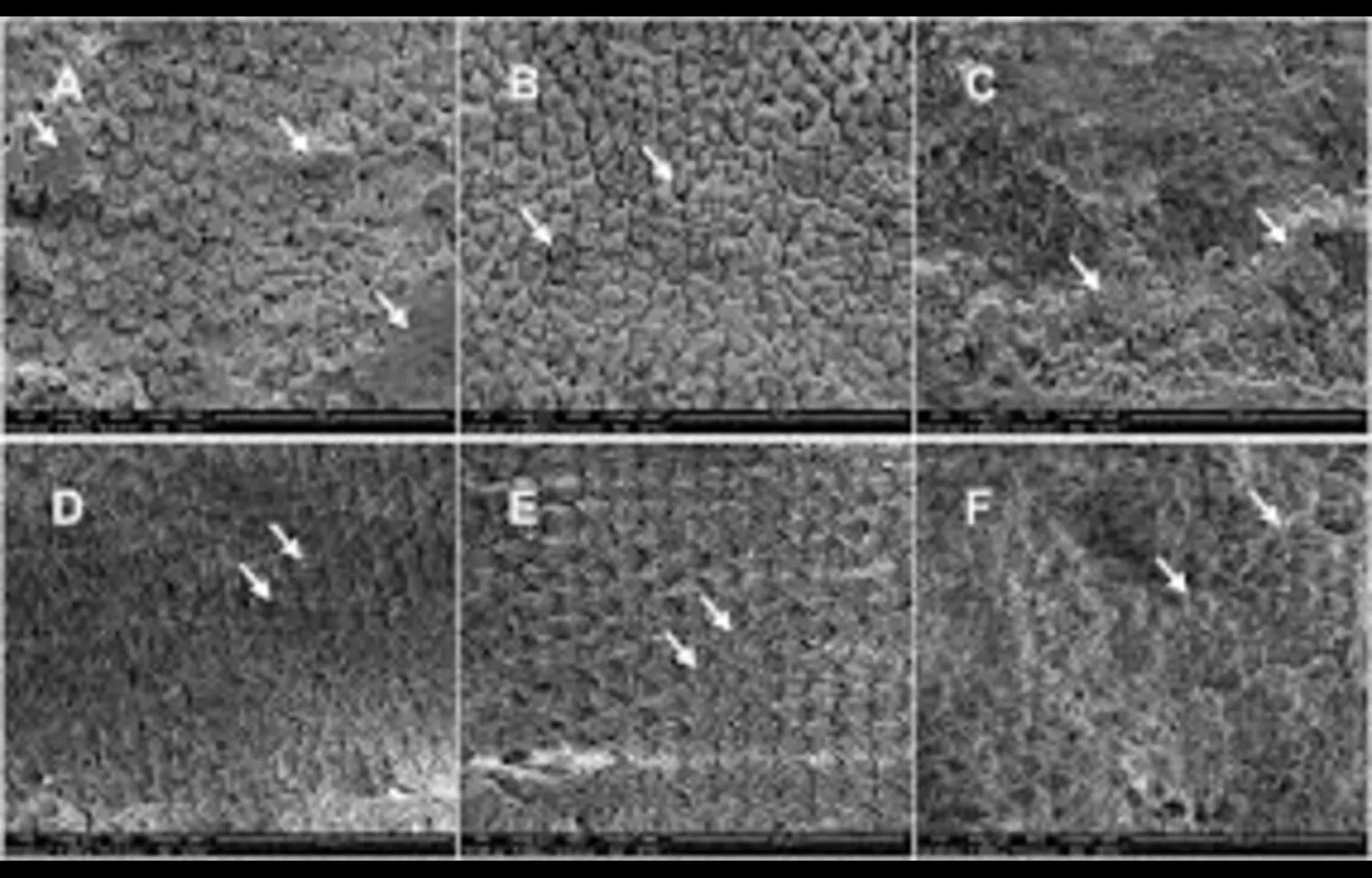
According to a new study use of an Er:YAG laser at 100 mJ/30 Hz combined with 60 seconds of acid etching produces excellent adhesion for attachments on fluorotic enamel — restoring bond strength to levels comparable with healthy enamel while avoiding over-etching damage. The researchers found that this approach avoids the risk of over-etching, which can damage enamel structure, while still achieving strong adhesion for orthodontic attachments. Dental fluorosis can alter the mineralization of enamel, making it more resistant to acid etching and reducing bond strength for orthodontic brackets or attachments. Standard etching protocols often fail to achieve optimal adhesion, and aggressive etching may compromise the enamel surface. In this study, the laser-plus-acid combination created an ideal surface texture for micromechanical interlocking without excessive enamel loss. The findings suggest that this protocol could offer clinicians a predictable and safe way to manage bonding challenges in patients with dental fluorosis. By fine-tuning the laser energy and etching duration, the researchers were able to overcome the adhesion limitations of fluorotic enamel while preserving tooth integrity. The authors note that further clinical trials are needed to confirm these in vitro results and evaluate long-term bond durability under oral conditions. If validated, this method could improve treatment outcomes in orthodontics, particularly in regions where fluorosis prevalence is high.

Reference:
Xia R, Lei J, Luo M, Xiao Y (2025) Effects of Er: YAG laser and acid etching on bond strength of clear aligner attachments to fluorotic enamel. PLoS One 20(8): e0328937. https://doi.org/10.1371/journal.pone.0328937

Keywords:

Er:YAG laser, dental fluorosis, acid etching, enamel bonding, orthodontics, attachment adhesion, surface preparation

Powered by WPeMatico

Iontophoresis and low-dye taping are effective conservative interventions for plantar fasciitis: study

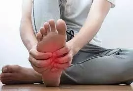
Plantar fasciitis (PF) is a frequent cause of heel pain, affecting approximately 10% of the population. Conservative treatments such as iontophoresis and low-dye taping (LDT) are widely used to alleviate symptoms, often providing short-term pain relief.

Aurora Castro‑Mendez et al conducted a systematic review to compare the efficacy of iontophoresis (with 5% acetic acid, 0.4% dexamethasone, dexamethasone and lidocaine, or placebo) versus low-dye taping (LDT) in treating plantar fasciitis. Additionally, it evaluated the combined effect of iontophoresis and LDT application.

A systematic search was conducted in Scopus, PubMed, Web of Science, CINAHL, and the Cochrane Library databases, following PRISMA guidelines and the Cochrane Handbook for Systematic Reviews of Interventions. Inclusion and exclusion criteria were predefined. Two independent reviewers screened and extracted data from eligible studies, assessing their quality. Included studies comprised randomized controlled trials, non-randomized clinical trials, case–control studies, systematic reviews, and meta-analyses. The review protocol was registered with PROSPERO.

Key findings of the study were:

• Eight studies published between 1997 and 2018 were included, providing a moderate level of evidence.

• Both iontophoresis (with the specified agents) and low-dye taping, alone or combined, were associated with statistically significant reductions in pain scores compared to baseline.

The authors concluded – “Iontophoresis and low-dye taping are effective conservative treatments for reducing foot pain in adults with plantar fasciitis. However, evidence from this review suggests that the combined use of iontophoresis and LDT provides superior pain relief compared to either treatment alone. Among the substances used, 5% acetic acid appears to be the most effective when applied via iontophoresis.

Nevertheless, the current body of research is limited by heterogeneity in methodologies, small sample sizes, and short-term follow-up. Future studies should aim to standardize treatment protocols, use consistent outcome measures, and include larger populations with long-term follow-up to better assess the durability of therapeutic effects.”

Further reading:

Comparative Effectiveness of Iontophoresis vs. Low Dye Taping in Plantar Fasciitis: A Systematic Review

Aurora Castro‑Mendez et al

Indian Journal of Orthopaedics

https://doi.org/10.1007/s43465-025-01477-4

Powered by WPeMatico

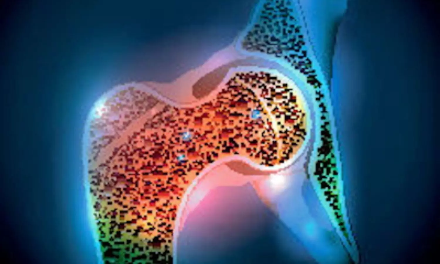
Fertility-Related Factors May Predict Osteoporosis Risk in Postmenopausal Women, claims research

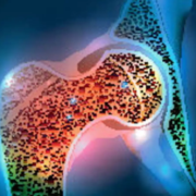
Researchers have discovered in a new study that infertility, miscarriage, stillbirth, and having fewer children might raise the risk of osteoporosis among women undergoing natural menopause. The large international study concluded that these reproductive factors for health are independently linked to bone loss in older age, and that age at natural menopause doesn’t entirely account for these associations. These results indicate that reproductive history may be an important factor in the early recognition of women who are at high risk for osteoporosis, and in steering preventive measures to minimize long-term morbidity. The study was conducted by LIANG Chen and fellow researchers published in the journal of Fertility and Sterility.

This research, undertaken under the auspices of the International Collaboration for a Life Course Approach to Reproductive Health and Chronic Disease Events (InterLACE), combined data from 141,222 naturally postmenopausal women from five cohorts. It was designed to test two overarching hypotheses: whether reproductive difficulties like infertility, miscarriage, stillbirth, and low parity are related to elevated risk of osteoporosis, and whether menopausal age might explain these relationships.

This pooled analysis employed retrospective and prospective data from five cohorts from around the world who belonged to the InterLACE consortium. Excluding women who had undergone unnatural menopause and those for whom reproductive history, osteoporosis status, and covariates were incomplete, the researchers included only those women who met these criteria. Major exposures to infertility, miscarriage, stillbirth, and parity were reported by participants themselves.

Diagnoses of osteoporosis were derived from various sources of data, such as surveys, hospital discharge records, death registers, primary care data, and records of drug use. Cox regression models were used by researchers to calculate hazard ratios (HRs) and 95% confidence intervals (CIs) for the association between factors related to fertility and osteoporosis risk. Models were also adjusted for key covariates including race, age, education, smoking, alcohol consumption, BMI, physical activity, and age at menarche, with a further step adjusting for age at natural menopause as a time-varying covariate.

Key Findings

Modest but persistent elevated risks of osteoporosis in women with unfavorable reproductive histories were found by the study:

  • Infertility history was linked with a 16% increased risk of osteoporosis (HR=1.16, 95% CI: 1.13–1.19).

  • Women with three or more miscarriages had 17% increased risk (HR=1.17, 95% CI: 1.05–1.30).

  • Those who had stillbirth had 14% increased risk (HR=1.14, 95% CI: 1.10–1.17).

  • No live births: HR=1.20 (95% CI: 1.15–1.25)

  • One live birth: HR=1.15 (95% CI: 1.14–1.16)

This big international study verifies that infertility, habitual miscarriage, stillbirth, and low parity are associated with higher risk of osteoporosis in naturally postmenopausal women. These relationships remained significant even after controlling for age at menopause, indicating an independent contribution of reproductive factors in bone health. Physicians can think of these reproductive markers as preliminary indicators for osteoporosis risk assessment and individualized prevention measures.

Reference:

Chen, L. I. A. N. G., Chung, H.-F., Anderson, D. J., VAN DER Schouw, Y. T., Avis, N. E., Karvonen-Gutierrez, C. A., Dobson, A. J., & Mishra, G. D. (2025). The association of infertility, miscarriage, stillbirth, and parity with osteoporosis: a pooled analysis of five cohort studies. Fertility and Sterility. https://doi.org/10.1016/j.fertnstert.2025.07.382

Powered by WPeMatico

Ureteroscopy and Shockwave Lithotripsy exhibit Similar kidney stone Clearance: JAMA

A new study published in the Journal of American Medical Association found that despite shockwave lithotripsy and ureteroscopy having comparable kidney stone clearance rates, patients felt that shockwave lithotripsy was a superior overall experience.

Despite the ambiguity and equal weight of guidelines for shockwave lithotripsy and ureteroscopy, ureteroscopy is the most common treatment for kidney and ureteral stones in children and adolescents. Thus, to assess stone clearance and patient-reported outcomes following ureteroscopy or shockwave lithotripsy, this study was carried out in children and adolescents.

The patients were recruited for this nonrandomized clinical research at 31 US and Canadian medical facilities between March 16, 2020, and July 31, 2023. The patients with ureteral stones, kidney stones, or both who were between the ages of 8 and 21 were included. The last day of follow-up was October 15, 2023.

6 (±2) weeks following surgery, the main result was the stone removal measured by standardized ultrasonography. Stone clearance was assessed per kidney or ureter using logistic regression, and estimated stone clearance rates were produced for each operation using inverse probability weighting and random intercepts each location.

A total of 1,142 patients (690 females [60.4%]) with a median age of 15.6 years (IQR, 12.6-17.3 years) were included in this research. Regarding race and ethnicity, 884 patients (77.4%) were White, 130 patients (11.4%) were Hispanic, and 41 patients (3.6%) were Black.

A median stone size of 6.0 mm (IQR, 4.0-9.0 mm) was found in 1069 kidneys or ureters treated by 124 urologists using ureteroscopy and shockwave lithotripsy (n = 953 and 189 patients), respectively.

Almost, 841 operations for 767 patients (80.4%) undergoing ureteroscopy and 6 procedures for 5 patients (2.6%) undergoing shockwave lithotripsy involved the placement of ureteral stents at the time of index surgery.

In 105 patients who had shockwave lithotripsy (67.5% [95% CI, 61.0%-74.1%]) and 474 patients who had ureteroscopy (71.2% [95% CI, 63.8%-78.5%]), stone removal was achieved; however, this difference was not statistically significant (risk difference, 3.6% [95% CI, −6.2% to 13.5%]).

One week following surgery, ureteroscopy caused more urinary symptoms (symptom score difference, 3.9 [95% CI, 1.2-6.7]) and discomfort interference (T-score difference, 5.0 [95% CI, 2.3-7.8]) than shockwave lithotripsy.

In the week after surgery, ureteroscopy patients missed more coursework (risk difference: 21.3% [95% CI: 9.7%-32.8%]) and caregivers missed more work (risk difference: 23.0% [95% CI: 11.0%-35.0%]). Overall, improved patient-reported outcomes were linked to shockwave lithotripsy. 

Reference:

Tasian, G. E., Chu, D. I., Nelson, C. P., DeFoor, W. R., Ziemba, J. B., Huang, J., Luan, X., Kurtz, M., Ching, C. B., Dangle, P., Schaeffer, A. J., Sturm, R., Wu, W., Bayne, C., Fernandez, N., Chua, M. E., DeMarco, R., Ellsworth, P., Augelli, B., … Meenakshi-Sundaram, B. (2025). Ureteroscopy vs shockwave lithotripsy to remove kidney stones in children and adolescents: A nonrandomized clinical trial: A nonrandomized clinical trial. JAMA Network Open, 8(8). https://doi.org/10.1001/jamanetworkopen.2025.25789

Powered by WPeMatico

tsRNAs emerge as powerful non-invasive biomarkers in liquid biopsy, reveals research

A new review highlights the transformative potential of transfer RNA-derived small RNAs (tsRNAs) in the field of liquid biopsy, emphasizing their role as promising biomarkers for a wide range of diseases, particularly cancers. These non-coding RNA fragments, originating from mature or precursor tRNAs, are detectable in diverse body fluids-including blood plasma, saliva, urine, semen, and cerebrospinal fluid-and exhibit remarkable stability, even in challenging biological environments.

The article presents tsRNAs as a rising star in precision medicine, with significant diagnostic and prognostic value. Found in both plasma and serum, tsRNAs offer disease-specific expression patterns and correlate with key clinical features such as tumor stage, metastasis, and patient survival. In breast cancer, certain circulating tsRNAs are associated with improved prognostic accuracy compared to traditional markers, while in lung cancer, tsRNA signatures have helped differentiate between early and advanced stages. tsRNAs have also shown promise in gastrointestinal, ovarian, pancreatic, renal, and bladder cancers, as well as in conditions like nonalcoholic fatty liver disease, systemic lupus erythematosus, infertility, and neurodegenerative disorders.

A notable advantage of tsRNAs is their presence across multiple biofluids, enabling non-invasive sampling that is more accessible and less risky than tissue biopsies. This versatility opens up opportunities for early detection, disease monitoring, and treatment stratification. Importantly, tsRNAs demonstrate unique expression profiles distinct from similar small RNA classes like miRNAs, reinforcing their biological relevance and clinical utility.

The article also discusses challenges such as inconsistent nomenclature, variation in sample preparation, and the need for optimized detection platforms like PANDORA-seq. It calls for standardized analytical frameworks to enable consistent cross-study comparisons and emphasizes the importance of accounting for age-related and tissue-specific variation in tsRNA expression.

Reference:

Qinglin Wang, Zehao Pan, Si Liang, Yuanjian Shi, Gaochao Dong, Lin Xu, Qixing Mao, Feng Jiang, Transfer RNA-derived small RNAs (tsRNAs): A rising star in liquid biopsy, Genes & Diseases, https://doi.org/10.1016/j.gendis.2025.101608.

Powered by WPeMatico

Losing weight before IVF may increase chance of pregnancy: Study

A systematic review and meta-analysis of randomized controlled trials (RCTs) assessed whether weight loss interventions before in vitro fertilization (IVF) improved reproductive outcomes. The review found that weight loss interventions before IVF could increase the chances of pregnancy, especially in unassisted conception, although the effect on live births was unclear. The findings are published in Annals of Internal Medicine.

Researchers from the University of Oxford reviewed 12 RCTs comprising 1,921 patients conducted between 1980 through 27 of May 2025. Inclusion criteria included studies conducted on women at least 18 years old with a BMI of 27 kg/m2 or greater who were seeking IVF with or without intracytoplasmic sperm injection treatment for infertility. Outcomes of interest were number of participants achieving pregnancy without IVF (unassisted pregnancy), with IVF (treatment-induced pregnancy), overall (unassisted plus treatment-induced) and those delivering a live infant.

The researchers found that participants were typically women in their early 30s with a median baseline BMI of 33.6 kg/m2. Weight loss interventions studied included low-energy diets, an exercise program accompanied by healthy eating advice, and pharmacotherapy accompanied by diet and physical activity advice. Overall, weight loss interventions before IVF were associated with greater unassisted pregnancy rates. Evidence was inconclusive on the effect of weight loss interventions on treatment-induced pregnancies. Evidence on the association between weight loss interventions before IVF and live births was uncertain, although there was moderate certainty of no association with pregnancy loss.

The findings suggest that weight loss interventions before IVF increase total pregnancies, mainly through an increase in unassisted pregnancy rates. However, further high-quality clinical trials testing different weight loss interventions, particularly those known to achieve greatest weight losses (e.g. low-energy total diet replacement programs) are needed.

Reference:

Moscho Michalopoulou, Susan Ann Jebb, Alice Hobson, et al. The Effect of Weight Loss Before In Vitro Fertilization on Reproductive Outcomes in Women With Obesity: A Systematic Review and Meta-analysis. Ann Intern Med. [Epub 12 August 2025]. doi:10.7326/ANNALS-24-01025.

Powered by WPeMatico

Double Relief: Study evaluates Synergistic Effects of Hydromorphone and Ropivacaine in Thoracoscopic Surgery

Recently published clinical trial aimed to evaluate the efficacy of hydromorphone as an adjunct to ropivacaine in serratus anterior plane block (SAPB) for postoperative analgesia and inflammatory response management in patients undergoing video-assisted thoracoscopic surgery (VATS).

Methodology

Patients aged 20-75 with American Society of Anesthesiologists classification I or II were randomized into three groups: Hydromorphone-Ropivacaine SAPB (HR), Ropivacaine SAPB (R), and control (C). Randomization used Excel-generated integers, ensuring blinding throughout the study. A total of 129 patients were identified, with 120 included in the final analysis. VAS scores were recorded at 2, 6, 12, 24, and 48 hours post-surgery, while inflammatory markers (CRP, IL-6, TNF-α) were measured preoperatively and at 24 and 48 hours post-surgery.

Key Findings

Postoperative Pain Relief: Group HR exhibited significantly lower VAS scores than Group C at all measured intervals, particularly at 6 hours post-surgery, where it scored 2.00 (IQR: 2.00) compared to Group C’s 3.00 (IQR: 3.00) (P < 0.001). The HR group showed enhanced pain control throughout the recovery period.

Opioid Consumption: Total opioid consumption and the need for rescue analgesia were significantly reduced in Groups R and HR when compared to Group C. Specifically, HR group had a lower requirement for patient-controlled intravenous analgesia (PCIA) activations (P < 0.001).

Inflammatory Response: The combination of hydromorphone and ropivacaine significantly attenuated postoperative inflammatory markers, with CRP levels being markedly lower in Group HR at 24 and 48 hours compared to Group C (P < 0.001).

Hemodynamic Stability: No significant intraoperative hemodynamic disturbances were attributed to the analgesic techniques, and major postoperative complications were absent.

Limitations

The study was conducted at a single center, potentially limiting the generalizability of findings. Long-term follow-up data was not collected, leaving the long-term effects of the hydromorphone-ropivacaine combination uncertain. Further investigation is needed to compare SAPB with traditional analgesic methods like thoracic epidural analgesia.

Conclusion

The addition of hydromorphone to ropivacaine in SAPB markedly improved postoperative pain management and reduced inflammatory responses in patients undergoing VATS, reinforcing its potential as an effective analgesic strategy. Future research should explore the long-term outcomes and optimal dosing strategies for hydromorphone in SAPB.

Key Points

Analgesic Efficacy: The combination of hydromorphone and ropivacaine in serratus anterior plane block (SAPB) resulted in significantly lower Visual Analog Scale (VAS) pain scores at all assessed time intervals compared to the control group, with the most notable improvement observed at 6 hours post-surgery.

Reduced Opioid Use: Patients receiving the hydromorphone-ropivacaine combination demonstrated a statistically significant reduction in total opioid consumption and need for rescue analgesia, specifically showing fewer activations of patient-controlled intravenous analgesia (PCIA) compared to the control group.

Inflammatory Marker Reduction: The treatment group receiving hydromorphone and ropivacaine showed a marked decrease in inflammatory markers, such as C-reactive protein (CRP), at 24 and 48 hours post-surgery, indicating an enhanced anti-inflammatory response.

Safety Profile: The analgesic techniques employed did not result in significant intraoperative hemodynamic disturbances, and no major postoperative complications were reported, suggesting a favorable safety profile for the combination therapy.

Study Limitations: Generalizability may be constrained as the trial was conducted at a single center, and the absence of long-term follow-up data raises questions about the sustained effects of the hydromorphone-ropivacaine combination.

Future Research Directions: Further studies are recommended to assess long-term outcomes associated with hydromorphone in SAPB and to investigate optimal dosing strategies while potentially comparing SAPB with established analgesic methods such as thoracic epidural analgesia.

Reference –

Xuefeng Chen et al. (2025). The Impact Of Hydromorphone Combined With Ropivacaine In Serratus Anterior Plane Block On Postoperative Pain In Patients Undergoing Video-Assisted Thoracoscopic Pulmonary Lobectomy: A Randomized, Double-Blind Clinical Trial. *BMC Anesthesiology*, 25. https://doi.org/10.1186/s12871-025-03101-2.

Powered by WPeMatico

A Gentle Approach: Study Evaluates Efficacy of Medical Management for Missed Miscarriage

Recent study evaluates the efficacy of mifepristone and misoprostol as a medical management option for missed miscarriage, aiming to identify significant risk factors affecting treatment success. Missed miscarriage, identified when a nonviable pregnancy is retained in the uterus, commonly leads to management via expectant, medical, or surgical means, with medical management increasingly favored for its safety and cost-effectiveness.

Research Methodology

The retrospective case-control study analyzed 163 patients diagnosed with missed miscarriage, employing logistic regression to isolate factors influencing treatment success. The primary outcome was the spontaneous passage of the gestational sac within 24 hours of administering misoprostol, emphasizing the efficiency of the medical regimen.

Key Findings

Findings indicate several demographic and clinical variables significantly correlate with medication management outcomes. High gravidity (more than three pregnancies) increased the likelihood of unsuccessful treatment by 3.67 times, while a history of parity and prior miscarriage escalated risks by 2.29 times and 2.09 times, respectively. Additionally, prior uterine surgery, particularly cesarean sections, was associated with a 2.94-fold increase in unsuccessful management. Importantly, the necessity for additional misoprostol doses did not improve success rates, suggesting that the administration dosage may not be the central determinant of efficacy.

Environmental Factors

Meteorological factors were also assessed, revealing that average visibility over four days was linked to treatment outcomes, stressing an intriguing interplay between environmental conditions and medical efficacy. Initial univariable regressions flagged visibility as statistically significant, which multivariable analysis later affirmed as independent of gestational age, reinforcing the need for personalized treatment considerations.

Discussion of Efficacy

Despite mifepristone and misoprostol’s reported efficacy, with previous literature indicating success rates of 83% to 93.3%, results found significant variability due to varying clinical factors. The study posits that not only previous obstetric history but also environmental exposure may influence uterine responsiveness, suggesting further exploration is warranted in broader populations.

Limitations and Future Research

Limitations include the retrospective nature of the study and the potential exclusion of other relevant factors like socioeconomic status and individual lifestyle variables that could confound results. Recommendations for future research emphasize the necessity of prospective multicenter trials with larger sample sizes to validate these findings, aiming toward enhancing clinical guidelines to optimize individualized management strategies for patients facing missed miscarriage.

Key Points

– -Efficacy of Medical Management-: The study assesses mifepristone and misoprostol as a medical management approach for missed miscarriage, favorably contrasted against expectant and surgical methods, emphasizing their safety profile and cost-effectiveness when managing nonviable pregnancies retained in the uterus.

– -Demographic and Clinical Correlates-: Logistic regression analysis on 163 diagnosed cases identified significant risk factors that negatively impacted treatment outcomes, including high gravidity (>3 pregnancies), history of parity, and prior miscarriage, increasing unsuccessful treatment probabilities by 3.67, 2.29, and 2.09 times, respectively. Additionally, prior uterine surgeries, notably cesarean sections, raised the odds of unsuccessful management by 2.94 times.

– -Efficacy of Misoprostol Dosage-: The requirement for additional doses of misoprostol was not associated with improved success rates, challenging the assumption that dosage quantity is the principal factor affecting the efficiency of the medical regimen for missed miscarriage.

– -Influence of Environmental Conditions-: Analysis of environmental factors, specifically average visibility over a four-day period, was shown to correlate with treatment outcomes, suggesting a previously unexamined interaction between meteorological conditions and uterine response to medical management.

– -Variability in Success Rates-: Despite documented success rates of mifepristone and misoprostol ranging from 83% to 93.3%, significant variability was observed based on the demographic and clinical characteristics of the patients, indicating that individualized treatment protocols may be necessary to improve efficacy.

– -Study Limitations and Research Directions-: The retrospective nature limits data interpretation and possible confounding effects of omitted variables such as socioeconomic status and lifestyle. Future research should focus on prospective multicenter trials with larger populations to further corroborate the findings and refine clinical guidelines for the management of missed miscarriage.

Reference –

Huiyuan Wang et al. (2025). Factors Related To Successful Medication Management With Mifepristone And Misoprostol In Missed Miscarriage: A Retrospective Case-Control Study. *BMC Pregnancy And Childbirth*, 25. https://doi.org/10.1186/s12884-025-07662-5.

Powered by WPeMatico

IPC 304 A only applicable when doctors commit rash or negligent act: HC relief to doctor

Ernakulam: Quashing the criminal proceedings against a doctor under Section 304 A of the Indian Penal Code, 1860, the Kerala High Court recently observed that a medical practitioner would only be liable under this section if he had committed a rash or negligent act that resulted in the patient’s death.

“The law is thus well laid down that a medical practitioner can be prosecuted for medical negligence only if the procedure/treatment adopted by him is contrary to the general and approved practice. Moreover, for attracting the offence under Section 304A, the doctor should have committed a rash or negligent act,” observed the HC bench comprising Justice V.G. Arun.

“Here it is essential to note that, as far as rash acts are concerned, the criminality lies in running the risk of doing such an act with recklessness or indifference as to the consequences. Criminal negligence on the other hand is the gross and culpable neglect or failure to exercise that reasonable and proper care and precaution to guard against injury, either to the public generally or to an individual in particular which, having regard to all the circumstances out of which the charge has arisen, it was the imperative duty of the accused to have adopted. For attracting the offence under Section 304A, the death must also be the direct or proximate result of the rash or negligent act of the accused,” it further noted.

The plea was filed by the accused, an on-call Medical Officer, seeking quashment of criminal proceedings for medical negligence. The matter concerned a remand prisoner who suffered head injuries after a fall back in January 2021. Even though initially he was treated at local hospitals, due to a lack of beds, he was referred to the Government Medical College Hospital, Kottayam. 

After he suffered another seizure, the patient was referred for a neurosurgery consultation, and the Senior Resident examined him and informed the accused doctor about his condition over the phone. The accused doctor advised a CT scan and pre-operative investigations to be prepared for an emergency surgery.

The next day, when the accused’s duty ended at 8:00 a.m., another doctor (co-accused) assumed the charge as the on-call Medical Officer. It was alleged that even though an emergency surgery was advised after the worsening scan results, the procedure was delayed due to the unavailability of the operating theatre. The patient died in the afternoon on the same day, while awaiting surgery.

Consequently, an FIR was registered, and the patient’s death led to public protests alleging custodial torture, promoting a Crime Branch investigation. An Expert Panel was constituted to investigate the complaints against the doctors, and the panel concluded that the patient had not received reasonable medical care while undergoing the treatment. Accordingly, the accused doctors were booked under Section 304 A IPC.

Challenging this, the petitioner doctor filed the plea before the High Court. The accused’s counsel contended that even if the prosecution allegations were accepted, the offence under Section 304-A IPC would not be attracted because the golden hour, as far as a person with seizure was concerned, was six hours and the inordinate delay in providing proper treatment and medication worsened the patient’s condition.

It was argued that by the time the accused doctor was informed about the patient, already 18 hours had elapsed after the first CT-scan, and it was also the peak pandemic period, and the Government advisories prevented the accused from examining the patient physically. Further, the counsel submitted that in the appeal filed by the accused against the Expert Panel Report, the State Level Apex Expert Committee held that there was no gross and culpable negligence by the doctors at the Medical College in giving treatment to the patient.

On the other hand, the Public Prosecutor submitted that the matter was still being investigated and the investigating officer was not bound by the Apex Body’s findings. Therefore, if there were sufficient materials to prove the accused’s complicity, he could be prosecuted for the alleged offence.

While considering the matter, the HC bench opined that while negligence was an omission to do something which a reasonable man, guided upon those considerations which ordinarily regulated the conduct of human affairs, would do, or the doing of something which a prudent and reasonable man would not do, medical negligence occurred when a doctor breached his duty of care, causing harm to a patient.

The HC bench referred to the Supreme Court order in the case of Suresh Gupta (Dr.) v. Govt. of NCT of Delhi, where the Apex Court had held that “for fixing criminal liability on a doctor or a surgeon, the standard of negligence required to be proved must be so high as can be described as ‘gross negligence’ or ‘recklessness’. It was not merely lack of necessary care, attention and skill.”

The findings in the case of Suresh Gupta were affirmed by the Apex Court in the case of Jacob Mathew v. State of Punjab, where the Court observed that “A simple lack of care, an error of judgment or an accident, is not proof of negligence on the part of a medical professional. So long as a doctor follows a practice acceptable to the medical profession of that day, he cannot be held liable for negligence merely because a better alternative course or method of treatment was also available or simply because a more skilled doctor would not have chosen to follow or resort to that practice or procedure which the accused followed. When it comes to the failure of taking precautions, what has to be seen is whether those precautions were taken which the ordinary experience of men has found to be sufficient; a failure to use special or extraordinary precautions which might have prevented the particular happening cannot be the standard for judging the alleged negligence.”

Further referring to other judicial precedent, the HC bench observed that a medical practitioner can be prosecuted for medical negligence only if the procedure/treatment adopted by him is contrary to the general and approved practice. Moreover, for attracting the offence under Section 304A, the doctor should have committed a rash or negligent act.

The HC bench observed that in this case, the allegation against the petitioner doctor was about the failure to provide timely treatment.

 “Indisputably, there was delay in bringing the patient to the Medical College Hospital after the first seizure. Even after admitting the patient, petitioner was informed about his condition only by 05:20 am on 13.01.2021. Immediately the petitioner directed to conduct CT scan, since no decision could be taken based on the earlier CT scan, which was conducted many hours back and when the patient was in a much better condition. The CT scan report was received at 07:45 am and the petitioner advised for immediate surgery. The petitioner cannot also be attributed with gross negligence for his failure to examine the patient physically since the Covid related SoPs prevented such examination. Further, the petitioner’s duty time ended by 8 am and the delay in conducting the surgery occurred due to nonavailability of operation theatre. Pertinent in this context to note that the Expert Panel had only opined that the patient had not received reasonable standard of care while under treatment in Government Medical College, Kottayam, without naming any particular doctor. Even that opinion of the Expert Panel has lost its relevance in view of the conclusive unanimous opinion of the State Level Apex Body that there was no gross and culpable negligence from the part of the treating doctors for giving treatment to *** (patient).”

Accordingly, the bench observed that the criminal proceedings against the accused doctor were liable to be quashed. It stated,

“In the light of the above undisputed facts, and in view of the law laid down by the Apex Court, it can unhesitatingly be held that the prosecution of the petitioner for the offence under Section 304A amounts to an abuse of process of court. As held by the Supreme Court in State of Haryana and Others v. Bhajan Lal and Others [1992 SCC (Cri) 426], an FIR is liable to be quashed when the allegations, even if accepted in their entirety, do not make out the ingredients for constituting the offence alleged against the accused.”

“For the aforementioned reasons, the Crl.M.C is allowed. FIR and all further proceedings against the petitioner in Crime No.10 of 2021 of the Crime Branch, CU-II Unit, Ernakulam are quashed,” the HC bench further noted.

To view the order, click on the link below:

https://medicaldialogues.in/pdf_upload/kerala-hc-ipc-304-a-298145.pdf

Also Read: Some doctors disregard human life, maligning noble profession: HC refuses to quash FIR under IPC 304A against Specialist

Powered by WPeMatico