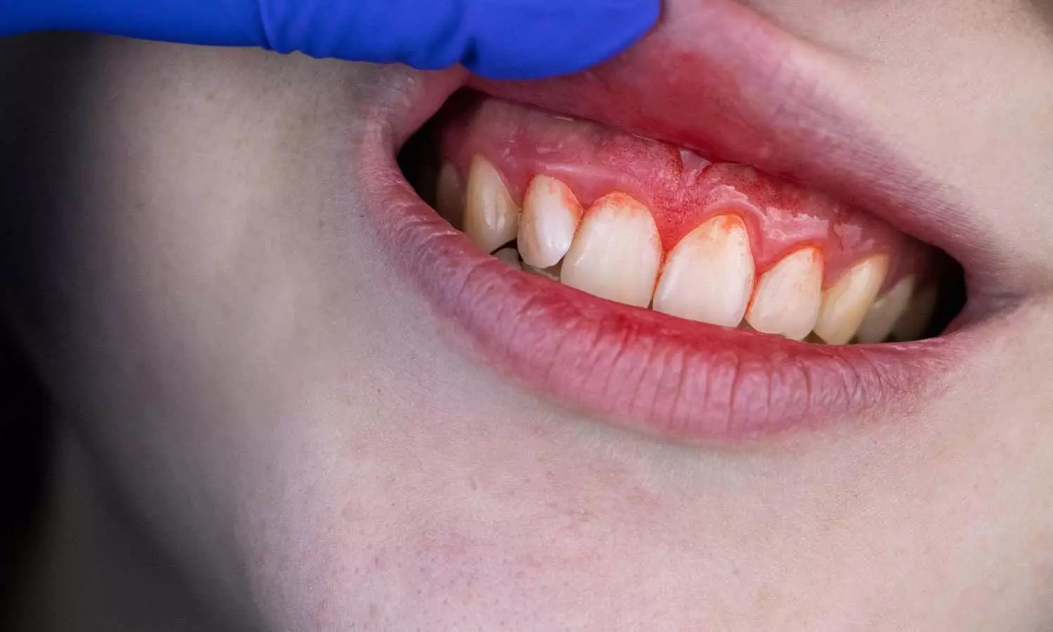
Gum Disease Linked to Increased Risk of Gestational Diabetes: Study

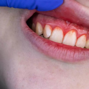
A study published in BMC Oral Health suggests that women of childbearing age with gum disease may face a higher risk of developing gestational diabetes. Researchers observed a dose-dependent relationship between the severity of gum inflammation measured by clinical attachment level and the occurrence of gestational diabetes, highlighting the key role of inflammation in this association. The study was conducted by Jing Cheng and colleagues.

The study drew a large cohort of 5,283 women in the age group of 20 to 44 years, drawn from NHANES III and NHANES 2009–2014 data sets. These subjects were categorized based on their diabetes status as non-diabetes, diabetes mellitus (DM), and gestational diabetes mellitus (GDM). Periodontal status of the participants was measured by both categorical diagnosis of periodontitis as well as continuous variables such as clinical attachment level (CAL).

Multivariate logistic regression analyses were utilized to compare the associations between periodontal measures and status of diabetes. Linear regression models were applied to examine associations with hemoglobin A1c (HbA1c). Mediation analysis was also used to evaluate if inflammatory biomarkers such as white blood cell count (WBC), monocyte count, and monocyte-lymphocyte ratio (MLR) were mediators of the relationship between periodontitis and GDM.

Key Findings

• Women with periodontitis had 1.68 times the risk of having GDM as women without periodontitis (OR = 1.68, 95% CI: 1.14–2.50).

• Increased clinical attachment level (CAL) was correlated with a 50% increased risk of GDM with every 1-mm increase (OR = 1.50, 95% CI: 1.25–1.81).

• HbA1c was positively correlated with both periodontitis (Beta = 0.19, SE = 0.06, p < 0.01) and CAL (Beta = 0.06, SE = 0.03, p < 0.001).

• White blood cell count mediated 5.02%

• Monocyte count mediated 5.88%

• Monocyte-lymphocyte ratio mediated 5.20%

The research concluded that periodontitis was highly related to gestational diabetes mellitus risk among women of childbearing age. Systemic inflammation, as indicated by heightened white blood cell count, monocyte count, and monocyte-lymphocyte ratio, seemed to partially mediate this relationship. The findings are in favor of incorporating periodontal screening in prenatal care to possibly eliminate the risk of GDM through early detection and intervention of oral inflammation.

Reference:

Cheng, J., Jiang, Q., Liu, Y. et al. Mediating role of systemic inflammation on the association between periodontitis and gestational diabetes mellitus: a cross-sectional study. BMC Oral Health 25, 1179 (2025).https://doi.org/10.1186/s12903-025-06541-x

Powered by WPeMatico

New approach to kidney transplant matching could lead to better long-term outcomes: Study

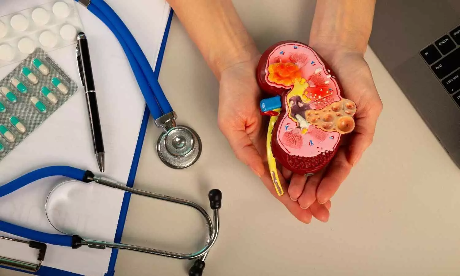
Researchers at the University of Pittsburgh have identified a new way of predicting whether a kidney donor and recipient are a good match for transplantation.

The findings, published today in Science Translational Medicine, could complement existing methods to identify patients at higher risk of rejecting a new kidney and who may benefit from additional immunosuppression to reduce that risk.

“The dream of any kidney transplant surgeon is one transplant for life,” said senior author Aravind Cherukuri, M.D., Ph.D., assistant professor of medicine, surgery and immunology at Pitt, co-director of clinical research at the Thomas E. Starzl Transplantation Institute and nephrologist at UPMC. “That goal has not been achieved yet, but even small advances in prolonging the life of a transplant would be amazing because it will reduce the need for organs and the number of people on waitlists.”

Before a kidney transplant, clinicians determine whether donors and recipients are a good match by evaluating their human leukocyte antigens (HLA). According to Cherukuri, HLA is the genetic fingerprint of the transplanted organ and allows the immune system to distinguish self from non-self. When T cells recognize foreign HLA molecules on a transplanted organ, they initiate immune responses that can lead to inflammation, scarring and eventual graft failure.

“HLA matching is the main way of risk stratifying when it comes to kidney transplant,” said Cherukuri. “Even with a good HLA match, recipients must take immunosuppressant drugs for the rest of their lives to reduce the chance of rejection. However, there has been stagnation in the field in terms of new tools to identify rejection risk and development of new therapies to reduce that risk.”

The new study describes a novel tool to help stratify rejection risk in the form of a cell surface receptor called SIRP-alpha, found on innate immune cells called monocytes.

The research builds on a 2017 Science Immunology study by co-senior author Fadi Lakkis, M.D., now a professor in the Division of Nephrology in the Department of Medicine at Stanford University, who did this research while at Pitt and UPMC, which found that SIRP-alpha is involved in distinguishing self from non-self in mice.

Now, Cherukuri, Lakkis and their team have found evidence that similar SIRP-alpha mismatches contribute to transplant rejection, premature scarring and graft failure in humans and suggest that SIRP-alpha matching between donors and recipients could be an important clinical tool for risk stratification and eventually improving transplant outcomes.

The researchers started by establishing the link between SIRP-alpha mismatch and worse rejection in mice and then screened thousands of human genomes from publicly available repositories. They categorized the 10 most commonly prevalent SIRP-alpha gene variants into two groups, which they named A and B.

Then they compared 455 donor-recipient pairs from transplants performed at UPMC and showed that when the SIRP-alpha type did not match — that is, when one had A and one had B — there was increased early acute graft rejection, more scarring and worse long-term transplant outcomes compared to when they matched.

They also validated these findings in an independent cohort of 258 kidney transplant donor-recipient pairs from Northwestern University.

The researchers say that SIRP-alpha testing could complement, not replace, HLA matching in kidney transplantation.

“Testing for SIRP-alpha type could provide an extra armament in the armory of physicians when it comes to stratifying rejection risk for transplant patients,” he said. “It could help us identify a subset of patients who could benefit from an enhanced immunosuppressant regime. This is exciting because it’s one step forward in the direction of personalized immunosuppression.”

Cherukuri and his team now plan to investigate whether therapeutics that target monocytes, such as corticosteroids, could improve kidney transplant outcomes when donors and recipients have SIRP-alpha mismatches.

Reference:

Daqiang Zhao et al. ,Donor-recipient mismatch at the SIRPA locus adversely affects kidney allograft outcomes.Sci. Transl. Med.17,eady1135(2025).DOI:10.1126/scitranslmed.ady1135

Powered by WPeMatico

University of Houston optometry researcher provides new hope for injured corneas

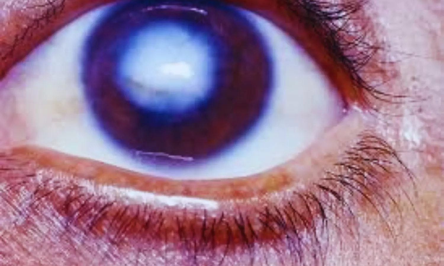
Consider the cornea: the eye’s transparent, dome-shaped surface that provides most of its focusing power and serves as a protective barrier. It directs light onto the retina for sharp vision and shields the eye from dust, debris and harmful UV rays. That’s a lot to ask from a structure made of cells and proteins that’s about two-thirds the size of a dime, yet it does its job brilliantly.

Until it is damaged. Then vision can blur, distort-or be lost entirely. And the cornea is definitely prone to damage.

“When the cornea gets injured, it often heals with scar tissue and abnormal blood vessels, both of which can cause permanent vision loss,” said Tarsis G. Ferreira, assistant professor at the UH College of Optometry. “Unfortunately, there are few treatments for this, and in severe cases, patients need a corneal transplant.”

That dilemma led Ferreira to begin developing a treatment for corneal scarring. He is supported in his work with a five-year grant for $2.2 million from the National Institutes of Health. The treatment focuses on a natural protein in the cornea, named decorin, which plays an essential role in keeping the cornea clear and healthy. It does this by blocking another protein called TGF-β1 (transforming growth factor beta 1), which triggers the scarring process after injury.

“Our goal is to engineer a better version of decorin that more strongly blocks TGF-β1 and another protein receptor that promotes blood vessel growth (VEGFR2), to prevent both scarring and unwanted blood vessels from forming,” said Ferreira.

Natural decorin does that, but the protein is hard to produce. Ferreira has built a version of decorin, which he named mini-dec, that binds better to the two targets and is easy to produce and deliver as eyedrops.

“We hope this new therapy could soon be used to treat eye injuries and surgeries, helping people heal without losing vision,” said Ferreira. The decorin-based drugs will be simple to use and have the potential for treating corneal injuries in the clinic.”

Powered by WPeMatico

Sharp Rise in Nicotine Pouch Ingestions Among Young Children Despite Overall Decline: Study

Nicotine ingestions among young children declined from 2015 to 2023. However, a study in published in Pediatrics journal reported a dramatic increase (over 700%) in nicotine pouch ingestions from 2020 to 2023. In light of this, experts urge parents to store nicotine products in locked, child-proof containers to prevent access.

Numerous items that contain nicotine, such as gum, e-liquids, lozenges, pouches, powder or granules, pills, capsules, or caplets, as well as regular cigarettes, are linked to the frequent exposure of children to nicotine. Investigating the features and patterns of nicotine ingestions among young children submitted to US poison centres is the aim of this study.

From 2010 to 2023, researchers examined accidental, single-substance nicotine intakes in children under the age of six that were reported to the National Poison Data System, as a part of the primary investigative method of this study.

Of the 134,663 cases of nicotine intake in children under the age of six that were reported to US poison centers, the majority occurred in children under the age of two (76.2%), in boys (55.5%), or at home (98.5%). from 2010 and 2015, the rate of nicotine ingestions rose by 59.4% (P <.001), and from 2015 and 2023, it decreased by 34.1% (P <.001).

The main factor influencing this rate pattern was the rate of consumption of liquid nicotine, which rose by 450.0% between 2010 and 2015 (P <.001) and subsequently fell by 45.2% between 2015 and 2023 (P <.05). Nearly, 81.3% did not obtain treatment at a medical facility, and the majority of nicotine ingestions were linked to either no impact (36.7%) or a slight effect (19.6%). There were 39 ingestions with serious consequences and two fatalities, whereas 1.2% of ingestions had moderate effects.

Almost, 0.5% of ingestions required medical admission, whereas 15.9% of ingestions were treated, assessed, and released. Between 2020 and 2023, the rate of nicotine pouch ingestions rose by 763.1%. When compared to all product formulations combined, nicotine pouches had a higher likelihood of being linked to a significant medical result (odds ratio [OR]: 1.53, 95% CI: 1.10–2.13) or a hospital admission (OR: 2.03, 95% CI: 1.31–3.15).

Overall, serious consequences, including 2 fatalities, did occur even though the majority of nicotine ingestions in children under the age of 6 were linked to minimal or clinically minor effects. The rise and relative seriousness of nicotine pouch ingestions serve as a reminder of the public health issues associated with the changing market for oral nicotine products. 

Source:

Olivas, M., Hays, H. L., Kistamgari, S., Rine, N. I., Zhu, M., Yang, J., & Smith, G. A. (2025). Nicotine ingestions among young children: 2010-2023. Pediatrics, e2024070522. https://doi.org/10.1542/peds.2024-070522

Powered by WPeMatico

Imposter Syndrome among Orthopaedic Surgery residents extremely common and disproportionately affects female residents: study

Imposter syndrome is a psychological phenomenon in which individuals experience persistent self-doubt and feelings of inadequacy despite achieving objective measures of success. This phenomenon is associated with lower job satisfaction, increased rates of burnout, and reduced productivity.

Harsh Wadhwa et al conducted a study to determine the prevalence of imposter syndrome among orthopaedic surgery residents and determine whether specific individual or residency characteristics predict higher rates of imposter syndrome.

The study was performed at the Department of Orthopaedic Surgery, Stanford University Medical Center, Stanford, California.

An anonymous survey was distributed to residents at 7 ACGME-accredited orthopaedic surgery residencies by the authors. Respondents provided deidentified demographic data and completed the Clance Imposter Phenomenon Scale (CIPS) assessment. CIPS scores were used to categorize imposter syndrome severity as none to mild, moderate, significant, or intense. Differences were calculated and compared using t-test and x2 analyses. Multivariable regression analysis was used to identify predictors of increased symptom severity.

The key findings of the study were:

• One hundred orthopaedic surgery residents completed the survey.

• Forty percent of respondents were female, and the mean age was 30.6 ± 2.8 years.

• The prevalence of significant or intense imposter syndrome was 73%.

• CIPS scores and significant or intense imposter syndrome did not vary by age, gender, sexual orientation, race/ethnicity, level of training, program region, research year, non-MD degrees, and Step 1, Step 2, or Orthopaedic In-Training Examination (OITE) scores on univariable analyses.

• On multivariable analysis, female residents were 5.64 (OR = 5.64 [1.04-30.63]) times more likely to have significant or intense imposter syndrome (p = 0.045).

• Residents at western programs were 0.13 (OR = 0.13 [0.02-0.90]) times as likely to experience significant or intense imposter syndrome (p = 0.039).

• Women (estimate = 8.72 [1.41-16.04]; p = 0.0201) and those with OITE scores in the 0 to 25th (estimate = 11.92 [0.29- 23.54]; p = 0.0446) and 51st-75th (estimate = 12.73 [2.79-22.67]; p = 0.0128) percentiles had higher CIPS scores.

The authors concluded that – “Imposter syndrome is highly prevalent among orthopaedic surgery residents, with significant or intense symptoms affecting 73% of those surveyed. Female gender and lower OITE scores were associated with increased imposter syndrome severity, while training at an orthopaedic surgery residency program in the West was associated with decreased imposter syndrome. Additional research is needed to identify effective interventions to address imposter syndrome and minimize its effects on individuals’ careers. Implementing focused initiatives to address imposter syndrome may help alleviate burnout and promote both career fulfillment and professional growth.”

Further reading:

Imposter Syndrome Among Orthopaedic Surgery Residents is Extremely Common and Disproportionately Affects Female Residents

Harsh Wadhwa et al

JBJS Open Access 2025:e24.00132.

http://dx.doi.org/10.2106/JBJS.OA.24.00132

Powered by WPeMatico

HC Allows B.Pharm Graduates to Apply for Pharmacist Posts, Calls Diploma-Only Clause Arbitrary

Raipur: In an interim order, the Chhattisgarh High Court has allowed candidates holding a Bachelor of Pharmacy (B.Pharm) degree to apply for the post of Pharmacist (Grade-II) advertised by the National Health Mission (NHM). The Court took strong exception to the exclusion of degree holders in favour of diploma holders and described the restriction as potentially arbitrary and violative of constitutional rights under Articles 14 and 16.

The case, filed as WPS No. 8548 of 2025, was heard by a division bench comprising Chief Justice Ramesh Sinha and Justice Bibhu Datta Guru. The petition was filed by 11 candidates, including Rahul Verma and others, challenging the advertisement dated 30.06.2025 issued by the NHM, which limited eligibility for the pharmacist post strictly to diploma holders in pharmacy.

Counsel for the petitioners, Harshwardhan Parganiya, argued that the exclusion of B.Pharm degree holders, despite their higher qualification and valid registration with the Pharmacy Council, was arbitrary and unconstitutional. He contended that the Service Recruitment Rules, 2013 (Entry No. 08 to Schedule-III) prescribing only a diploma in pharmacy as the required qualification violated Articles 14 and 16 by failing the test of intelligible differentia and rational nexus.

He further submitted, “The impugned advertisement and the relevant provisions… by treating ‘Diploma’ as the sole or ‘prescribed’ qualification rather than the minimum qualification, amount to a manifestly arbitrary classification… violating the fundamental rights of the petitioners guaranteed under Articles 14 and 16 of the Constitution of India.”

To support his argument, he relied on the Supreme Court’s judgment in Puneet Sharma & Others v. Himachal Pradesh State Electricity Board Ltd. (C.A. No. 1318-1322 of 2021), where degree holders were allowed to participate in recruitment despite diploma-specific criteria.

The learned Advocate General Prafull N. Bharat, appearing for the State and some health authorities, submitted that he had not yet received instructions in the matter and sought time to file a reply. Other counsels representing CGVYAPAM (Respondent No. 2 – Chhattisgarh Professional Examination Board Vyapam) and National Health Mission (Respondent No. 3) were also present.

The Court, considering their presence, dispensed with formal notice and granted, “four weeks’ time to file their respective returns and thereafter, two weeks’ further time to the petitioners to file rejoinder, if so advised.”

Recognising the urgency, since the last date for submission of online applications was 5 p.m. on 25.07.2025—the Court directed the authorities to immediately allow eligible B.Pharm candidates to submit their applications.

The bench observed;

“The respondent- State is directed to issue necessary instruction to the respondent-CGVYAPAM who has to conduct the examination, to make necessary changes/amendments in the online portal for the intending candidates / participants of the examination in question for submission of their online forms and the petitioners are at liberty to submit their forms in the online portal, however, their participation shall be subject to the final out come of the present petition.”

Importantly, the Court clarified that:

“It is made clear that this order would operate in rem and not in personam and would be applicable to all the similarly situated candidates having Degree in Bachelor of Pharmacy (В. Pharma) or any other equivalent or a higher qualification, subject to the condition that they fulfill other requisite criteria as may be prescribed by the respondent Department in the advertisement in question.”

The Court also directed the Advocate General to inform CGVYAPAM of the order so the portal could be updated and the deadline extended if necessary.

The matter is now listed for final hearing after six weeks.

To view the original judgement, click on the link below:

https://indiankanoon.org/doc/156508648/

Powered by WPeMatico

Alcohol alters gene function in differentiating cells of the embryo, reveals research

During the tightly regulated gastrulation, embryonic cells differentiate into the three germ layers – endoderm, mesoderm, and ectoderm – which eventually give rise to all tissues and organs. The late, renowned developmental biologist Lewis Wolpert once stated: “It is not birth, marriage, or death, but gastrulation which is truly the most important time in your life.” Gastrulation occurs during the fifth week of pregnancy, a time when many women are not yet aware that they are pregnant.

According to estimates by the Finnish Association on Intellectual and Developmental Disabilities, 600–3,000 children are born in Finland each year with permanent damage caused by alcohol, but due to the challenges of diagnosis, the true number is unknown.

Researchers at the University of Helsinki, in collaboration with the University of Eastern Finland, have now examined the effects of alcohol on this difficult-to-study stage of human development.

In the study, pluripotent embryonic stem cells were differentiated into the three germ layers in culture dishes. The cells were exposed to two different concentrations of alcohol: the lower exposure corresponded to less than one per mille, while the higher exceeded three per mille. The researchers then investigated the effects of alcohol on gene expression, epigenetic markers regulating gene activity, and cellular metabolism.

Stronger alcohol exposure caused more changes than the lower dose, and a dose-response relationship was observed in both gene activity and metabolism. The most significant metabolic changes were detected in the methionine cycle of the cells.

“The methionine cycle produces vital methyl groups in our cells, which attach to DNA strand and influence gene regulation. The observed changes confirm the importance of this epigenetic regulation in the disturbances caused by alcohol exposure,” the doctoral researcher Essi Wallén explains.

The First Neural Cells Are Most Sensitive to Alcohol

The most pronounced changes caused by alcohol exposure were seen in ectodermal cells, which give rise to the nervous system and the brain during development. It is well-known that prenatal alcohol exposure is one of the most significant causes of neurodevelopmental disorders.

“Many of the developmentally important genes altered in this study have previously been linked to prenatal alcohol exposure and its associated features, such as defects in heart and corpus callosum development, as well as holoprosencephaly, a failure of the forebrain to divide properly,” says Adjunct Professor Nina Kaminen-Ahola, who led the study.

According to the study, some of the developmental disorders caused by alcohol may arise during the very first weeks of pregnancy, when even minor changes in gene function may influence the course of development. However, further research is needed to clarify how well the cell model and alcohol concentrations correspond to actual exposure in humans.

This research is part of a broader project investigating the mechanisms by which alcohol affects early development and later health. Prenatal alcohol exposure causes a range of developmental disorders collectively referred to as fetal alcohol spectrum disorders (FASD).

Reference:

E. Wallén, Effects of alcohol on the transcriptome, methylome and metabolome of in vitro gastrulating human embryonic cells, Disease Models & Mechanisms, https://doi.org/10.1242/dmm.052150

Powered by WPeMatico

Thyroid Dysfunction Common After Radiotherapy in Head and Neck Cancer Patients, Study Warns

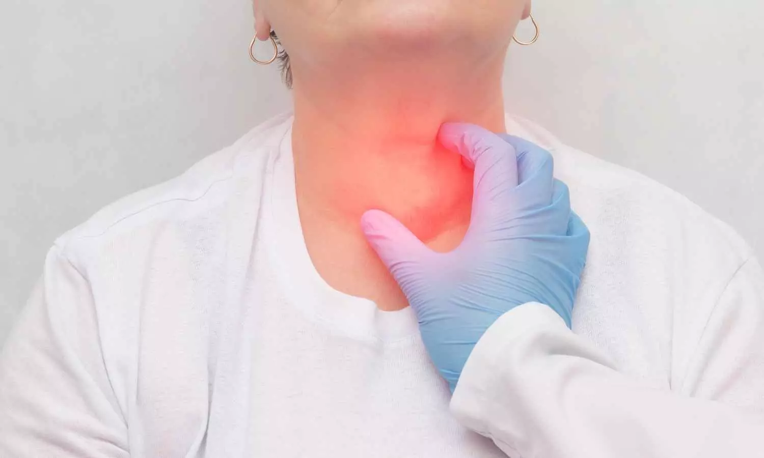
Brazil: A recent study published in Head & Neck highlights the increased risk of thyroid dysfunction in patients with head and neck squamous cell carcinoma (HNSCC) following external radiotherapy (RT). The study found that more than half (54.7%) of HNSCC patients who underwent RT developed thyroid dysfunction (TD), with 53.7% experiencing long-term complications.

“Subclinical hypothyroidism emerged as the most prevalent form, progressing to overt hypothyroidism in 21% of cases. The risk of TD was notably higher in patients with type 2 diabetes mellitus (T2DM), metastatic lymph node involvement, and those who did not undergo tumor resection, highlighting the importance of regular thyroid monitoring after RT,” the researchers reported.

Radiotherapy, a cornerstone in the management of HNSCC, is known to impact the thyroid gland due to its anatomical proximity to the treatment field. Hypothyroidism remains one of the most common adverse effects, often manifesting months or even years after therapy. Considering this, Denise Engelbrecht Zantut-Wittmann, Department of Internal Medicine, School of Medical Sciences, Endocrinology Division, University of Campinas, São Paulo, Brazil, and colleagues aimed to assess thyroid dysfunction in patients with head and neck squamous cell carcinoma undergoing external radiotherapy.

For this purpose, the researchers examined early and long-term thyroid dysfunction in 285 patients with head and neck squamous cell carcinoma who received radiotherapy either alone or in combination with radiochemotherapy and/or surgery.

The study led to the following findings:

  • 156 patients (54.7%) experienced thyroid dysfunction (TD) during follow-up, with 153 patients (53.7%) experiencing long-term TD.
  • Subclinical hypothyroidism (SCH) was the first detected TD in 43.5% of cases.
  • Among those with SCH, 68.5% had persistent SCH, 21% progressed to overt hypothyroidism, 0.8% developed central hypothyroidism, and 9.7% returned to euthyroidism.
  • The average time to first TD detection was 7.2 months after radiotherapy (RT).
  • Subclinical thyrotoxicosis was identified at an average of 3.85 months post-RT, while SCH was detected at 17.77 months.
  • Risk factors for TD included type 2 diabetes mellitus (DM2), metastatic lymph node involvement, and the absence of tumor resection.
  • All patients with thyroid-stimulating hormone (TSH) levels ≥7.5 mIU/mL developed primary hypothyroidism or SCH.
  • At long-term follow-up, 19.5% of patients with TSH levels <7.5 mIU/mL remained euthyroid.

The authors emphasize the importance of regular thyroid function monitoring in patients with head and neck squamous cell carcinoma after radiotherapy, particularly those with type 2 diabetes mellitus, metastatic lymph node involvement, or lack of surgical tumor resection.

They concluded that the high incidence of thyroid dysfunction in these patients necessitates early detection through routine assessments, enabling timely intervention and reducing the risk of complications associated with hypothyroidism. They noted that integrating thyroid monitoring into post-treatment care protocols could significantly enhance long-term outcomes and improve the quality of life for HNSCC survivors.

Reference:

Fiore, A. R., Lourenço, G. J., Pereira, E. B., Passos Lima, C. S., & Zantut-Wittmann, D. E. Early and Long-Term Thyroid Dysfunction in Patients With Head and Neck Squamous Cell Carcinoma After External Radiotherapy: Clinicopathological Risk Factors. Head & Neck. https://doi.org/10.1002/hed.28150

Powered by WPeMatico

Innovative imaging tool could improve diagnosis and treatment of hearing loss, states research

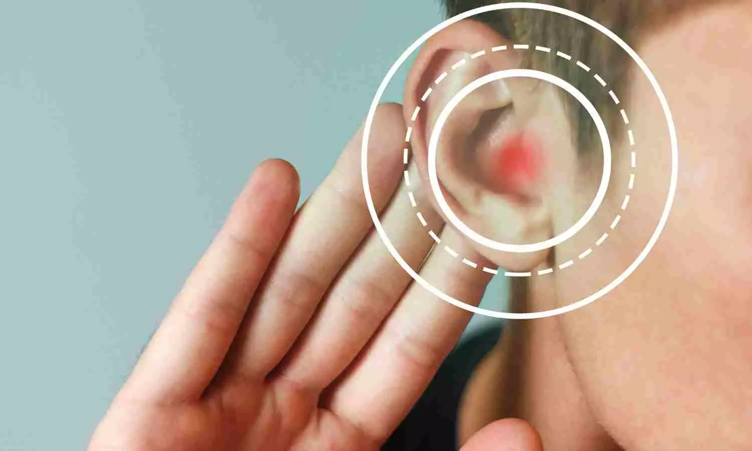
Optical coherence tomography (OCT), a tool routinely used to diagnose and plan treatment for eye diseases, has now been modified to collect images of the inner ear. A proof-of-concept study led by researchers at the Keck School of Medicine of USC found that OCT imaging can measure fluid levels in the inner ear, which correlate with a patient’s degree of hearing loss. The findings were just published in Science Translational Medicine.

“These findings are exciting because hearing loss can happen very suddenly, and we often don’t know why. OCT offers a way to explore the underlying cause and potentially guide treatment,” said senior author John Oghalai, MD, professor and chair in the Caruso Department of Otolaryngology – Head and Neck Surgery and the Leon J. Tiber and David S. Alpert Chair in Medicine at the Keck School of Medicine.

A sudden loss of hearing, sometimes accompanied by vertigo, happens in Ménière’s disease, cochlear hydrops and other ear conditions. One hallmark of these diseases is an imbalance of fluids in the inner ear, but measuring fluid balance is a challenge. The best available technology, magnetic resonance imaging (MRI), lacks the resolution needed to reliably diagnose or guide treatment.

OCT offers a quicker, more accurate and less expensive way to see inner ear fluids, hair cells and other structures relevant to diagnosing and treating hearing loss. With funding from USC and the National Institutes of Health, Oghalai, Brian Applegate, PhD, professor of otolaryngology – head and neck surgery, and ophthalmology at the medical school and of biomedical engineering and electrical and computer engineering at USC Viterbi School of Engineering, and their team tested the new tool in 19 patients undergoing ear surgery for various conditions. They found that OCT could reliably detect fluid imbalance in the inner ear, which was correlated with the severity of a patient’s hearing loss. Although the tool is currently limited to use during surgery, the researchers are working to adapt it for application in the clinic.

The latest findings build on the team’s earlier work, where they used OCT to collect images of the cochlea in awake animals for the first time. With improvements, the tool could help clinicians more quickly find the cause of hearing loss, determine how to treat it, and even support efforts to develop new therapies to restore hearing, Oghalai said.

Measuring fluid balance

OCT uses light waves to scan tissue and create 3D images, similar to the way ultrasound produces images using sound waves.

The Keck School of Medicine team used the tool to scan the inner ears of 19 patients undergoing ear surgery. Six patients had normal inner ear function, four had Ménière’s disease, and nine had vestibular schwannoma (a benign tumor on a nerve that connects the inner ear to the brain). During surgery, a thick outer bone known as the mastoid was temporarily removed, allowing researchers to use OCT to collect images of the fluid compartments in the inner ear.

OCT images showed that patients with Ménière’s disease or vestibular schwannoma had higher levels of a fluid called endolymph, compared to those with normal inner ear function. Increased endolymph levels were linked to greater hearing loss, indicating that measuring these fluid levels could help predict the severity of symptoms.

“We’ve known for a long time that endolymph is related to hearing loss, but until now, measuring it in a living patient has been a major challenge,” Oghalai said.

A faster way to help

OCT can assist surgeons, including by helping to avoid damage to delicate ear structures and distinguishing brain tumors from healthy tissue. To this end, the researchers are working to develop a smaller, more affordable version of the tool that they plan to distribute and test with surgeons.

But OCT can benefit many more people if it can be used to collect images of the inner ear while patients are awake in the clinic. To achieve this goal, Oghalai, Applegate and their team have received federal funding and a Nemirovsky Engineering and Medicine Opportunity (NEMO) Prize from USC. They are now working to improve the software and image-processing techniques to obtain clear images from patients without removing the mastoid bone.

OCT could offer major benefits over MRI because it is less expensive, faster and can be done back-to-back to test treatment effectiveness. For example, a provider could collect an image, give medication treatment for a fluid imbalance, then take another image 30 minutes later.

“Patient symptoms—sensations of hearing loss, pressure in the ear, or vertigo-can be affected by many unrelated factors such as mood, stress, nasal congestion and allergies. However, inner ear fluid levels reliably assess what is going on inside the inner ear,” Oghalai said. “We have lots of treatment options available, but it often takes many weeks to figure out what works for a given patient. This could be a faster way to help.”

If OCT can be used outside the operating room, it can also support the development of new treatments for hearing loss. Several gene therapies, which aim to regenerate lost hair cells in the inner ear, are currently in clinical trials. OCT may provide a fast and precise way to test the effectiveness of such therapies by collecting images of hair cells as they grow.

“OCT helped researchers develop treatments for retinal diseases, including macular degeneration, so we’re hoping it can have a similar impact for hearing loss,” Oghalai said.

Reference:

Kim W, Pan DW, Kim BJ, Yang Z, Moran Mojica M, Doherty JK, Shibata SB, Applegate BE, Oghalai JS. Human inner ear fluid imbalance detected by optical coherence tomography correlates with hearing loss. Sci Transl Med. 2025 Jul 23;17(808):eadv3783. doi: 10.1126/scitranslmed.adv3783. 
Powered by WPeMatico

Vaginal Progesterone for Preterm Birth Prevention Not Tied to Neuropsychological Outcomes in Dichorionic Twins: Study

Spain: Researchers have found in a new study that administration of 200 or 400 mg/day vaginal progesterone in late pregnancy for preterm birth prevention showed no impact on the cognitive or psychopathological outcomes of dichorionic twins at 6–9 years of age. The study was published online in the American Journal of Obstetrics and Gynecology.

Recent studies have shown that giving vaginal progesterone to women carrying twins with a short cervix can lower the chances of early preterm birth and reduce health problems in newborns. However, it is still unclear whether using progesterone during pregnancy has any long-term effects on the mental health and cognitive development of the twins as they grow older.

To bridge this knowledge gap, Alfredo PERALES, Department of Pediatrics, Obstetrics and Gynecology, University of Valencia, Valencia, Spain, and colleagues aimed to assess the mental health and thinking abilities of children aged 6 to 9 years, whose mothers had taken part in a study comparing two daily doses of vaginal progesterone (200 mg and 400 mg) with a placebo during twin pregnancies to prevent preterm birth.

For this purpose, the researchers conducted a follow-up of a multicenter, double-blind randomized trial with three groups—placebo, 200 mg/day, and 400 mg/day vaginal progesterone—given from 20 to 34 weeks of pregnancy or until delivery. At 6 to 9 years of age, children were assessed using the Child Behavior Checklist for behavioral and emotional issues, and Raven’s Colored Progressive Matrices for non-verbal intelligence. All participants and assessors were blinded to treatment allocation. Analyses were done intention-to-treat, with P < 0.05 considered statistically significant.

Key findings were as follows:

  • The study included 206 children born to 104 mothers, accounting for 35.4% of the original trial participants.
  • Of these, 75 children were exposed to 200 mg/day of vaginal progesterone, 63 to 400 mg/day, and 68 to placebo.
  • There were no notable differences in maternal background, pregnancy, or neonatal characteristics between participants and non-participants, or among the three groups.
  • Children exposed to progesterone (either dose) showed no significant differences in behavioral or emotional outcomes compared to those in the placebo group, based on the Child Behavior Checklist scores.
  • The average total scores on the Child Behavior Checklist were similar across groups: 31.08 in the 200 mg group, 37.48 in the 400 mg group, 34.00 in the combined group, and 34.60 in the placebo group.
  • The Raven’s test results, which measured non-verbal intelligence, showed slightly higher average percentiles in the progesterone groups (63.11 and 60.40) compared to the placebo group (59.40), but these differences were not statistically significant.

Overall, the study concluded that prenatal administration of vaginal progesterone—at either dosage—during the second half of pregnancy did not adversely influence the cognitive or psychological development of children born from dichorionic twin pregnancies.

“These findings provide reassuring evidence that the use of vaginal progesterone for preventing preterm birth in twin gestations does not carry long-term developmental risks for children, supporting its continued use in appropriate clinical scenarios,” the researchers wrote.

Reference:

PERALES, A., AVIÑO, J., DIAZ-MARTINEZ, A., CONDE-AGUDELO, A., ROMERO, R., ROJO, L., & SERRA, V. (2025). Prenatal exposure to vaginal progesterone for the prevention of preterm birth is not associated with abnormal psychopathological and cognitive profiles in dichorionic twins at 6 to 9 years of age: A follow-up study of a randomized controlled trial. American Journal of Obstetrics and Gynecology. https://doi.org/10.1016/j.ajog.2025.04.056

Powered by WPeMatico