Costoclavicular Technique for Perineural Catheter Insertion in brachial plexus block effectively controls pain after upper limb orthopedic procedures: Study

Even with advancements in perioperative pain management, patients who undergo upper limb orthopaedic surgeries often experience moderate pain once the effects of their initial nerve block wear off. Administering continuous perineural infusion following a single injection enhances postoperative pain relief and reduces the need for additional pain medication. Recent study compared the postoperative local anesthetic (LA) consumption and other outcomes between two different techniques (costoclavicular and lateral sagittal) of inserting a perineural catheter for regional analgesia after upper limb orthopedic surgery.

Patient Selection and Perineural Catheter Technique

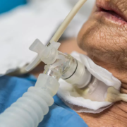
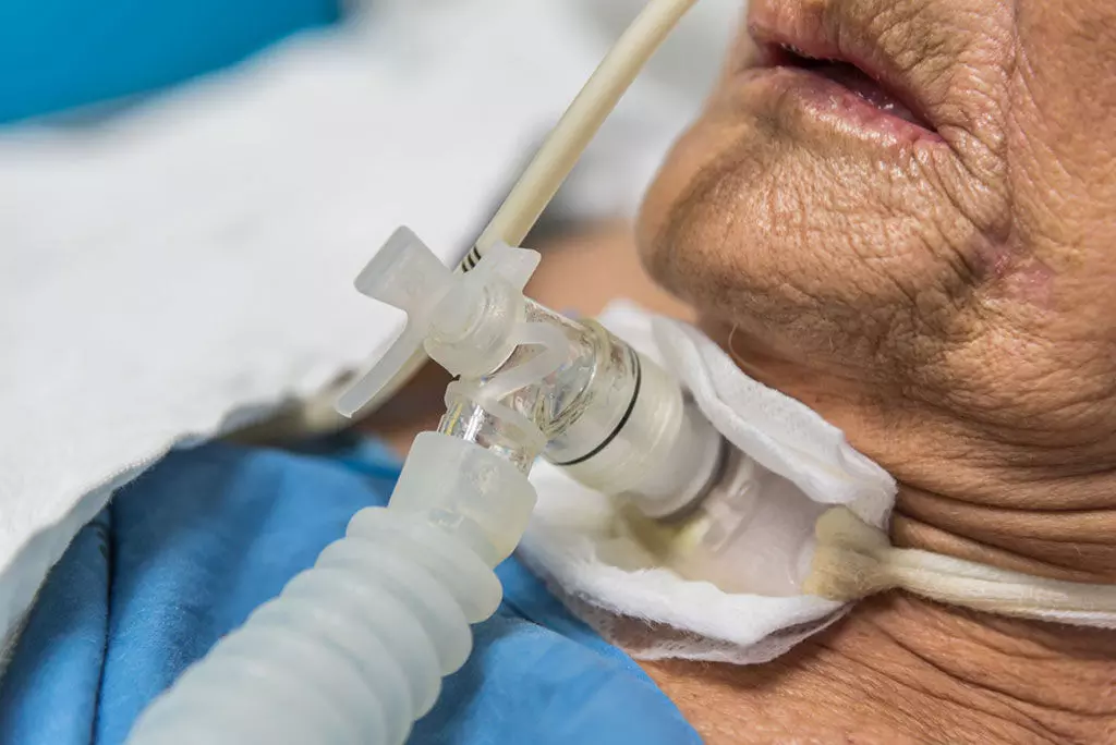
Eighty patients undergoing upper limb orthopedic surgery were randomly assigned to receive a perineural catheter via either the costoclavicular (CC) technique (Group CC) or the lateral sagittal (LS) technique (Group LS). Postoperatively, all patients received patient-controlled regional analgesia with 0.125% bupivacaine.

Results

The primary outcome was the 24-hour LA consumption. The results showed that patients in Group CC required significantly less LA over 24 hours compared to Group LS (83.35 ml vs 121.40 ml, p<0.001). Patients in Group CC also took a longer time to activate the patient-controlled analgesia (p=0.003) and reported better satisfaction scores (p=0.001) compared to Group LS. Pain scores were similar between the two groups.

Complications

One patient in Group CC experienced transient paresthesia which resolved on removal of the catheter. There were no other complications like vascular injury or LA toxicity in either group.

Conclusion

The study concluded that patients receiving postoperative analgesia via the costoclavicular perineural catheter technique required less 24-hour local anesthetic and reported better satisfaction compared to the lateral sagittal technique. The compact and consistent anatomy of the brachial plexus cords in the costoclavicular space likely contributed to the improved analgesic efficacy of this approach.

This randomized trial provides evidence that the costoclavicular technique for inserting perineural catheters can be an effective method for providing prolonged postoperative analgesia after upper limb orthopedic procedures.

Key Points –

The key points from the provided research paper are:

1. The study compared the postoperative local anesthetic (LA) consumption and other outcomes between two different techniques (costoclavicular and lateral sagittal) of inserting a perineural catheter for regional analgesia after upper limb orthopedic surgery.

2. Eighty patients were randomly assigned to receive a perineural catheter via either the costoclavicular (CC) technique (Group CC) or the lateral sagittal (LS) technique (Group LS), and all patients received patient-controlled regional analgesia with 0.125% bupivacaine postoperatively.

3. The primary outcome was the 24-hour LA consumption, and the results showed that patients in Group CC required significantly less LA over 24 hours compared to Group LS (83.35 ml vs 121.40 ml, p<0.001). Patients in Group CC also took a longer time to activate the patient-controlled analgesia (p=0.003) and reported better satisfaction scores (p=0.001) compared to Group LS.

4. Pain scores were similar between the two groups.

5. One patient in Group CC experienced transient paresthesia which resolved on removal of the catheter, and there were no other complications like vascular injury or LA toxicity in either group.

6. The study concluded that the costoclavicular perineural catheter technique can be an effective method for providing prolonged postoperative analgesia after upper limb orthopedic procedures, likely due to the compact and consistent anatomy of the brachial plexus cords in the costoclavicular space.

Reference –

Mondal S, Sinha C, Kumari P, Kumar A, Kumar A, Agarwal P. Costoclavicular versus lateral sagittal infraclavicular brachial plexus block for postoperative analgesia in patients undergoing upper limb orthopaedic surgery: A randomized controlled trial. Indian J Anaesth 2024;68:693‑8.

Powered by WPeMatico

Long-term exposure to PPIs and oral anticoagulants linked to increased risk of iron deficiency anaemia: Study

Long-term exposure to PPIs and oral anticoagulants linked to increased risk of iron deficiency anaemia suggests a study published in the BMJ Open.

A study was done to estimate the strength of association between exposure to selected classes of prescribed medications and the risk of developing iron deficiency anaemia (IDA), specifically considering oral anticoagulants (OACs), antidepressants, antiplatelet agents, proton pump inhibitors (PPIs) and non-steroidal anti-inflammatories. A case-control study involving the analysis of community repeat prescriptions among subjects referred with IDA, and unmatched controls referred as gastroenterology fast-tracks for other indications. Multivariable logistic regression modelling was used to calculate ORs for the association between IDA presentation and each medication class, adjusted for age, sex and coprescribing. For those classes showing significance, it was also used to calculate risk differences between those in the IDA group with or without haemorrhagic lesions on investigation. Results: A total of 1210 cases were analysed-409 in the IDA group, and 801 in the control group. Significant associations were identified between presentation with IDA and long-term exposure to PPIs (OR 3.29, 95% CI: 2.47 to 4.41, p<0.001) and to OACs (OR 2.04, 95% CI: 1.29 to 3.24, p=0.002). IDA was not associated with long-term exposure to any of the other three drug classes. In contrast to the relationship with PPIs, the association with OACs was primarily in the IDA sub-group with haemorrhagic lesions. Long-term exposure to PPIs and OACs are independently associated with the risk of developing IDA. There are grounds for considering that these associations may be causal, though the underlying mechanisms probably differ.

Reference:

Prabhu K, Warricker F, Almilaji O, Williams E, Snook J. Role of prescribed medication in the development of iron deficiency anaemia in adults-a case-control study. BMJ Open Gastroenterol. 2024;11(1):e001305. Published 2024 Jun 26. doi:10.1136/bmjgast-2023-001305

Keywords:

Long-term, exposure, PPIs, oral anticoagulants, increased risk, iron deficiency anemia, study

Powered by WPeMatico

Novel treatment improves embryo implantation and live birth rates in infertile women undergoing IVF and ICSI: Study

New research has demonstrated the effectiveness of a first-in-class oral, non-hormonal drug in increasing embryo implantation, pregnancy and live birth rates among infertile women who are undergoing in vitro fertilization (IVF) or intracytoplasmic sperm injection (ICSI). The findings, presented today at the ESHRE 40th Annual Meeting in Amsterdam, represent a significant step toward the first therapeutic tool to increase embryo implantation and live birth rate success.

Worldwide, one in six people of reproductive age experience infertility in their lifetime. Over 3 million IVF cycles are performed annually and yet, despite advancing IVF technologies, embryo implantation failure remains a significant challenge.

In response to this unmet need, researchers have unveiled the promising findings of their Phase 2 clinical trial, OXOART2. This randomised, double-blind, placebo-controlled trial conducted across 28 centres in Europe evaluated OXO-001, a first-in-class oral drug that acts directly on the endometrium (inner lining of the uterus) to improve embryo implantation and pregnancy rates.

The OXOLIFE exploratory subset study analysed 96 women aged up to 40 years old who underwent a single embryo transfer, 42 receiving placebo and 54 receiving a daily dose of OXO-001. Treatment began one menstrual cycle before the embryo transfer cycle and continued until five weeks after the transfer.

Statistically significant improvements were observed in biochemical pregnancy rates-an early detection of pregnancy-with rates of 75.9% in the OXO-001 group compared to 52.4% in the placebo group. Clinically relevant improvements were also seen in clinical pregnancy rates (foetal heartbeat 5 weeks after embryo transfer), and in ongoing pregnancy rates (10 weeks post-embryo transfer), being a +14.3 absolute increase (50.0% for OXO-001 vs. 35.7% for placebo) and a +10.6 absolute increase (46.3% for OXO-001 vs 35.7% for placebo) respectively.

Most importantly, there was an absolute increase of +6.9 in live birth rates (42.6% for OXO-001 vs. 35.7% for placebo).

Dr. Agnès Arbat, OXOLIFE’s CEO and CMO, comments, “From scientific societies, key opinion leaders, clinicians and patients, we know that an absolute increase of more than 5 percentage points in ongoing pregnancy is considered clinically meaningful. We observed an increase higher than +9, giving renewed hope to patients and the scientific community. We look forward to advancing this promising treatment through the next phases of clinical development.”

The occurrence of side effects was similar in both groups. The most common side effects were headaches, nausea, vomiting, gastrointestinal issues, and dizziness, most of which were mild to moderate. More importantly, in the six-month follow-up, the babies indicated good development with no differences with placebo. Overall, OXO-001 was well tolerated, with high rates of compliance.

Dr. Ignasi Canals, CSO of OXOLIFE adds, “We are thrilled with the results of this trial, which highlight OXO-001’s potential to become the first therapeutic treatment to increase embryo implantation success, with a non-hormonal drug using a new mechanism of action, acting directly on the endometrium.”

Professor Dr Karen Sermon, Chair of ESHRE, explains “Despite continuous developments in ovarian stimulation, embryo manipulation and culture, improving live birth rates in medically assisted reproduction has been incremental at best. A jump of nearly 7% is very good news for our patients, and hopefully this can be confirmed in larger patient groups.”

The study abstract will be published in Human Reproduction, one of the world’s leading reproductive medicine journals.

Reference:

Novel treatment improves embryo implantation and live birth rates in infertile women undergoing IVF and ICSI, European Society of Human Reproduction and Embryology, Meeting: ESHRE 40th Annual Meeting.

Powered by WPeMatico

Phospholipid curcumin Meriva Promising in Treatment of Nonalcoholic Steatohepatitis and Kidney Disease: Study

Researchers recently have found that Meriva, a phospholipid formulation of curcumin, is both safe and effective in improving liver histology, kidney disease, and metabolic profiles in patients with nonalcoholic steatohepatitis (NASH). This study was published in the journal Hepatology  by Musso G. and colleagues. This study could lead to improved treatment options for NASH patients, who face increased risks of liver-related and kidney morbidity.

Nonalcoholic steatohepatitis (NASH) is a progressive liver disease that poses serious health risks, including liver fibrosis and chronic kidney disease (CKD). Traditional treatments have limited efficacy, prompting the search for alternative therapies. Meriva®, known for its enhanced systemic absorption and delivery of curcumin, has shown potential anti-inflammatory and metabolic benefits, making it a candidate for NASH treatment.

In a double-blind trial, 52 patients with biopsy-confirmed NASH were randomized to receive either 2 grams of Meriva® per day or a placebo for 72 weeks. The study’s primary endpoint was the resolution of NASH without worsening fibrosis. Secondary endpoints included improvement in liver fibrosis, regression of significant fibrosis and CKD, and enhancements in renal, glucose, lipid, and inflammatory parameters. The study also explored Meriva®’s effect on hepatic activation of Nuclear Factor (NF)-kB, a pro-inflammatory transcription factor targeted by curcumin.

Fifty-one patients completed the trial, with significant differences observed between the Meriva® and placebo groups:

  • 62% of patients on Meriva® achieved NASH resolution compared to 12% on placebo (RR=5.33, 95% CI=1.76–12.13; p=0.003).

  • 50% of patients on Meriva® showed at least a one-stage improvement in liver fibrosis, versus 8% on placebo (RR=6.50, 95% CI=1.63-21.20; p=0.008).

  • 42% of Meriva® patients experienced regression compared to none on placebo (RR=18.01, 95% CI=1.43-36.07; p=0.02).

  • 50% of patients on Meriva® saw regression in CKD versus none on placebo (RR=10.71, 95% CI=1.94-17.99; p=0.004).

Meriva® significantly improved various metabolic and inflammatory markers:

  • eGFR: Improved by +3.59 mL/min/1.73 m²/year (p=0.009).

  • Fasting Glucose: Decreased by 17 mg/dL (95% CI=−22, −12).

  • HbA1c: Reduced by 0.62% (95% CI=−0.87%, −0.37%).

  • LDL-C: Lowered by 39 mg/dL (95% CI=−45, −33).

  • Triglycerides: Decreased by 36 mg/dL (95% CI=−46, −26).

  • HDL-C: Increased by 10 mg/dL (95% CI=+8, +11).

  • The inhibition of hepatic NF-kB activity was a predictor of NASH resolution (AUC=0.90, 95% CI=0.84-0.95) and fibrosis improvement (AUC=0.89, 95% CI=0.82-0.96).

  • Adverse events were rare, mild, and evenly distributed between the Meriva® and placebo groups, confirming the safety and tolerability of Meriva® over the 72-week period.

This study demonstrates that Meriva® is a promising therapeutic option for NASH patients, providing significant benefits in liver histology, kidney disease, and metabolic health, likely through the inhibition of NF-kB. Further research and larger trials are warranted to confirm these findings and potentially expand the use of Meriva® in clinical practice.

Reference:

Musso, G., Pinach, S., Mariano, F., Saba, F., De Michieli, F., Framarin, L., Berrutti, M., Paschetta, E., Parente, R., Lizet Castillo, Y., Leone, N., Castellino, F., Cassader, M., & Gambino, R. (2024). Effect of phospholipid curcumin Meriva® on liver histology and kidney disease in nonalcoholic steatohepatitis a randomized, double-blind, placebo-controlled trial. Hepatology (Baltimore, Md.), 10.1097/HEP.0000000000000937. https://doi.org/10.1097/hep.0000000000000937

Powered by WPeMatico

Low-level laser therapy effectively improve skin elasticity and wrinkles, claims study

Skin aging and photoaging can manifest at a relatively early age which pose significant aesthetic challenges. The common signs of skin aging include wrinkles, dyspigmentation and decreased elasticity. A recent study published in the Journal of Cosmetic Dermatology explores the effectiveness and safety of low-level laser therapy (LLLT) for skin rejuvenation by presenting a potential breakthrough in cosmetic dermatology.

The study involved 30 Syrian female patients aged between 25 and 50. These patients were diagnosed with moderate to severe melasma and wrinkles. They were divided into two groups where each participant received LLLT with a wavelength of 660 nm by emitting a continuous wave. The power density and dose applied were 15.6 mW/cm² and 3 J/cm², respectively, over an effective laser area of 32 cm². The evaluations were conducted before, during and after 12 treatment sessions by utilizing photographs and several diagnostic tools, including the modified Melasma Area Severity Index (MASI), the Pinch test and Fitzpatrick’s classification of facial wrinkling at baseline. The primary objective of this study evaluated the effectiveness and safety of LLLT for skin rejuvenation in women suffering from moderate to severe melasma and wrinkles.

The findings of this study were strong by indicating significant improvement in skin rejuvenation post-treatment, with statistical significance (p < 0.05). The patients reported marked improvements in the skin elasticity and wrinkle reduction. Additional outcome measures, like the patient satisfaction scores, reflected high levels of satisfaction with the treatment. Also, no adverse effects or re-pigmentation were observed during the study.

The study concluded that LLLT is a safe therapeutic option for treating melasma by enhancing skin elasticity and reducing wrinkles. These findings support the integration of LLLT into cosmetic and dermatological treatment and follow-up programs. The non-invasive nature of LLLT when combined with its effectiveness positions it as a valuable tool in the ongoing battle against skin aging.

These outcomes point towards the significant improvements observed in skin texture and appearance illuminate the potential of LLLT as a rooted option in cosmetic treatments, this could revolutionize current approaches to skin rejuvenation. The absence of adverse effects further highlights its safety and could make it a crucial option for long-term use. Overall, further studies could expand on different demographics and varying degrees of skin conditions. Long-term studies could also provide insights into the sustainability of LLLT benefits over time. Thereby, integrating LLLT with other skin care treatments could improve its efficacy for comprehensive skin rejuvenation protocols.

Source:

Shurrab, K., & Alzghayar, J. N. (2024). Low‐level laser therapy for skin rejuvenation: A safe and effective solution baked by data and visual evidence. In Journal of Cosmetic Dermatology. Wiley. https://doi.org/10.1111/jocd.16404

Powered by WPeMatico

UNC researchers identify potential treatment for Angelman syndrome

 Angelman syndrome is a rare genetic disorder caused by mutations in the maternally-inherited UBE3A gene and characterized by poor muscle control, limited speech, epilepsy, and intellectual disabilities. Though there isn’t a cure for the condition, new research at the UNC School of Medicine is setting the stage for one.

Ben Philpot, PhD, the Kenan Distinguished Professor of Cell Biology and Physiology at the UNC School of Medicine and associate director of the UNC Neuroscience Center, and his lab have identified a small molecule that could be safe, non-invasively delivered, and capable of “turning on” the dormant paternally-inherited UBE3A gene copy brain-wide, which would lead to proper protein and cell function, amounting to a kind of gene therapy for individuals with Angelman syndrome.

“This compound we identified has shown to have excellent uptake in the developing brains of animal models,” said Philpot, who is a leading expert on Angelman syndrome. “We still have a lot of work to do before we could start a clinical trial, but this small molecule provides an excellent starting point for developing a safe and effective treatment for Angelman syndrome.”

But these results, which were published in Nature Communications, mark a major milestone in the field, according to Mark Zylka, W.R. Kenan Jr. Distinguished Professor of Cell Biology and Physiology at the UNC School of Medicine and Director of the UNC Neuroscience Center. No other small molecule compound has yet to show such promise for Angelman, he added.

Unlike other single-gene disorders such as cystic fibrosis and sickle-cell anemia, Angelman syndrome has a unique genetic profile. Researchers have found that children with the conditions are missing the maternally-inherited copy of the UBE3A gene, while the paternally-inherited copy of the UBE3A gene remains dormant in neurons, as it does in neurotypical individuals. Typically, UBE3A helps regulate the levels of important proteins; missing a working copy leads to severe disruptions in brain development.

For reasons that aren’t fully clear, the paternal copy of UBE3A is normally “turned off” in neurons throughout the entire brain. Thus, when the maternal copy of the UBE3A gene is mutated, this leads to a loss of UBE3A protein in the brain. Philpot and other researchers have theorized that turning on the paternal copy of UBE3A could help treat the condition.

Hanna Vihma, PhD, a postdoctoral research fellow in the Philpot lab and first author on the study, and colleagues screened more than 2,800 small molecules from a Pfizer chemogenetic library to determine if one could potently turn on paternal UBE3A in mouse models with Angelman syndrome.

Researchers genetically modified mouse neural cells with a fluorescent protein that glows when the paternal UBE3A gene is turned on. After treating the neurons with more than 2,800 small molecules for 72 hours, researchers compared their thousands of treated cells against those treated with topotecan, a known small molecule that can turn on paternal UBE3A but lacked therapeutic value in animal models of the condition.

(S)-PHA533533, a compound that was previously developed as an anti-tumor agent, caused neurons to express a fluorescent glow that rivaled that induced by topotecan, meaning that its effect was potent enough to successfully turn on paternal UBE3A. Researchers were able to confirm the same results using induced pluripotent stem cells derived from humans with Angelman syndrome, indicating that this compound has clinical potential.

Additionally, researchers observed that (S)-PHA533533 has excellent bioavailability in the developing brain, meaning it travels to its target with ease and sticks around. This is notable in that previous genetic therapies for Angelman syndrome have had more limited bioavailability.

“We previously showed that topotecan, a topoisomerase inhibitor, had very poor bioavailability in mouse models,” said Vihma. “We were able to show that (S)-PHA533533 had better uptake and that the same small molecule could be translated in human-derived neural cells, which is a huge finding. It means it, or a similar compound, has true potential as a treatment for children.”

Although (S)-PHA533533 shows promise, researchers are still working to identify the precise target inside cells that causes the desired effects of the drug. Philpot and colleagues also need to conduct further studies to refine the medicinal chemistry of the drug to ensure that the compound – or another version of it – is safe and effective for future use in the clinical setting.

“This is unlikely to be the exact compound we would take forward to the clinic,” said Philpot. Along with medicinal chemists in the lab of Jeff Aubé, PhD, the Philpot lab is working to identify similar molecules with improved drug properties and safety profiles. “However, this gives us a compound that we can work with to create an even better compound that could be moved forward to the clinic.”

Reference:

Vihma, H., Li, K., Welton-Arndt, A. et al. Ube3a unsilencer for the potential treatment of Angelman syndrome. Nat Commun 15, 5558 (2024). https://doi.org/10.1038/s41467-024-49788-8.

Powered by WPeMatico

Study evaluates Early vs. Late Tracheostomy and Bacterial Infections in COVID-19 ICU Patients

The COVID-19 global pandemic had a substantial effect on healthcare, particularly in critical care settings. Recent study examined the impact of early versus late tracheostomy on bacterial infections in COVID-19 patients requiring mechanical ventilation in the intensive care unit (ICU). The researchers conducted a retrospective single-center study at the Royal Medical Services Military Hospital, including COVID-19 patients who underwent tracheostomy and were admitted to the ICU from March 2020 to March 2022.

Comparison of Early versus Late Tracheostomy

The primary objective was to compare the incidence of ventilator-associated pneumonia between patients who underwent early tracheostomy (within 14 days) and late tracheostomy (more than 14 days after starting mechanical ventilation). The secondary objective was to assess the time required to wean from mechanical ventilation between the two groups.

The study included 36 patients, with no statistically significant differences in baseline characteristics between the early and late tracheostomy groups. The findings showed no significant association between the timing of tracheostomy and the incidence of bacterial infections, complications, or outcomes. Mortality was higher in the early tracheostomy group, with nine deaths compared to seven in the late tracheostomy group, but the difference was not statistically significant.

Association with Specific Bacterial Infections

Interestingly, the study found that infection with Acinetobacter baumannii was associated with a statistically significant lower mortality rate, with 75% of affected patients surviving post-tracheostomy. In contrast, infections with other bacteria, such as Pseudomonas aeruginosa, Stenotrophomonas maltophilia, and Klebsiella pneumonia, did not show significant differences in survival rates.

Conclusion and Implications

The researchers concluded that the timing of tracheostomy does not significantly impact the incidence of bacterial pneumonia or other complications in COVID-19 patients requiring mechanical ventilation. These results support the need for personalized decision-making when conducting tracheostomies. Further research is necessary to determine the impacts of tracheostomy timing on patient outcomes more definitively.

Key Points

The 3 key points of the research article are:

1. The study examined the impact of early versus late tracheostomy on bacterial infections in COVID-19 patients requiring mechanical ventilation in the intensive care unit (ICU). The primary objective was to compare the incidence of ventilator-associated pneumonia between patients who underwent early tracheostomy (within 14 days) and late tracheostomy (more than 14 days after starting mechanical ventilation). The secondary objective was to assess the time required to wean from mechanical ventilation between the two groups.

2. The findings showed no significant association between the timing of tracheostomy and the incidence of bacterial infections, complications, or outcomes. Mortality was higher in the early tracheostomy group, but the difference was not statistically significant. Interestingly, the study found that infection with Acinetobacter baumannii was associated with a statistically significant lower mortality rate, while infections with other bacteria, such as Pseudomonas aeruginosa, Stenotrophomonas maltophilia, and Klebsiella pneumonia, did not show significant differences in survival rates.

3. The researchers concluded that the timing of tracheostomy does not significantly impact the incidence of bacterial pneumonia or other complications in COVID-19 patients requiring mechanical ventilation. These results support the need for personalized decision-making when conducting tracheostomies, and further research is necessary to determine the impacts of tracheostomy timing on patient outcomes more definitively.

Reference –

Alkoheji H, Alabbasi L, Aldoseri M S, et al. (July 03, 2024) Clinical Outcomes of Early vs. Late Tracheostomy in Ventilated COVID-19 Patients. Cureus 16(7): e63757. DOI 10.7759/cureus.63757

Powered by WPeMatico

IL-1β and IL-6 levels linked to development of obsessive compulsive disorder, suggests study

A new study published in the Public Library of Science unveiled a link between serum IL-1β and IL-6 levels and the etiology of obsessive-compulsive disorder (OCD). Obsessive-compulsive disorder equates to a global prevalence of 2% to 3%. The hallmarks of OCD are ritualized, repeated behavioral or mental acts (compulsions) that follow recurrent, unwelcome, intrusive thoughts, feelings, urges or ideas (obsessions) that magnify concerns about danger, harm, or cleanliness. The biological, genetic, environmental and immunological variables are only a few of the many etiological elements that contribute to OCD. And now, SSRIs, TCAs, SNRIs and dopamine antagonists are available as treatments for OCD. In addition to these pharmaceutical treatments, cognitive-behavioral therapy is often used as the non-pharmacological treatment for OCD. There may be a possible connection between the pathophysiology of OCD and immunological dysregulation, according to the outcomes of this study. 

30 age- and sex-matched healthy controls (HCs) and 50 OCD patients were enrolled in this study where a licensed psychiatrist used the Diagnostic and Statistical Manual for Mental Health Disorders, Fifth Edition (DSM-5) criteria to diagnose OCD patients and evaluate HCs. After applying the Yale-Brown Obsessive Compulsive Scale (Y-BOCS), this research assessed the severity of OCD. Also, ELISA kits were used to test serum levels of IL-1β and IL-6 using the recommended procedures.

The key findings of this study were:

The findings demonstrated that the serum IL-1β levels of OCD patients were much higher than the of healthy controls.

Also, the serum IL-6 levels in OCD patients were considerably higher than in healthy controls.

The Y-BOCS scores of OCD patients showed a positive correlation with both cytokines which suggested a potential involvement of both cytokines in the pathophysiology of this disorder.

The outcomes of this study highlight the complex connection between the pathophysiology of OCD and dysregulated inflammatory responses. The elevated levels of IL-1β and IL-6 in the serum showed a reliable relationship with OCD severity. Therefore, serum levels of IL-1β and IL-6 may provide useful blood-based biomarkers for estimating the risk of OCD. By expanding the existing knowledge of immune-mediated pathways in OCD, this research creates new opportunities for investigation and advancements in treatment.

Reference: 

Sarmin, N., Roknuzzaman, A. S. M., Sarker, R., Rashid, M.-, Hasan, A., Qusar, M. M. A. S., Kabir, E. R., Islam, Md. R., & Mahmud, Z. A. (2024). Exploring the role of interleukin-1β and interleukin-6 in the pathophysiology of obsessive-compulsive disorder. In M. Koronyo-Hamaoui (Ed.), PLOS ONE (Vol. 19, Issue 6, p. e0306125). Public Library of Science (PLoS). https://doi.org/10.1371/journal.pone.0306125

Powered by WPeMatico

Ultrathin Zirconia Laminate Veneers Fabricated Using Conventional and Speed Sintering have similar esthetics and strength: Study

Ultrathin Zirconia Laminate Veneers Fabricated Using Conventional and Speed Sintering have similar esthetics and strength suggests a new study published in the Journal of Prosthetic Dentistry. 

The application of highly translucent multilayered zirconia ceramic in minimally invasive esthetic dentistry allows the achievement of both esthetics and strength with minimal thickness. Clinical studies that have assessed the performance of zirconia ultrathin veneers sintered with conventional and speed procedures are lacking. This clinical study aimed to evaluate the effect of speed sintering processes on the translucency and clinical performance of zirconia laminate veneers. Four participants had their teeth restored with 32 ultra translucent zirconia laminate veneers. Based on the zirconia sintering procedure, the participants were randomly allocated into 2 groups. The teeth were prepared by selective reduction over trial restorations. Zirconia veneers were milled from multilayer zirconia blanks and sintered either by speed sintering or conventional sintering as specified by the manufacturer’s recommendations. The intaglio surface of the veneers was airborne-particle abraded with 50-μm aluminium-oxide, and the veneers were then adhesively bonded to the teeth with translucent light-polymerizing resin cement. The modified California Dental Association (CDA)/Ryge criteria were used to assess participants at baseline and every 3 months for 12 months: the translucency and the colour difference of the tooth before and after veneer restoration were evaluated. For statistical analysis, the Pearson chi-squared test, independent t-test, and paired t-test were used (α=.05).

Results: Translucency and colour differences were significantly higher in the conventionally sintered group (P<.05). No restorations were lost. No significant differences were found between the 2 groups in the CDA/Ryge criteria or colour parameter after follow-up intervals (P>.05). The primary qualitative changes observed at the final recall were marginal integrity and marginal discolouration. The colour match and zirconia surface were rated Alfa. After 1 year of follow-up, both conventional and speed-sintered ultrathin zirconia laminates showed satisfactory functional, esthetic, and colour stability outcomes.

Reference:

Yousry M, Hammad I, El Halawani M, Aboushelib M. Randomized clinical trial of zirconia laminate veneers sintered by using conventional versus speed process: 1-year follow-up. J Prosthet Dent. 2024 May 31:S0022-3913(24)00352-4. doi: 10.1016/j.prosdent.2024.04.031. Epub ahead of print. PMID: 38824110.

Keywords:

Ultrathin, Zirconia, Laminate, Veneers, Fabricated, Conventional, Speed, Sintering, similar esthetics, strength, Yousry M, Hammad I, El Halawani M, Aboushelib M

Powered by WPeMatico

Artemisinins Show Promise in Treating PCOS Through LONP1-CYP11A1 Interaction: Study

China: Recent research has unveiled a potential new avenue for treating polycystic ovarian syndrome (PCOS) using artemisinins, compounds traditionally known for their anti-malarial properties. A study published in Science has shed light on how artemisinins may ameliorate PCOS by influencing the LONP1-CYP11A1 interaction, a critical pathway involved in hormonal regulation.

Early results in human and animal models suggest that artemisinins might target all aspects of PCOS by suppressing ovarian androgen synthesis, researchers reported.

The study revealed that artemisinins reduced blood testosterone levels and cystic ovarian follicles in PCOS-like mice and improved litter size and embryo implantation in PCOS-like rats. Similarly, artemisinin treatment decreased levels of 2 blood markers of PCOS and reduced ovarian cysts in 19 human participants with the condition. The majority of participants (63%) also regained normal menstrual cycles.

PCOS, a common endocrine disorder affecting women of reproductive age, is characterized by irregular menstrual cycles, excess androgen production, and ovarian cysts. Current treatments primarily focus on managing symptoms such as infertility, insulin resistance, and hormonal imbalance. However, the underlying mechanisms driving PCOS have remained elusive until now. Current treatment options like birth control pills and metformin tend to target only specific symptoms of the condition.

Despite the high prevalence of PCOS, specific pharmacologic intervention for PCOS is challenging. In the study, Yang Liu, Fudan University, Shanghai, China, and colleagues identified artemisinins as anti-PCOS agents.

The finding demonstrated artemisinin derivatives’ efficacy in alleviating PCOS symptoms in both rodent models and human patients, curbing hyperandrogenemia by suppressing ovarian androgen synthesis.

The following were the key findings of the study:

  • Artemisinins promoted cytochrome P450 family 11 subfamily A member 1 (CYP11A1) protein degradation to block androgen overproduction.
  • Mechanistically, artemisinins directly targeted lon peptidase 1 (LONP1), enhanced LONP1-CYP11A1 interaction, and facilitated LONP1-catalyzed CYP11A1 degradation.
  • Overexpression of LONP1 replicated the androgen-lowering effect of artemisinins.

Artemisinins directly bind to LONP1, initiating the interaction between LONP1 and CYP11A1, promoting CYP11A1 degradation, subsequently inhibiting ovarian androgen synthesis, and curbing PCOS. Contrarily, the androgenic inducer disrupts LONP1-CYP11A1 interaction and aggravates PCOS.

“Overall, our research underscores the promising potential of artemisinins as effective medications for the holistic management of PCOS.,” the researchers wrote.

“This discovery reveals a newly identified interaction between LONP1 and CYP11A1, which artemisinins amplify to regulate androgen synthesis, suggesting new approaches for intervening in PCOS by targeting the LONP1-CYP11A1 interaction,” they concluded.

Reference:

Liu Y, Jiang JJ, Du SY, Mu LS, Fan JJ, Hu JC, Ye Y, Ding M, Zhou WY, Yu QH, Xia YF, Xu HY, Shi YJ, Qian SW, Tang Y, Li W, Dang YJ, Dong X, Li XY, Xu CJ, Tang QQ. Artemisinins ameliorate polycystic ovarian syndrome by mediating LONP1-CYP11A1 interaction. Science. 2024 Jun 14;384(6701):eadk5382. doi: 10.1126/science.adk5382. Epub 2024 Jun 14. PMID: 38870290.

Powered by WPeMatico