Rapid Initiation Procedure may Improve XR-Naltrexone Efficacy among Opioid Use Disorder: JAMA

A recent study has revealed a promising approach to expanding access to effective treatment against opioid use disorder (OUD). The research compared two procedures for initiating extended-release (XR)-naltrexone which is a key medication in OUD management. The major findings of the study were published in the Journal of American Medical Association.

This study was conducted at six community-based inpatient addiction treatment units to address a significant barrier to XR-naltrexone implementation and the need for patients to undergo opioid withdrawal prior to treatment initiation. This requirement often discourages individuals from seeking XR-naltrexone therapy.

The study spanned from March 16, 2021 to September 21, 2022 and focused on comparing the effectiveness of the standard procedure (SP) with a novel approach termed the rapid procedure (RP). As per XR-naltrexone package instructions, the SP involved a multi-day buprenorphine taper followed by an opioid-free period. The RP condensed this process into a single day by utilizing low doses of oral naltrexone and adjunctive medications to manage withdrawal symptoms.

The results from the study encompassed a total of 415 participants with OUD and unveiled promising findings. The RP demonstrated noninferiority to the SP with a higher rate of successful XR-naltrexone initiation (62.7% vs. 35.8%, respectively). Also, withdrawal severity did not significantly differ between the two procedures.

The implications of these findings suggest that the RP could significantly enhance access to XR-naltrexone treatment by streamlining the initiation process and could potentially revolutionize OUD management. Despite requiring more intensive medical supervision and safety monitoring, the RP offers a time-saving alternative that could make XR-naltrexone a more feasible option for patients with OUD.

The study emphasized the importance of these results which suggest that rapid initiation holds great promise in overcoming barriers to XR-naltrexone adoption. This can aid in improving the patient outcomes and ultimately manage the opioid crisis more effectively by simplifying the treatment process. The key findings of this study marks a major leap ahead in the quest for innovative solutions to address OUD. Overall, the adoption of rapid initiation procedures could represent a crucial point in offering renewed optimism in the battle against OUD.

Reference:

Shulman, M., Greiner, M. G., Tafessu, H. M., Opara, O., Ohrtman, K., Potter, K., Hefner, K., Jelstrom, E., Rosenthal, R. N., Wenzel, K., Fishman, M., Rotrosen, J., Ghitza, U. E., Nunes, E. V., & Bisaga, A. (2024). Rapid Initiation of Injection Naltrexone for Opioid Use Disorder. In JAMA Network Open (Vol. 7, Issue 5, p. e249744). American Medical Association (AMA). https://doi.org/10.1001/jamanetworkopen.2024.9744

Powered by WPeMatico

Rare case of Intramuscular endometriosis of forearm: a report

Endometriosis is a disorder that commonly affects females of reproductive age and is defined as the presence of endometrial glands or stroma outside the uterine cavity. Patients typically present with cyclical pain during menses. Endometriosis can be characterized as endopelvic or extra pelvic depending on the sites involved.

Smith et al reported a case of a 40-year-old, right-hand dominant, female who presented with a painful mass in her right proximal forearm. She was ultimately diagnosed with intramuscular endometriosis and underwent surgical excision.

A 40-year-old, right-hand-dominant, female (G3P3003) presented to the Orthopedic Department with an 18-month history of an intermittently painful mass in the medial aspect of her right forearm. The mass would become increasingly tender and firm in the days before menses and during menstruation. Her past medical history was notable for relapsing remitting multiple sclerosis (MS), hypertension, dyslipidemia, anxiety, and morbid obesity. She denied any excessively heavy or painful menses.

Physical examination revealed a small palpable mass in the right flexor pronator musculature and was tender to palpation. Previous imaging, including radiographs of the right forearm demonstrated no acute osseous or soft tissue abnormality. Magnetic resonance imaging (MRI) without intravenous contrast showed an oval intramuscular mass in the anterior compartment of the proximal forearm, measuring approximately 2.2 × 2.1 × 3.0 cm. The mass demonstrated heterogenous T2 signal and cystic changes. There was no invasion of deeper musculature or fascia.

On ultrasound imaging, the mass was circumscribed and hypoechoic. The mass also demonstrated internal doppler flow. Unfortunately, the ultrasound guided core needle biopsy was nondiagnostic. Therefore, the patient elected to undergo open biopsy with frozen section diagnosis, which was consistent with endometriosis.

The patient was referred to Obstetrics/Gynecology for further evaluation and to discuss treatment options. Additional history obtained at that time revealed that she had delivered all three of her children by uncomplicated Cesarean section. Physical examination did not result in any concern for pelvic endometriosis. Dienogest, a progestin-based medication, was recommended with the goal of suppressing the proliferation of the endometrium to decrease symptoms. As the patient was still working to achieve remission of her MS with medications, she did not wish to add another medication at that time and declined the progestin medication.

Approximately 18 months after initial presentation to the orthopedic oncology clinic, the patient’s symptoms worsened with pain now radiating into the elbow, distal forearm, and hand. She elected to trial Dienogest; however after several months, this did not improve her symptoms. After discussion with the orthopedic surgeon, the patient ultimately decided to undergo surgical excision of the mass. At 6 months postoperatively, the patient remains significantly improved compared to pre-resection. She does have very mild soreness at the forearm at the time of menstruation but states it does not bother her.

The authors commented – “In summary, we report an extremely rare case of intramuscular endometriosis in the proximal forearm. Extremity endometriosis may be included in the differential diagnosis of soft tissue tumors when symptoms of pain or a palpable mass occur in the extremities of women of reproductive age, particularly those masses with a cyclic pattern, which coincides with the menstrual cycle.”

Further reading:

Intramuscular endometriosis of the forearm: a case report

Kira L. Smith et al

Skeletal Radiology

https://doi.org/10.1007/s00256-024-04648-w

Powered by WPeMatico

Risk Factors for Retinopathy among long term Hydroxychloroquine users identified in JAMA study

USA: Hydroxychloroquine (HCQ), a medication commonly prescribed for autoimmune conditions such as rheumatoid arthritis and lupus, has garnered attention for its therapeutic benefits and potential ocular side effects. In a comprehensive study published in JAMA Network Open, researchers have delved into the risk factors associated with hydroxychloroquine retinopathy and its subtypes, shedding light on crucial considerations for patients and healthcare providers.

In the cohort study of 4677 long-term hydroxychloroquine users with masked adjudication of retinopathy outcomes, female sex, increasing age, chronic kidney disease (CKD) stage 3 or greater, and tamoxifen use were each associated with a higher risk of hydroxychloroquine retinopathy.

“This study suggests considering these factors when making decisions about hydroxychloroquine dosing and screening for hydroxychloroquine retinopathy,” the researchers wrote.

Hydroxychloroquine retinopathy is a rare but serious complication characterized by damage to the retina, the light-sensitive tissue at the back of the eye. Long-term use of HCQ has been implicated in the development of retinopathy, which can lead to irreversible vision loss if left unchecked. Understanding the risk factors associated with this condition is paramount for early detection and intervention. Current guidelines recommend limiting the dose and screening annually for retinopathy among all long-term users, but individual patient factors may be linked with retinopathy risk.

Against the above background, April M. Jorge, Division of Rheumatology, Allergy, and Immunology, Massachusetts General Hospital, Boston, and colleagues aimed to identify risk factors beyond hydroxychloroquine dose and duration of use for hydroxychloroquine retinopathy.

For this purpose, the researchers conducted a cohort study of 4677 patients in the Kaiser Permanente Northern California integrated health network who initiated hydroxychloroquine, continued treatment, and underwent retinopathy screening following five years of use, from 1997 to 2020, with up to 15 years of follow-up.

Candidate risk factors included age at hydroxychloroquine initiation, race and ethnicity, sex, CKD, indications, diabetes, liver disease, tamoxifen use, and medications that interact with hydroxychloroquine metabolism. Hydroxychloroquine dose was evaluated from pharmacy dispensing records.

Incident hydroxychloroquine retinopathy was adjudicated from the masked review of guideline-recommended screening studies and categorized as a parafoveal or pericentral pattern. Potential risk factors for hydroxychloroquine retinopathy within 15 years of initiation were assessed.

The study led to the following findings:

  • Among 4677 long-term hydroxychloroquine users (mean age at initiation, 52.4 years; 82.9% women), 125 patients developed hydroxychloroquine retinopathy within 15 years (102 parafoveal, 23 pericentral).
  • Older age at the time of hydroxychloroquine initiation was associated with retinopathy risk, with adjusted hazard ratios (HRs) of 2.48 for those aged 45 to 54 years, 3.82 for those aged 55 to 64 years, and 5.68 for those aged 65 years or older compared with those younger than 45 years.
  • Retinopathy risk was higher among females than males (HR, 3.83), among patients with CKD stage 3 or greater (HR, 1.95), and among individuals with tamoxifen use (HR, 3.43).
  • There was a higher likelihood of pericentral retinopathy among Asian patients (HR, 15.02) and Black patients (HR, 5.51) compared with non-Hispanic White patients.

The findings suggest that female sex, increasing age, tamoxifen use, and CKD stage 3 or greater were associated with a higher risk of hydroxychloroquine retinopathy, whereas male sex and being younger than 45 years at hydroxychloroquine initiation were associated with a lower risk. Ethnicity and race were also associated with the pattern of retinopathy.

“These factors should be incorporated into hydroxychloroquine dosing decisions,” the researchers wrote.

In conclusion, the study on risk factors for hydroxychloroquine retinopathy and its subtypes represents a significant advancement in our understanding of this sight-threatening complication. As the medical community continues to refine strategies for optimizing HCQ therapy, personalized risk assessment and vigilant monitoring remain crucial pillars of patient care, ensuring that the benefits of treatment outweigh the risks to ocular health.

Reference:

Jorge AM, Melles RB, Marmor MF, Zhou B, Zhang Y, Choi HK. Risk Factors for Hydroxychloroquine Retinopathy and Its Subtypes. JAMA Netw Open. 2024;7(5):e2410677. doi:10.1001/jamanetworkopen.2024.10677

Powered by WPeMatico

What are risk factors for Vascular Calcification in Peritoneal Dialysis Patients?

China: Vascular calcification, a common complication in patients undergoing peritoneal dialysis (PD), has emerged as a significant determinant of long-term prognosis. A recent analysis published in BMC Nephrology has shed light on the multifaceted factors influencing vascular calcification in PD patients and their profound impact on overall health outcomes.

Comorbid diabetes, longer dialysis time, comorbid hypertension, and serum sclerostin (SOST) are risk factors for vascular calcification in peritoneal dialysis patients. Additionally, longer dialysis time, abdominal aortic calcification (AAC), and comorbid diabetes are associated with an increased risk of all-cause mortality in PD patients.

Peritoneal dialysis, a life-saving therapy for individuals with end-stage renal disease (ESRD), involves peritoneal membrane use as a natural filter to remove waste products and excess fluid from the body. While PD offers several advantages over hemodialysis, such as greater flexibility and preservation of residual renal function, it is not without its complications. 

Vascular calcification, the abnormal calcium deposition in the blood vessels, is increasingly recognized as a prevalent complication in PD patients. This process, similar to the hardening of arteries seen in atherosclerosis, contributes to the development of cardiovascular disease and is associated with increased morbidity and mortality.

Against the above background, Zhongxin Li, Capital Medical University, Beijing, China, and colleagues aimed to investigate the influencing factors of vascular calcification in peritoneal dialysis patients and its relationship with long-term prognosis.

For this purpose, the researchers conducted a retrospective cohort study including chronic kidney disease (CKD) patients undergoing PD at the Peritoneal Dialysis Center of Beijing Luhu Hospital, Capital Medical University, from January 2019 to March 2019.

Clinical laboratory and demographic data, including serum sclerostin (SOST), phosphate (P), calcium (Ca), intact parathyroid hormone (iPTH), and serum albumin (ALB) levels, were collected. Abdominal aortic calcification (AAC) was evaluated using an abdominal lateral X-ray examination to determine vascular calcification occurrence, and based on the results, patients were divided into the AAC group and the Non-AAC group.

The study included a total of 91 patients. The AAC group comprised 46 patients, while the Non-AAC group consisted of 45 patients.

The researchers reported the following findings:

  • The AAC group had significantly older patients than the non-AAC group and longer dialysis times.
  • Multivariable logistic regression analysis indicated that risk factors for vascular calcification in PD patients included dialysis time, diabetes, hypertension, and SOST.
  • Kaplan–Meier survival analysis showed that the AAC group had a significantly higher mortality rate than the non-AAC group (χ2 = 35.993).
  • Multivariable Cox regression analysis revealed that dialysis time, diabetes, and AAC were risk factors for all-cause mortality in peritoneal dialysis patients.

The findings suggest that comorbid diabetes, longer dialysis duration, SOST, and comorbid hypertension are risk factors for vascular calcification in PD patients. The study showed that sclerostin is an independent risk factor for vascular calcification in PD patients.

“There is a need for further research to elucidate the specific mechanisms by which Sclerostin impacts vascular calcification and to evaluate the efficacy of interventions targeting Sclerostin in clinical practice,” the researchers wrote.

Reference:

Cao, Q., Shi, Y., Liu, X. et al. Analysis of factors influencing vascular calcification in peritoneal dialysis patients and their impact on long-term prognosis. BMC Nephrol 25, 157 (2024). https://doi.org/10.1186/s12882-024-03582-2

Powered by WPeMatico

Inpatient IBD Pain Management Protocol Reduces Opioid Use: Study

In a recent trial, opioid usage was reduced among hospitalised patients with inflammatory bowel disease who were treated with a new pain management practice that included scheduled nonopioid drugs modified depending on pain intensity.

The strategy, created by experts at Cedars-Sinai in Los Angeles, was also shown to be more successful at reducing pain than usual therapy that involves opioids, according to a recently published study.

Opioids are often used to alleviate pain, which is a primary symptom for the majority of hospitalised IBD patients. However, there are serious hazards associated with these medications, including as infection, readmission to the hospital, overdose, and accidental diversion.

Patients with IBD are more likely than those with any other chronic GI illness to be prescribed opioids, despite the associated dangers. Even though many IBD patients in hospitals will need opioid medication for pain management, nonopioid analgesic alternatives must be investigated in order to reduce related dangers and enhance patients’ long-term pain management.

The Proactive Analgesic Inpatient Narcotic-Sparing (P.A.I.N.-Sparing) protocol was created by Dr. Berry and his co-investigators after a study of the literature on pain management and investigation into analgesic regimens used in other illnesses comparable to IBD. The strategy depends on regularly scheduled nonopioid painkillers that are adjusted based on the intensity of a patient’s discomfort (Figure). Patients may, however, ask to be prescribed opioids if their breakthrough pain is out of control.

Mild Pain (VRS 1-3)

• PO Acetaminophen 1,000 mg q8h

Moderate Pain (VRS 4-6)

• PO Acetaminophen 1,000 mg q8h

• PO Gabapentin 400 mg QHS (hold for sedation)

• PO Lorazepam 1 mg q8h PRN (hold for sedation)

• PO Celecoxib 200 mg on day 1, then 100 mg BID (4 days total – automatic end date)

• Sequential compression device + SQ heparin (auto order)

Severe Pain (VRS 7 to >10)

• PO Acetaminophen 1,000 mg q8h

• PO Gabapentin 600 mg QHS (hold for sedation)

• PO Lorazepam 2 mg q8h PRN (hold for sedation)

• PO Celecoxib 400 mg on day 1, then 100 mg BID (4 days total – automatic end date)

• Sequential compression device + SQ heparin (auto order)

Breakthrough Pain

• PO Tramadol 50 mg q6h PRN

• IVPB Morphine 2 mg q4h PRN (opiate-naive)

• IVPB Morphine 4 mg q4h PRN (opiate-tolerant – may adjust)

• They randomly assigned 33 IBD patients to receive either the P.A.I.N. protocol or standard treatment with opioids in order to evaluate the effectiveness of the P.A.I.N.-Sparing procedure.

The intervention group used significantly less opioids (average daily morphine milligramme equivalents, 11.8±15.3 vs. 30.9±42.2; P=0.027) and reported significantly lower pain scores (3.02±0.90 vs. 4.29±0.81; P=0.059) than the other group. However, both groups reported significantly reduced pain from admission to discharge.

Patients who followed the regimen also had a substantially greater step count on day 4 in the hospital (2,330±1,709 vs. 1,050±1,214; P=0.014) in terms of mobility.

They provide evidence that “a proactive pain control bundle for hospitalised patients with IBD can reduce the use of opioids, which are associated with so much potential for harm, while also improving pain and leading to overall better outcomes, including physical activity in the hospital.”

“Offers a useful guide to proactively manage pain for patients hospitalised with IBD,” said the report.

From hospital admission to release, the P.A.I.N.-sparing strategy used in this research shows a practical and systematic way to assess, evaluate, and manage pain management.

It’s important to note that the intervention protocol only included pharmacologic interventions. However, the researchers also suggested that nonpharmacologic strategies that are presently being researched, like virtual reality and biofeedback, could be included into a proactive strategy to reduce pain and discomfort in IBD patients who are hospitalised.

Reference –

Berry, S.K., Takakura, W., Patel, D. et al. A randomized controlled trial of a proactive analgesic protocol demonstrates reduced opioid use among hospitalized adults with inflammatory bowel disease. Sci Rep 13, 22396 (2023). https://doi.org/10.1038/s41598-023-48126-0

L

Powered by WPeMatico

Prophylactic Role of Perioperative Corticosteroids for Postoperative Neurocognitive Dysfunction- metaanalysis

Recent research paper titled “Effect of prophylactic corticosteroids on postoperative neurocognitive dysfunction in the adult population: An updated systematic review, meta‑analysis, and trial sequential analysis of randomised controlled trials” aimed to evaluate the prophylactic role of perioperative corticosteroids for postoperative neurocognitive dysfunction (PNCD) in adult surgical patients. The systematic review and meta‑analysis included 15 randomized controlled trials (RCTs) comprising 15,398 patients. The primary outcome assessed was the incidence of PNCD within 1 month, while secondary outcomes included the use of antipsychotic medications for treatment, postoperative infection, and hospital length of stay.

Findings and Conclusions

The findings from the meta-analysis revealed that the incidence of PNCD was significantly lower in the corticosteroid group than in the control group, with a pooled odds ratio of 0.75 (95% CI 0.58, 0.96; P = 0.02; I2 = 66%). The results of the trial sequential analysis showed the clinical benefit of corticosteroids in preventing PNCD, though the requisite information size was considered inadequate. Subgroup analysis supported the prophylactic effect of corticosteroids on delirium prevention, but not on delayed neurocognitive recovery. The meta-analysis concluded that corticosteroids demonstrated statistically significant protective effects on the incidence of PNCD. However, the researchers emphasized the need for further studies to confirm the protective role of corticosteroids for preventing PNCD.

Study Limitations and Recommendations

The paper provided detailed information on the methods used for the systematic review and meta-analysis, the specific outcomes measured, the results obtained from the analysis, and the conclusions drawn based on the findings. The researchers also discussed the limitations of the study, including clinical heterogeneity in outcomes, the lack of standardized screening criteria, and the limited evidence from RCTs targeting high-risk patients. Additionally, the paper emphasized the need for bundled approaches targeting multiple pathways of neurological injury during the perioperative period to effectively prevent PNCD in high-risk patient populations. Overall, the paper provided comprehensive insights into the potential prophylactic role of corticosteroids in preventing PNCD in adult surgical patients.

Key Points

1. The research paper conducted a systematic review and meta-analysis of 15 randomized controlled trials involving 15,398 adult surgical patients to evaluate the prophylactic role of perioperative corticosteroids for preventing postoperative neurocognitive dysfunction (PNCD). The primary outcome assessed was the incidence of PNCD within 1 month post-surgery, and secondary outcomes included the use of antipsychotic medications for treatment, postoperative infection, and hospital length of stay.

2. The findings from the meta-analysis indicated that the incidence of PNCD was significantly lower in the corticosteroid group compared to the control group, with a pooled odds ratio of 0.75 (95% CI 0.58, 0.96; P = 0.02; I2 = 66%). The trial sequential analysis also supported the clinical benefit of corticosteroids in preventing PNCD, although the requisite information size was considered inadequate. Subgroup analysis demonstrated the prophylactic effect of corticosteroids on delirium prevention but not on delayed neurocognitive recovery. The researchers concluded that corticosteroids showed statistically significant protective effects on the incidence of PNCD, but they emphasized the need for further studies to confirm their role in preventing PNCD.

3. The study discussed various limitations, including clinical heterogeneity in outcomes, the lack of standardized screening criteria, and the limited evidence targeting high-risk patients. The researchers recommended the need for bundled approaches targeting multiple pathways of neurological injury during the perioperative period to effectively prevent PNCD in high-risk patient populations. Overall, the paper provided comprehensive insights into the potential prophylactic role of corticosteroids in preventing PNCD in adult surgical patients.

Reference –

Singh NP, Makkar JK, Goel N, Karamchandani K, Singh M, Singh PM. Effect of prophylactic corticosteroids on postoperative neurocognitive dysfunction in the adult population: An updated systematic review, meta analysis, and trial sequential analysis of randomised controlled trials. Indian J Anaesth 2024;68:517-26.

Powered by WPeMatico

Rare case unravels uncommon Hematological Complication of typhoid Fever: A report

India: In a medical anomaly that has puzzled experts, a recent
case report has unveiled an unusual hematological complication associated with
typhoid fever, shedding new light on the complex manifestations of this
infectious disease. This discovery underscores the importance of vigilant
monitoring and comprehensive assessment in managing typhoid fever cases,
especially in regions where the disease remains endemic.

Typhoid fever, caused by the bacterium Salmonella enterica
serotype Typhi, is a systemic illness characterized by fever, abdominal pain,
and gastrointestinal symptoms. While complications such as intestinal
perforation and encephalopathy are well-documented, hematological abnormalities
beyond leukopenia and thrombocytopenia are relatively rare.

The case report published in the Journal of the Association of Physicians of
India
 describes the case of a young female
who presented with complaints of severe left upper quadrant pain after being
diagnosed with typhoid fever.

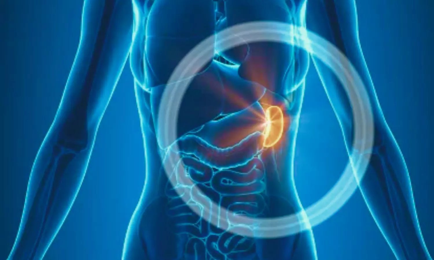
Computed tomography (CT) showed
multiple wedge-shaped splenic infarcts. She was treated with antibiotics and
was also started on antiplatelets. She completely recovered with this
management, and antiplatelets were tapered off on subsequent visits.

The case is of a 25-year-old female
patient with no known comorbidities who was referred to a hospital with complaints
of high-grade fever for 9 days, which was associated with loose stools. She was
diagnosed with typhoid fever from the referring hospital and was started on ceftriaxone.
She then started complaining of chest pain, left-sided abdominal pain, and
breathlessness. She had a family history of rheumatoid arthritis in her mother.

On examination,
she was oriented and conscious, febrile, and had a temperature of 104°F. Her
abdominal examination showed tenderness over the left hypochondrium, and her
respiratory system showed signs of left-sided pleural effusion.

She was admitted,
and baseline blood investigations were conducted. Her blood culture confirmed
the diagnosis of Salmonella Typhi, which was sensitive to the
antibiotic. Her serology for other tropical illnesses, such as dengue, malaria,
leptospirosis, and scrub typhus, was negative.

On further
evaluation, an abdomen ultrasound showed hepatomegaly, mild splenomegaly with an evolving abscess of size 8 × 7 cm. It was followed by contrast-enhanced
computed tomography (CBCT) of the chest and abdomen, which revealed multiple
wedge-shaped splenic infarcts and mild pleural effusion. The echocardiogram was
unremarkable, and the thrombophilia panel was within normal range.

She
was treated with antibiotics, nonsteroidal anti-inflammatory drugs, and fluid
resuscitation and was put on oral antibiotics on discharge. A rheumatologist and hematologist took an opinion to exclude hypercoagulable states,
blood-borne malignancy, autoimmune diseases, and collagen vascular diseases.

She
was initiated on antiplatelets and continued for 6 months. Her follow-up
ultrasound of the abdomen revealed a normal splenic echotexture and size.

“Splenic infarction is an unusual complication of typhoid
fever, and there are only a few literature mentioned for the same,” the
researchers wrote. “Common presentation is left-sided abdomen pain and
tenderness. CT scan is the imaging modality of choice for suspected patients.”

“It is also crucial to exclude other common causes of
splenic infarction. We have found a benefit with antiplatelet
therapy and supportive treatment in our patient,” they concluded.

Reference:

John S, V A. An Unusual Hematological Complication of
Typhoid Fever Case Report. J Assoc Physicians India 2024;72(5):101-102.

Powered by WPeMatico

Submit Data on Paracetamol-Tapentadol Combination: CDSCO Panel tells MSN Laboratories

New Delhi: In response to the proposal presented by MSN Laboratories for the fixed-dose combination (FDC) drug Paracetamol plus Tapentadol Hydrochloride tablet, the Subject Expert Committee (SEC) functional under the Central Drug Standard Control Organisation (CDSCO) has opined the firm to submit the literature demonstrating safety profile, adverse effect profile, drug abuse potential and international approval status.

This came after MSN Laboratories presented the proposal before the committee for the fixed-dose combination (FDC) drug Paracetamol IP 325mg plus Tapentadol Hydrochloride IP 50mg (as extended-release) tablet.

Tapentadol is an opioid used to manage severe pain that has not responded to non-opioid medications, and for which opioid analgesic therapy is appropriate. Tapentadol is a centrally-acting synthetic analgesic with a dual mechanism of action. It is a mu-opioid receptor agonist that also inhibits norepinephrine reuptake.

Tapentadol is indicated for the management of acute pain severe enough to require an opioid analgesic and for which alternative treatments are inadequate. Due to the risks of addiction, drug abuse, and drug misuse, tapentadol is reserved for patients for whom alternative treatment options are unavailable

Paracetamol/acetaminophen is one of the most popular and most commonly used analgesic and antipyretic drugs around the world, available without a prescription, both in mono- and multi-component preparations.

It is recommended as a first-line treatment of pain associated with osteoarthritis. The mechanism of action is complex and includes the effects of both the peripheral (COX inhibition), and central (COX, serotonergic descending neuronal pathway, L-arginine/NO pathway, cannabinoid system) antinociception processes and “redox” mechanism.

Paracetamol is a well-tolerated drug and produces few side effects from the gastrointestinal tract, however, despite that, every year, has seen a steadily increasing number of registered cases of paracetamol-induced liver intoxication all over the world.

At the recent SEC meeting for Analgesic and Rheumatology held on 2nd May 2024, the expert panel reviewed the proposal presented by drug major MSN Laboratories for the fixed-dose combination (FDC) drug Paracetamol IP 325mg plus Tapentadol Hydrochloride IP 50mg (as extended-release) tablet.

After detailed deliberation, the committee opined that:

1. The firm should present Literature demonstrating a better safety profile and less chance of dependence on the proposed FDC.

2. The firm should present Literature demonstrating lesser adverse event/ side effect profile with the proposed FDC.

3. The firm should present more data on the drug abuse potential of Tapentadol.

4. The firm should present the International approval status of the proposed FDC.

Accordingly, the expert panel suggested that the firm should submit the above data to CDSCO for further review by the committee.

Powered by WPeMatico

SR, Specialist, Super Specialist Post At ESIC Delhi: Check Out Walk In Interview Details Here

New Delhi: The Employees State Insurance Hospital (ESIC Hospital Okhla), New Delhi has released Vacancies for Senior Resident posts on a Contractual basis in different departments in this institute.

ESI is a statutory body constituted under an Act of Parliament (ESI Act, 1948) and works under the administrative control of the Ministry of Labour and Employment, Government of India.

ESIC Hospital Delhi Vacancy Details:

Total no of Vacancies: 19

Senior Resident:- 13

The Vacancies are in the Departments of – Anesthesia & Critical Care, Casualty, Pulmonary Medicine, Dermatology, ENT, Gynaecology, Pathology, Radiology, Biochemistry.

Specialist:- 05

The Vacancies are in the Departments of – Anaesthesia, General Medicine, Pulmonary Medicine, and Radiology.

Super Specialist:- 01

The Vacancies are in the Departments – Medical Oncology.

Date for Walk in Interview: 16th May 2024.

Venue: ESIC Hospital, Okhla, New Delhi.

Reporting time 9:00 AM to 11:00 AM.

For more details about Qualifications, Age, Pay Allowance, and much more, click on the given link:

How to apply?

How to apply:- Interested candidates may appear for interview with attested photocopies of Matriculation certificate, proof of age, Caste Certificate, MBBS, Diploma/Degree certificate, attempt certificate of MBBS & Diploma/PG, valid DMC/MCl/State Registration Certificate & Experience Certificate, along with originals. Candidates should also bring two recent passport size photographs along with proof of residence. Fee: For General & OBC Candidates fee is Rs. 300/- and for SC/ST is Rs. 75/- in the form of Demand Draft payable at ‘New Delhi ‘ in favour of ‘ESI Fund A/c no. 01’. There is no fee for women & PWD Candidates.

(i) Identity Proof [PAN Card, Passport, Driving License, Voter Card, Aadhar Card etc.]

(ii) Address Proof [Ration Card, Passport, Driving License, Aadhar Card etc.]

(iii) Certificate showing Date of Birth [10th Certificate/Birth Certificate].

(iv) Two Recent passport size photographs.

(v) Photocopies of Aadhar Card/PAN Card and Bank Passbook [At the time of joining for only selected candidates] .

(vi) Class 10th& 12th Mark Sheets or Certificates.

(vii) MBBS, MD/MS/DNB/DM and Experience certificate whichever is applicable as per NMC guidelines.

(viii) Registration with Medical Council of India/ State Medical Council [For Medical Candidates]

(ix) Internship Completion Certificate.

(x) Reservation category Certificate [OBC/SC/ST/PH/EWS] in the format prescribed by Govt. of India.

Also Read:80 SR Post Vacancies At AIIMS Raipur: Check Walk In Interview Details Here

Powered by WPeMatico

NEIGHRIHMS To Start to Training Faculty Members for New Medical College

Meghalaya- To provide a much-needed boost to local health manpower in Meghalaya, the North Eastern Indira Gandhi Regional Institute of Health and Medical Sciences (NEIGHRIHMS) has offered to assist the Meghalaya government in developing trained faculty for the new state-owned medical college.

This was announced by Dr C Daniala, during the 23rd Scientific Conference-cum-General Body Meeting of the Meghalaya Medical Services Association (MMSA), which was concluded on Saturday. Dr C Daniala is the medical superintendent of NEIGRIHMS and also the president of the Indian Medical Association (IMA) Meghalaya chapter.

We can provide a helping hand in fulfilling the desire of the state to have trained faculties so that they are qualified to teach in accordance with the norms laid down by the National Medical Commission (NMC) which is the supreme power of medical education in the country”, said Daniala.

Emphasising the importance of a medical college in the state, Mr Daniala further said that a medical college is a need of the hour in Meghalaya. Although it is extremely necessary for the state to have its own medical college, especially when one looks at the reservation policy of the Centre, the Shillong Times reports.

Having a state-run medical college would mean a lion’s share of MBBS seats for the aspiring medical students of the state. The decision to set up the medical college is a bit late but nevertheless is going to benefit the state,” Dr Daniala added.

In addition, NEIGHRIHMS has also recently started a Regional Training Center for Faculty Development Program which is the first of its kind in the North East. This program has been set up by the National Medical Commission (NMC). Referring to this, Daniela said that this program will help the state to meet the NMC criteria for running medical colleges. This will also ensure that all personnel receiving training are recognised by the Commission.

He said, “I am also happy to know that the state government recently started two-year diploma post-graduate courses in Anesthesiology and Radiology in Shillong Civil Hospital”.

Powered by WPeMatico