From Complexity to Clarity: Study Sheds Light On The Rise of Graphical Abstracts and Infographics in Scientific Research

India: A recent review published in Apollo Medicine delves into the significance of graphical abstracts (GAs) and infographics (IGs) in enhancing the dissemination of research. These tools have revolutionized how scientific findings are communicated in the digital age by converting complex data into visually appealing summaries.

The study highlights that GAs and IGs not only make research more accessible to scientists and the general public but also improve comprehension, engagement, and retention of information. Using strategic layouts, appropriate colors, and interactive elements, these visuals distill essential details without overwhelming viewers with technical jargon.

In the ever-evolving landscape of scientific communication, GAs and IGs have emerged as transformative tools that bridge the gap between intricate research findings and diverse audiences. Historically, visual elements have played a crucial role in science, as demonstrated by Charles Darwin’s evolutionary trees. Over time, these visual representations have evolved into sophisticated and engaging formats, reshaping how scientific data is shared and understood.

Against the above background, the researchers examined the evolution, impact, and design principles of these tools in scientific communication, emphasizing their importance in the digital age.

The study’s lead author Dr. Madhan Jeyaraman, Department of Orthopaedics, ACS Medical College and Hospital, Dr MGR Educational and Research Institute, Chennai, Tamil Nadu, India, speaking to Medical Dialogues, emphasized the critical role of these tools, stating, “Our study reveals that graphical abstracts and infographics are pivotal in transforming complex scientific data into visually engaging and easily understandable formats. By bridging the gap between intricate research findings and diverse audiences, these visual tools enhance comprehension, engagement, and retention of information. This advancement significantly contributes to scientific communication by making research more accessible and appealing in today’s digital landscape.”

Dr. Jeyaraman provided practical recommendations for researchers aspiring to incorporate GAs and IGs into their work effectively. These include focusing on clarity and simplicity, utilizing specialized software such as Adobe Illustrator or Canva, and collaborating with professional designers to elevate the quality of visuals. He also urged adherence to best practices like hierarchical structuring, clear labeling, and the thoughtful use of colors to maximize impact.

The review further explored how journals and publishers could promote the adoption of these tools. Suggestions included offering standardized templates, conducting training sessions, and showcasing successful examples where GAs and IGs significantly enhanced research visibility.

While the study emphasizes the transformative potential of these visual tools, it also acknowledges certain limitations. Co-author Dr. (Prof) Raju Vaishya, Department of Orthopaedics and Joint Replacement Surgery, Indraprastha Apollo Hospitals, New Delhi, India, highlights the need for empirical research to evaluate their long-term impact on public engagement and scientific literacy. Future directions include exploring the role of emerging technologies like Augmented Reality (AR) and Virtual Reality (VR) in creating immersive scientific experiences.

“Unique to this study is its comprehensive analysis across multiple scientific disciplines, coupled with actionable insights derived from examples in Apollo Medicine. This dual focus offers valuable guidance for both academic and public audiences,” Dr. Vaishya remarked.

Dr. Jeyaraman emphasized the importance of maintaining scientific integrity while creating GAs and IGs. He added, “Our study also highlights the promising role of Artificial Intelligence (AI) in automating and enhancing the creation of these visuals, as well as the potential of AR and VR to offer immersive scientific experiences. Ethical considerations and standardized guidelines will further strengthen the reliability and impact of visual science communication.”

Dr. Jeyaraman concluded, “Embracing GAs and IGs represents a significant step forward in making scientific research more accessible, engaging, and impactful for a broad range of audiences.”

Reference:

Jeyaraman, M., Jeyaraman, N., Ramasubramanian, S., Vaish, A., & Vaishya, R. (2024). Decoding Research with a Glance: The Power of Graphical Abstracts and Infographics. Apollo Medicine. https://doi.org/10.1177/09760016241281426

Powered by WPeMatico

Novel Obesity Drug Induces Weight Loss Up to 22% with No Safety Concerns in clinical trial

In a phase 1b/2a trial subcutaneous amycretin demonstrated a safety profile similar to other incretin-based drugs . Adults with obesity experienced greater weight loss with 1.25 mg, 5 mg, and 20 mg amycretin compared to placebo.A novel GLP-1 and amylin receptor agonist, amycretin, showed a safety profile comparable to other incretin-based therapies while delivering substantial weight loss in adults with obesity. These findings come from top-line results of a phase 1b/2a clinical trial.

Amycretin (Novo Nordisk) is a once-weekly subcutaneous drug in development for treating overweight individuals, obesity, and type 2 diabetes. The study enrolled 125 adults with overweight or obesity in a five-part trial, with treatment-emergent adverse events as the primary endpoint.

A three-part proof-of-concept study compared amycretin’s weight loss effects against a placebo, demonstrating its potential as a promising treatment option for obesity management.

The trial was a combined single ascending dose, multiple ascending dose and dose-response trial investigating three different maintenance doses with a total treatment duration of up to 36 weeks.

The primary endpoint was treatment emergent adverse events. The safety profile of amycretin was consistent with incretin-based therapies. The most common adverse events with amycretin were gastrointestinal and the vast majority were mild to moderate in severity.

When evaluating the effects of treatment if all people adhered to treatment1 from a mean baseline body weight of 92.7 kg, people treated with amycretin achieved an estimated body weight loss of 9.7% on 1.25mg (20 weeks), 16.2% on 5mg (28 weeks) and 22.0% on 20mg (36 weeks). People treated with placebo experienced an estimated 1.9%, 2.3% and 2.0% body weight gain, respectively.

“We are very encouraged by the subcutaneous phase 1b/2a results for amycretin in people living with overweight or obesity,” said Martin Lange, executive vice president for Development at Novo Nordisk. “The results seen in the trial support the weight lowering potential of this novel unimolecular GLP-1 and amylin receptor agonist, amycretin, that we have previously seen with the oral formulation.”

Based on the results, Novo Nordisk is now planning further clinical development of amycretin in adults with overweight or obesity.

About amycretin

Amycretin is a unimolecular long-acting GLP-1 and amylin receptor agonist under development by Novo Nordisk, to provide an efficacious and convenient treatment for adults with overweight or obesity and as a treatment for adults with type 2 diabetes. Amycretin is developed for oral and subcutaneous administration.

About the Phase 1b/2a subcutaneous amycretin trial

The trial was a randomised, placebo-controlled and double-blinded study assessing the safety, tolerability, pharmacokinetics, and proof-of-concept after subcutaneous administration of amycretin in people with overweight or obesity. The trial was conducted in 5 parts: A single ascending dose (Part A) for determination of pharmacokinetics and starting dose for the first multiple dose cohort in which the safety and tolerability were explored using dose escalation until 36 weeks of total treatment duration (Part B). Lastly, in the proof-of-concept part, body weight loss was explored for up to 36 weeks of dosing by escalating to dose levels of 1.25 mg, 5 mg, and 20 mg, respectively, dosed for 12 weeks (Part E, D and C).

Powered by WPeMatico

Raised stress hyperglycaemia ratio associated with increased mortality risk after MI and ischaemic stroke: Study

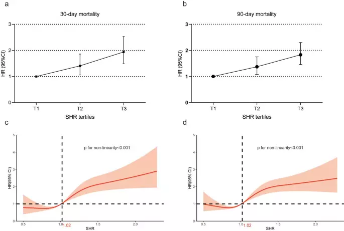
Raised stress hyperglycaemia ratio is  associated with increased mortality risk after MI and ischaemic stroke suggests a study published in the Cardiovascular Diabetology.

A raised stress hyperglycaemia ratio (SHR) has been associated with all-cause mortality and may better discriminate than an absolute glucose value. The aim of this meta analysis and systematic review is to synthesise the evidence assessing the relationship between the SHR and all-cause mortality across three common cardiovascular presentations.

They undertook a comprehensive search of Medline, Embase, Cochrane CENTRAL and Web of Science from the date of inception to 1st March 2024, and selected articles meeting the following criteria: studies of patients hospitalised for acute myocardial infarction, ischaemic stroke or acute heart failure reporting the risk (odds ratio or hazard ratio) for all-cause mortality associated with the SHR. A random effects model was used for primary analysis.

Subgroup analysis by diabetes status and of mortality in the short and long term was undertaken. Risk of bias assessment was performed using the Newcastle Ottawa quality assessment scale. Results: A total of 32 studies were included: 26 studies provided 31 estimates for the meta-analysis.

The total study population in the meta analysis was 80,010. Six further studies were included in the systematic review. Participants admitted to hospital with cardiovascular disease and an SHR in the highest category had a significantly higher risk ratio of all-cause mortality in both the short and longer term compared with those with a lower SHR (RR = 1.67 [95% CI 1.46–1.91], p < 0.001). This finding was driven by studies in the myocardial infarction (RR = 1.75 [95% CI 1.52–2.01]), and ischaemic stroke cohorts (RR = 1.78 [95% CI 1.26–2.50]). The relationship was present amongst those with and without diabetes (diabetes: RR 1.49 [95% CI 1.14–1.94], p < 0.001, no diabetes: RR 1.85 [95% CI 1.49–2.30], p < 0.001) with p = 0.21 for subgroup differences, and amongst studies that reported mortality as a single outcome (RR of 1.51 ([95% CI 1.29–1.77]; p < 0.001) and those that reported mortality as part of a composite outcome (RR 2.02 [95% CI 1.58–2.59]; p < 0.001). On subgroup analysis by length of follow up, higher SHR values were associated with increased risk of mortality at 90 day, 1 year and > 1year follow up, with risk ratios of 1.84 ([95% CI 1.32–2.56], p < 0.001), 1.69 ([95% CI 1.32–2.16], p < 0.001) and 1.58 ([95% CI 1.34–1.86], p < 0.001) respectively.

A raised SHR is associated with an increased risk of all-cause mortality following myocardial infarction and ischaemic stroke. Further work is required to define reference values for the SHR, and to investigate the potential effects of relative hypoglycaemia. Interventional trials targeting to the SHR rather than the absolute glucose value should be undertaken.

Reference:

Esdaile, H., Khan, S., Mayet, J. et al. The association between the stress hyperglycaemia ratio and mortality in cardiovascular disease: a meta-analysis and systematic review. Cardiovasc Diabetol 23, 412 (2024).https://doi.org/10.1186/s12933-024-02454-1

Powered by WPeMatico

Zydus Lifesciences Chairman Pankaj R Patel conferred with Padma Bhushan

Ahmedabad: Pankaj R. Patel, the Chairman of Zydus Lifesciences Limited, has been conferred with the Padma Bhushan by the Government of India.

Expressing his gratitude on being bestowed with one of India’s highest civilian honours, Mr. Pankaj R. Patel, Chairman, Zydus Lifesciences said, “I would like to thank the Government of India for this great honour and humbly accept this recognition. It’s a journey that started over seven decades ago when my father started out as an entrepreneur to contribute to nation building and making India self- reliant in lifesciences. This commitment to science, health and innovation always inspired me. I am fortunate to have 27000 people at Zydus as a part of this journey working on putting India at the forefront of innovation with pathbreaking discoveries that bridge unmet healthcare needs. I consider myself extremely honoured to be a part of these exciting, transformative years for the Indian Lifesciences Industry. We are taking a leap into the future with the convergence of health, digital advancements and technology. Innovation from India will bring critical access to affordable healthcare and empower people to lead healthier and more fulfilled lives.”
Mr. Pankaj Patel is the Chairman of Zydus Lifesciences, a discovery-driven, global Lifesciences company with operations in more than 80 countries worldwide. The Zydus Group is an innovative, global lifesciences company that discovers, develops, manufactures, and markets a broad range of healthcare therapies. 
With a Masters in Pharmaceutics and Pharmaceutical Technology from the L.M. College of Pharmacy and an alumni of the Indian Institute of Management (IIM), Ahmedabad, he was always driven by a passion to become a pharma entrepreneur. Mr. Patel combines both research and techno-commercial expertise. He has published over 100 research papers in peer reviewed journals and is a co-inventor in more than 64 patents.
Mr. Pankaj Patel is on the board of several prestigious institutions, including the Reserve Bank of India in.
He is also the Chairperson of the Board of Governors of IIM Ahmedabad and Chairman of IIM Udaipur and Invest India. He also officiates on the board of several Not-for-Profit & charitable institutions. Mr. Patel is the Executive Chairman of the Gujarat Cancer Society and Chairman of the Gujarat Cancer and Research Institute, a Regional Cancer Centre and one of the largest cancer centres of India 
He also officiates as the Chairman of the Deaf and Mute School, Ahmedabad. He is also the Chairman on the Board of Zydus Foundation which has set up Zydus Hospital and Medical College at Dahod. Mr. Patel supports the Group’s CSR initiatives in the field of education, pharmaceutical technology and innovation, grassroot innovation, sustainable livelihoods, water conservation, skill development for women from economically disadvantaged groups, and also specially abled children and Green Gujarat initiatives for sustainability.

Powered by WPeMatico

Glucagon Sensitivity Similar in Individuals With and Without Type 1 Diabetes: Study Finds

Denmark: Research published in The Journal of Clinical Endocrinology & Metabolism explored metabolic responses to glucagon stimulation in individuals with and without type 1 diabetes. The study observed comparable responses in both groups, except for heightened sensitivity to low-dose glucagon among individuals with diabetes. Moreover, recent episodes of hypoglycemia did not influence the metabolic effects induced by glucagon.

The metabolic effects of glucagon beyond its role in promoting endogenous glucose production in type 1 diabetes are poorly understood. Therefore, Rui She, Department of Clinical Medicine, University of Copenhagen, Copenhagen, Denmark, and colleagues aimed to evaluate the metabolic responses to glucagon stimulation in individuals with type 1 diabetes and to determine whether recent hypoglycemia influences glucagon sensitivity.

For this purpose, the researchers enrolled 29 participants, including 19 individuals with type 1 diabetes and 10 healthy controls. They underwent a hyperinsulinemic-euglycemic clamp procedure with five stepwise ascending glucagon injections, during which metabolic responses were evaluated before and after each injection.

The study led to the following findings:

  • The incremental area under the curve (AUC) for plasma glucose concentration during glucagon injections was similar between individuals with type 1 diabetes and healthy controls.
  • During the first injection of 10 μg glucagon, individuals with type 1 diabetes showed a higher incremental AUC for plasma glucose concentration than healthy controls.
  • Plasma concentrations of non-esterified fatty acids, glycerol, and metabolites from metabolomic analyses were comparable between the groups.
  • In participants with type 1 diabetes, recent hypoglycemia did not affect the metabolic responses to glucagon.

“These findings provide a clearer understanding of how glucagon functions in type 1 diabetes and offer a foundation for future research into therapeutic approaches. As researchers continue to explore the nuanced role of glucagon, these insights may contribute to improved treatment strategies for individuals with type 1 diabetes, aiming to balance metabolic processes more effectively,” the researchers wrote.

“Overall, metabolic responses to glucagon stimulation in individuals with type 1 diabetes are comparable to those in healthy controls. However, participants with type 1 diabetes showed increased glucose production at the lowest glucagon dose, suggesting heightened sensitivity to low-dose glucagon,” they concluded.

Reference:

She, R., Suvitaival, T., Andersen, H. U., Hommel, E., Nørgaard, K., & Wojtaszewski, J. F. Metabolic Effects of Glucagon Stimulations in Type 1 Diabetes and Healthy Controls. The Journal of Clinical Endocrinology & Metabolism. https://doi.org/10.1210/clinem/dgaf030

Powered by WPeMatico

Vitamin D supplementation could lead to better IBS management with low risk of adverse events: Study

Vitamin D supplementation could lead to better IBS management with a low risk of adverse events suggests a new study published in the Advances in Clinical and Experimental Medicine.

Qi, Shuang,Vitamin D, supplementation, better, IBS, management, low risk, adverse events, Advances in Clinical and Experimental MedicineVitamin D supplementation could offer irritable bowel syndrome (IBS) patients significant improvements in terms of symptom severity and overall quality of life (QoL). Yet, the potential benefits and risks associated with vitamin D supplementation still require additional investigation.
They aimed to evaluate the impact of vitamin D supplementation on IBS using a systematic review and meta-analysis. A comprehensive search was carried out utilizing 4 electronic databases (PubMed, Embase, Scopus, and Cochrane Library) to identify articles published in English-language peer-reviewed journals.
he odds ratios (ORs), risk ratios (RRs) and mean differences (MDs), along with their respective 95% confidence intervals (95% CIs), were computed. Heterogeneity was evaluated using the appropriate p-value and Cochrane Q and I2 statistics. For the analysis, RevMan 5.3 was utilized.
RESULTS: Nine randomized controlled trials involving a total of 780 participants were included in this study. Vitamin D supplementation, in adolescents and young adults with IBS, improves the IBS symptoms severity score, QoL and serum 25(OH)D levels compared to controls. We obtained an OR of 2.34 (95% CI: 1.56-3.50) for change in the IBS severity scoring system (IBS-SSS), OR = 2.51 (95% CI: 1.71-3.70) for change in QoL, low risk of any adverse events (RR 0.49 (95% CI: 0.35-0.69)), and substantial changes in serum 25(OH)D level (MD = 11.29 (95% CI: 7.13-15.45)). Results were statistically significant (p < 0.05). Vitamin D supplementation could lead to better IBS management with a low risk of adverse events.

Reference:

Qi, Shuang, et al. “Impact of Vitamin D Supplementation On Symptom Severity and Quality of Life in Patients With Irritable Bowel Syndrome: a Meta-analysis.” Advances in Clinical and Experimental Medicine : Official Organ Wroclaw Medical University, 2025.

Powered by WPeMatico

Micronutrient deficient Levels tied to Increased Migraine Frequency and Severity in Children: Study

Researchers have found in a new study that In pediatric migraine patients, lower levels of vitamin D, vitamin B12, and ferritin were associated with increased attack frequency and prolonged duration of migraine episodes.

The new study published in the Journal of Child Neurology highlighted the potential connection between micronutrient deficiencies and the frequency and severity of migraines in pediatric patients. The findings suggest that certain micronutrient levels (vitamin D, vitamin B12, and ferritin) might play a significant role in influencing the nature and management of migraines in young patients.

This research reviewed 171 children between February and December 2022 who were diagnosed with migraines at 2 hospitals. The study population included children with a mean age of 14.3 years. This research analyzed serum levels of key micronutrients and compared them against migraine attack frequency, duration, and the use of prophylactic treatments. Sharp differences were observed in nutrient levels across these parameters.

One of the standout findings was the relationship between vitamin D levels and migraine attack frequency. Children experiencing 1 to 3 migraine attacks per month had a median 25-hydroxyvitamin D level of 15.6 μg/L, significantly higher than the 11.9 μg/L observed in children suffering from more than three attacks monthly. This difference was statistically significant (P = .005), suggesting that higher vitamin D levels may be associated with fewer migraine episodes.

Vitamin B12 levels were also found to correlate with migraine attack duration. Children whose migraines lasted between 2 to 6 hours had a median vitamin B12 level of 386 ng/L, whereas the children with longer migraines (12-24 hours) had a lower median level of 343.5 ng/L (P = .048). These results imply that vitamin B12 deficiencies could contribute to prolonged migraine episodes in children.

Also, the study highlighted a marked association between ferritin levels and the use of prophylactic migraine treatments. The patients not on prophylactic medication had a median ferritin level of 34 μg/L when compared to 24 μg/L among those receiving preventive therapies (P = .019). While this finding may warrant further investigation, it raises questions about the potential influence of ferritin levels on treatment efficacy or selection.

Overall, the findings of this study emphasize the importance of evaluating serum micronutrient levels as part of a comprehensive approach to managing pediatric migraines. Clinicians may have an opportunity to reduce the frequency and severity of migraines in children by addressing deficiencies and potentially improving their quality of life.

Source:

Akif Özdemir, F. M., & Çelik, H. (2025). Assessment of micronutrient levels in pediatric migraine patients. Journal of Child Neurology. https://doi.org/10.1177/08830738241304868

Powered by WPeMatico

Timing and Extent of Tangential Excision may significantly impact Burn Patient Outcomes, reveals study

A new study published in the Annals of Surgery identified the optimal timing and surgical approaches for the first tangential excision in patients with severe burns covering ≥70% of their total body surface area (TBSA). This research analyzed data from 185 patients injured in the Kunshan factory explosion, the findings have significant implications for burn care management by potentially improving survival outcomes for the most critical cases. 

Tangential excision is the gold-standard surgical procedure for full-thickness burns, involves the removal of burned tissue to minimize infection risks and improve healing. While early intervention is widely accepted as beneficial, the timing and scope of this procedure remain debated for patients with burns exceeding 70% TBSA.

The findings suggest that performing the first tangential excision more than 3 days after the burn significantly increased mortality risk, with a hazard ratio of 2.37. The patients undergoing excision of areas exceeding 48% TBSA during the initial procedure had a hazard ratio of 2.41 for decreased survival.

These findings were validated through Kaplan-Meier survival analysis, which confirmed that delayed surgeries and excessively large excision areas were associated with a higher incidence of complications and poorer overall survival. External validation using a 10-year dataset of 144 severely burned patients reinforced these outcomes.

Also, this study underlined the importance of wound coverage methods. The patients with greater use of autografts (skin grafts from the patient’s own body) demonstrated significantly better survival rates when compared to those relying on temporary skin substitutes. However, the choice of coverage was influenced by factors like the severity of the injury, the availability of autologous skin, and resource constraints at the treatment center.

The subgroup analysis revealed that hemodynamic stability, hospital resources, and the availability of treatment modalities influenced the timing and surgical area. Tailored treatment approaches are recommended to address individual patient needs and the challenges posed by limited autologous skin or intensive care resources. Overall, this research highlighted the importance of initiating tangential excision within 3 days and limiting the excised area during the first surgery to under 48% TBSA for severely burned patients.

Source:

Huang, R., Yao, Y., Xie, S., Li, J., Zhang, W., Liu, Y., Xian, S., Sun, H., Wu, G., He, H., Li, L., Wu, X., Li, Y., Zhang, H., Lu, B., Zhou, J., Zhou, Y., Xu, D., Xia, Z., & Ji, S. (2025). Optimal timing and approaches for first tangential excision in patients with ≥70% TBSA burns: Insights from a retrospective cohort of a mass casualty incident. Annals of Surgery. https://doi.org/10.1097/sla.0000000000006626

Powered by WPeMatico

Chlorine dioxide-containing mouthwash may not impact frictional resistance or surface roughness of Orthodontic Archwire: Study

Chlorine dioxide-containing mouthwash may not impact frictional resistance or surface roughness of Orthodontic Archwire suggests a study published in the F1000Research.

They involve various types of mouthwashes which have been reported to cause alteration of mechanical properties of archwires. This study aimed to evaluate the effects of a new kind of chlorine-dioxide-containing mouthwash on the mechanical properties and surface morphology of stainless steel orthodontic archwires against the already prevalent chlorhexidine mouthwash in the market. Group A – Chlorhexidine mouthwash 0.2% (study), Group B – Chlorine Dioxide mouthwash (study), and Group C – Artificial Saliva (control). 42 specimens of 5 cm long 19×25 inch SS archwires were immersed in each group equally. Post immersion, the frictional force was analyzed in the universal testing machine for each group using custom-made acrylic jigs for 10 specimens. The remaining 4 specimens from each group were sent for surface morphology evaluation using an atomic force microscope. Results: Friction resistance evaluation for the archwires revealed a mean friction of 0.011 ± 0.0056 in Group A, 0.015 ± 0.0052 in Group B, and 0.010 ± 0.0067 in Group C. Results suggested that the static friction of Group C (control group) was found to be the least when compared with the experimental groups, although not producing statistically significant values. Surface roughness of archwires compared at a 10μm range revealed a mean roughness of 19.38 ± 0.82 in Group A, 25.39 ± 7.01 in Group B, and 16.65 ± 3.07 in Group C which shows there wasn’t any statistically significant difference in the mean roughness midst the three sets. Researchers concluded that Chlorine dioxide-containing mouthwash does not significantly impact the frictional resistance or surface roughness of stainless steel orthodontic archwires under the tested conditions. Chlorine dioxide may serve as a potential adjunct in orthodontic treatment. However, further research, particularly in vivo studies, is required to validate these findings and assess its long-term effects on orthodontic materials. Therefore Understanding the effects of Chlorine dioxide mouthwash on stainless steel archwires can aid in developing a mechanically efficient archwire-mouthwash combination for orthodontic treatment. Additionally, this study provides a foundation for future comparisons between Chlorine dioxide and the gold-standard Chlorhexidine mouthwash.

Reference:

Apte, Shivani, et al. “Impact of Chlorine Dioxide and Chlorhexidine Mouthwashes On Friction and Surface Roughness of Orthodontic Stainless Steel Wires: an In-vitro Comparative Study.” F1000Research, vol. 13, 2024, p. 1442.

Keywords:

Chlorine dioxide-containing, mouthwash, impact, frictional, resistance, surface, roughness, Orthodontic, Archwire, study, Apte, Shivani,F1000Research

Powered by WPeMatico

Muscle loss linked to increased dementia risk, claims study

Skeletal muscle loss is a risk factor for developing dementia, according to a study being presented today at the annual meeting of the Radiological Society of North America (RSNA).

Skeletal muscles make up about one-third of a person’s total body mass. They are connected to the bones and allow for a wide range of movements. As people grow older, they begin to lose skeletal muscle mass.

Because age-related skeletal muscle loss is often seen in older adults with Alzheimer’s disease (AD) dementia, this study aimed to examine whether temporalis muscle loss (a measure of skeletal muscle loss) is associated with an increased risk of AD dementia in older adults.

The temporalis muscle is located in the head and is used for moving the lower jaw. Studies have shown that temporalis muscle thickness and area can be an indicator of muscle loss throughout the body.

“Measuring temporalis muscle size as a potential indicator for generalized skeletal muscle status offers an opportunity for skeletal muscle quantification without additional cost or burden in older adults who already have brain MRIs for any neurological condition, such as mild dementia,” said the study’s lead author, Kamyar Moradi, M.D., postdoctoral research fellow in the Russell H. Morgan Department of Radiology and Radiological Sciences at Johns Hopkins University School of Medicine in Baltimore. “This is the first longitudinal study to demonstrate that skeletal muscle loss may contribute to the development of dementia.”

For the multidisciplinary research study, a collaboration between the radiology and neurology departments at Johns Hopkins Medical Institutions, Dr. Moradi and colleagues used baseline brain MRI exams from the Alzheimer’s Disease Neuroimaging Initiative cohort to quantify skeletal muscle loss in 621 participants without dementia (mean age 77 years).

The researchers manually segmented the bilateral temporalis muscle on MRI images and calculated the sum cross-sectional area (CSA) of these muscles. Participants were categorized into two distinct groups: large CSA (131 participants) and small CSA (488 participants). Outcomes included subsequent AD dementia incidence, change in cognitive and functional scores, and brain volume changes between the groups. Median follow-up was 5.8 years.

Based on their analysis, a smaller temporalis CSA was associated with a higher incidence risk of AD dementia. Furthermore, a smaller temporalis CSA was associated with a greater decrease in memory composite score, functional activity questionnaire score and structural brain volumes over the follow-up period.

“We found that older adults with smaller skeletal muscles are about 60% more likely to develop dementia when adjusted for other known risk factors,” said the study’s co-senior author and professor of neurology, Marilyn Albert, Ph.D.

According to Shadpour Demehri, M.D., co-senior author and professor of radiology, the study demonstrates that this muscle change can be opportunistically analyzed through any conventional brain MRI, even when conducted for other purposes, without incurring additional costs or burdens.

Dr. Albert pointed out that early detection through readily available brain MRI could enable timely interventions to address skeletal muscle loss, such as physical activity, resistance training and nutritional support.

“These interventions may help prevent or slow down muscle loss and subsequently reduce the risk of cognitive decline and dementia,” Dr. Demehri said.

Reference:

Muscle loss could increase dementia risk, Radiological Society of North America, Meeting: 110th Scientific Assembly and Annual Meeting of the Radiological Society of North America.

Powered by WPeMatico