Effective Treatment Options for Peri-Implant Mucositis: Laser vs. Ultrasonic Scaler
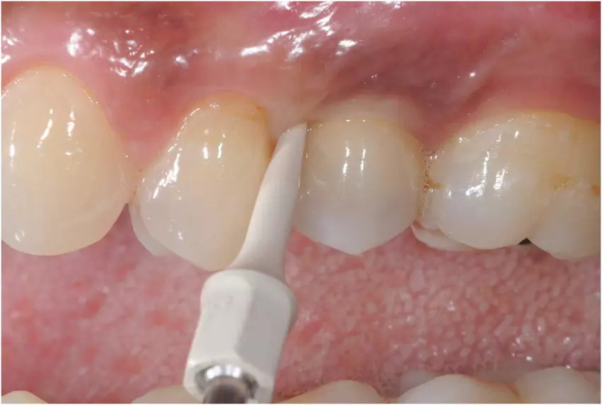
Researchers have found in a new study that peri-implant mucositis treatment yields positive results regardless of the method used. However, patients treated with the Er:YAG laser showed a greater reduction in diseased sites than those treated with an ultrasonic scaler after six months.

The study assessed the clinical outcomes following treatment of peri-implant mucositis using Er:YAG laser or an ultrasonic device over six months. Patients’ experience of pain, aesthetics, and Quality of life were further assessed. One dental implant, per the included patient, diagnosed with peri-implant mucositis underwent treatment with an Er:YAG laser (test) or an ultrasonic scaler (control) randomly.

Treatments were performed at baseline and months three and six. At each session, oral hygiene was instructed after plaque registration, and the patient was guided in proper cleaning techniques using a toothbrush and interproximal aids as needed. Full mouth bleeding on probing (FMBoP), full mouth plaque score (FMPS), implant bleeding on probing (BoP), implant mean graded bleeding (mBI), implant probing pocket depts (PPD), implant suppuration and bone levels were assessed. Oral health-related Quality of life (OHQoL) and visual analogue scales (VAS), which reflect aesthetic satisfaction and pain of the treatment, were also evaluated.

RESULTS: Forty-six patients were included. FMBoP was significantly reduced from 30.1 to 21.5% (test) (p < 0.001) respectively from 35.0% to 30% (control) (p < 0.01). FMPS showed significant reduction from 61.5 to 32.7% (test) (p < 0.001) and from 58.7 to 39.1% (control) (p < 0.001). At the implant, BoP reduced from 89.0 to 55.7% (test) (p < 0.001) respectively from 94.9 to 63.7% (control) (p < 0.001). mBI was reduced from 1.3 to 0.6 (test) (p < 0.01) and from 1.9 to 0.8 (control) (p < 0.001).

Distribution of “no bleeding” increased from 13 to 61% (test) (p < 0.05) and from 0 to 35% (control) (p < 0.05). At month three, statistically significant intergroup differences were shown for PPD ≥ 4 mm with 43.5% (test) respectively 73.9% (control) (p < 0.05). At month six, statistically significant intergroup differences were shown for FMBoP 21.5% (test) respectively 30% (control) (p < 0.05) and for plaque score at the implant 4.0% (test) respectively 26% (control) (p < 0.05). Less pain was reported in the laser group at three days 0.08 (test) respectively 0.2 (control) (p < 0.05).

Treatment of peri-implant mucositis was effective regardless of whether the treatment was performed with an Er:YAG laser or an ultrasonic scaler. Fewer diseased sites were diagnosed at six months following laser treatment.

Reference:

Bentsson, Viveca Wallin, et al. “Treatment of Peri-implant Mucositis Using an Er:YAG Laser or an Ultrasonic Device: a Randomized, Controlled Clinical Trial.” International Journal of Implant Dentistry, vol. 11, no. 1, 2025, p. 6.

Powered by WPeMatico

Hyperglycemia Identified as Rare Cause of Quadriplegia in Osmotic Demyelination Syndrome: Case Study

India: A recent case report published in the Journal of the Association of Physicians of India has highlighted hyperglycemia as a rare and unusual cause of quadriplegia, shedding light on the potential neurological complications of uncontrolled blood sugar levels.

Osmotic demyelination syndrome (ODS), particularly central pontine myelinosis (CPM), is typically associated with the rapid correction of hyponatremia, a condition in which sodium levels in the blood are dangerously low. However, according to the authors, the case marks the first known instance of ODS triggered by hyperglycemia, where both the pontine and extrapontine regions of the brain were affected.

Mahesh Shinde, Consultant Physician, Department of Medicine, Carewell Superspeciality Hospital, Aurangabad, Maharashtra, India, and colleagues describe the case of a young male who presented with acute-onset altered mentation, quadriplegia, and urinary retention. On examination, he was stuporous with a Glasgow Coma Scale (GCS) of 6/15 and signs of dehydration, including tachycardia. Neurologically, he exhibited hypotonic quadriplegia, absent reflexes, and normal-sized pupils that reacted to light. Investigations revealed significantly high blood sugar (550 mg/dL), cerebrospinal fluid (CSF) sugar (149 mg/dL), and urine sugar (3+), with normal cell count in CSF but elevated protein levels. The serum osmolality was 330 mOsm/kg H2O, and Electroencephalogram (EEG) showed mild to moderate dysfunction. Further tests indicated severe hyperglycemia with a hemoglobin A1c of 11%. Despite normal findings in initial imaging, including an MRI of the brain and spine, the patient’s condition worsened, requiring mechanical ventilation.

After one week, the patient’s clinical condition raised suspicion of pontine involvement, leading to a repeat MRI, which confirmed hyperintensity lesions in both pontine and extrapontine areas, indicating osmotic demyelination syndrome, including central pontine myelinosis and extrapontine myelinosis (EPM). Over the next three weeks, his mental state and limb power improved, and he was eventually weaned off the ventilator. After 35 days, he was discharged with antidiabetic treatment. On follow-up, he had fully recovered, except for reduced lower limb power and the presence of a urinary catheter.

In conclusion, the authors have presented a rare case of osmotic demyelination syndrome where both central pontine myelinosis and extrapontine myelinosis were identified as initial manifestations of hyperglycemia. This rare etiology and presentation, with its characteristic clinical and radiological findings, is seldom reported in the literature.

The authors emphasize that acute changes in osmolality and subsequent subacute changes can contribute to the development of ODS. Given this, hyperglycemia should be considered a potential cause of quadriplegia due to ODS, even in the absence of hyponatremia, diabetic ketoacidosis, or hyperosmolar hyperglycemic state. While there is no definitive treatment for ODS at present, the authors highlight that symptomatic management and comfort care have improved patient outcomes.

Reference:

Shinde M, Rathod V, Wahatule R, et al. Hyperglycemia as a Rare Cause of Quadriplegia: A Case Report. J Assoc Physicians India 2025;73(1):66-69.

Powered by WPeMatico

Tuberculosis Increases Risk of severe Aortic complications among patients with Takayasu Arteritis: Study

A recent study published in the journal of Advances in Rheumatology revealed a significant link between tuberculosis (TB) and more severe aortic complications in patients with Takayasu arteritis (TA), a rare inflammatory disease affecting large blood vessels. This research analyzed clinical and radiological data of patients treated at a public tertiary referral outpatient clinic in northeast Brazil over 5 years which uncovered notable differences in outcomes between patients with TA who had active or latent TB infections and those without TB.

The retrospective analysis included 50 patients diagnosed with TA between January 2017 and June 2022. Among them, 20 individuals (40%) were identified as having a history of TB, with 6 experiencing active TB and 14 showing evidence of latent TB infection (LTBI). This study compared the clinical presentations and radiological profiles of TA patients based on their TB status.

The key findings indicated that weight loss was significantly more prevalent among TA patients with TB (p=0.005). Furthermore, those with both conditions showed a higher occurrence of abdominal aortic involvement when compared to TA patients without TB (25% vs. 11.4%, p=0.013). The study also reported a markedly increased incidence of vascular dilations (p=0.009) and aneurysms (p=0.027) in the TB-affected group.

Also, other clinical parameters like comorbidities, medication use, inflammatory markers like erythrocyte sedimentation rate (ESR) and C-reactive protein (CRP) levels, showed no statistically significant differences between the 2 groups. However, the structural damage to the abdominal aorta, including dilations and aneurysms, was identified as a critical distinguishing factor in patients with a TB history. The study highlighted the overlapping histopathological and immunological characteristics of TA and TB, which may contribute to these differences. TB infections, whether active or latent, could exacerbate vascular inflammation and structural damage in TA patients.

These results emphasize the importance of screening for TB in individuals with TA and monitoring for potential vascular complications in those with a TB history. These findings could help guide treatment strategies and improve outcomes for this subset of TA patients. With TB still prevalent in many regions, especially in developing countries, these insights illuminate the need for a multidisciplinary approach to managing conditions like Takayasu arteritis that may interact with infectious diseases in complex ways. 

Source:

Souza Pedreira, A. L., de Oliveira Figueiroa, M. de L. C., Miranda, M. O., de Santana, A. R., Mattos, V. P., da Paz, A. S., Duran, C. C., & Santiago, M. B. (2024). Takayasu’s arteritis associated with tuberculosis: a retrospective study. Advances in Rheumatology (London, England), 64(1), 84. https://doi.org/10.1186/s42358-024-00424-5

Powered by WPeMatico

Two-dose regimen of vaccine offers substantial protection against Hepatitis E Outbreak, suggests study

A new study conducted during the first-ever reactive use of the Hecolin vaccine in South Sudan showed that a two-dose regimen of the vaccine offers substantial protection against Hepatitis E. The findings published in The Lancet Infectious Diseases suggested that the two-dose Hecolin vaccine could be a viable option for controlling outbreaks in resource-limited settings.

Hepatitis E is a major cause of acute viral hepatitis which disproportionately affects regions like Asia and Africa where genotypes 1 and 2 of the virus are prevalent. Despite a 3-dose recombinant vaccine (Hecolin) being licensed, its deployment during outbreaks has underwent logistical challenges due to the extended vaccination schedule. This study brings out the effectiveness of a shorter, two-dose regimen.

This case-control study was conducted during an outbreak from May to December 2022 in the Bentiu IDP camp. The study focused on individuals aged 16 to 40 years who were eligible for vaccination. Acute jaundice syndrome cases were screened, and confirmed hepatitis E cases were identified using RT-PCR or anti-HEV IgM ELISA testing. Each case was matched with 6 controls based on age, sex, pregnancy status, and residence. Vaccination status was verified through vaccination cards.

Of the total 859 suspected hepatitis E cases screened, 201 met the study criteria, and 21 were laboratory-confirmed as positive for the virus. Among confirmed cases, 48% were unvaccinated when compared to only 27% of their matched controls.

The key findings from the study revealed that the unadjusted two-dose vaccine effectiveness was around 67.8%. The effectiveness increased to 84% after adjusting for potential confounders. A secondary analysis using a test-negative design estimated the adjusted vaccine effectiveness to be as high as 89.4%. This method also addressed potential biases in health-care-seeking behaviors, which could underestimate vaccine efficacy.

While the sample size of the study was small, the findings indicate that the two-dose regimen of Hecolin offers robust protection against Hepatitis E genotype 1, with effectiveness estimates ranging between 84% and 89%. This provides strong data to support the use of a shorter vaccination schedule during outbreaks, especially in protracted emergency settings like the Bentiu camp. Also, this study pointed to the need for further research to optimize outbreak response strategies but provides a hopeful step toward controlling Hepatitis E in vulnerable populations.

Reference:

Nesbitt, R. C., Kinya Asilaza, V., Alvarez, C., Gitahi, P., Nkemenang, P., Duncker, J., Haile, M., Gakima, P., Wamala, J. F., Loro, F. B., Koyuncu, A., Biem, D., Albela, M., Rull, M., Gignoux, E., Rumunu, J., Eckerle, I., Ciglenecki, I., & Azman, A. S. (2025). The effectiveness of two doses of recombinant hepatitis E vaccine in response to an outbreak in Bentiu, South Sudan: a case–control and bias indicator study. The Lancet Infectious Diseases. https://doi.org/10.1016/s1473-3099(24)00657-1

Powered by WPeMatico

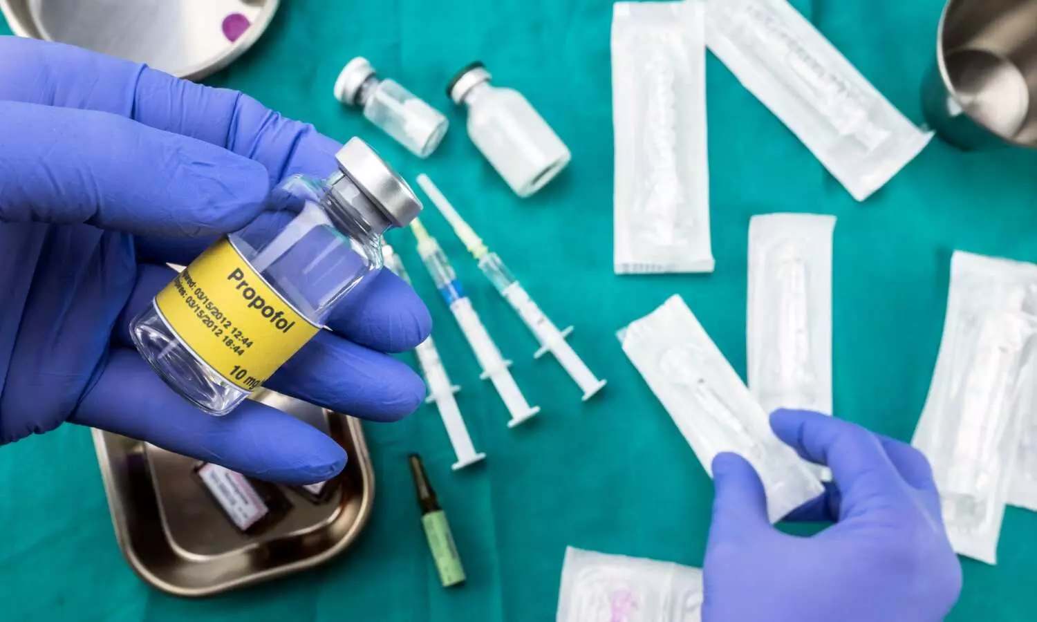
Optimizing Propofol Dosing for Liver Transplantation: Study suggests Phase-Specific Adjustments for Safe Anesthesia Management

Propofol has proven effective in liver transplantation procedures for both initiating and sustaining anesthesia. Compared to inhalational anesthetics, it offers superior hemodynamic stability. In addition to its anti-inflammatory and free radical scavenging attributes, propofol safeguards against ischaemia-reperfusion injury, offering protections to the liver, heart, kidneys, and nervous system. Recent study examined the changes in the target concentration of propofol required to maintain the bispectral index (BIS) between 40 and 60 during the three phases of liver transplantation. The researchers used a target-controlled infusion of total intravenous anesthesia (TCI-TIVA) and found significant differences in the mean target propofol concentration between the dissection, anhepatic, and neohepatic phases. The study was a prospective, observational pilot study conducted on 20 adult patients diagnosed with chronic liver disease who were scheduled for live-donor liver transplantation. After anesthesia induction and intubation, a BIS-guided propofol infusion was started using TCI-TIVA with an initial target plasma concentration (TPC) of 2.5 μg/mL. The TPC was decreased or increased by 0.2 μg/mL whenever the BIS values were persistently below 40 or above 60 for 15 minutes. The results showed that the mean TPC was significantly higher during the dissection phase [2.12 (SD: 0.63 μg/mL)] compared to the anhepatic [1.29 (SD: 0.65) μg/mL] and neohepatic [1.35 (SD: 0.54) μg/mL] phases (p<0.001). There was a significant difference between the dissection and anhepatic phases (mean difference: -0.87 μg/mL, 95% CI: -0.98, -0.75) and the dissection and neohepatic phases (mean difference: -0.77 μg/mL, 95% CI: -1.02, -0.53), but not between the anhepatic and neohepatic phases.

Dosage Comparison

The propofol dose was also significantly higher in the dissection phase compared to the anhepatic and neohepatic phases (p<0.001). The propofol’s mean TPC decreased to 61% and 63.7% of the dissection phase levels during the anhepatic and neohepatic phases, respectively.

Conclusion

The authors concluded that the propofol target concentration should be adjusted to 61% and 63.7% of the dissection phase levels during the anhepatic and neohepatic phases, respectively, to avoid overdosage and ensure optimal anesthesia management. The study highlights the importance of tailoring propofol dosing to the different phases of liver transplantation to maintain appropriate anesthetic depth.

Key Points

1. The study examined the changes in the target concentration of propofol required to maintain the bispectral index (BIS) between 40 and 60 during the three phases of liver transplantation using target-controlled infusion of total intravenous anesthesia (TCI-TIVA).

2. The study found significant differences in the mean target propofol concentration between the dissection, anhepatic, and neohepatic phases, with the mean target propofol concentration being significantly higher during the dissection phase compared to the anhepatic and neohepatic phases.

3. The propofol dose was also significantly higher in the dissection phase compared to the anhepatic and neohepatic phases, with the propofol’s mean target plasma concentration (TPC) decreasing to 61% and 63.7% of the dissection phase levels during the anhepatic and neohepatic phases, respectively.

4. The authors concluded that the propofol target concentration should be adjusted to 61% and 63.7% of the dissection phase levels during the anhepatic and neohepatic phases, respectively, to avoid overdosage and ensure optimal anesthesia management.

5. The study highlights the importance of tailoring propofol dosing to the different phases of liver transplantation to maintain appropriate anesthetic depth.

6. The study was a prospective, observational pilot study conducted on 20 adult patients diagnosed with chronic liver disease who were scheduled for live-donor liver transplantation.

Reference –

Garg N, Kalra Y, Panwar S, Arora MK, Dhingra U. Comparison of target concentration of propofol during three phases of live donor liver transplant surgery using a target‑controlled infusion of propofol total intravenous anaesthesia ‑ A prospective, observational pilot study. Indian J Anaesth 2024;68:971‑7

Powered by WPeMatico

Keto diet could one day treat autoimmune disorders, suggests research

Scientists have long suspected the keto diet might be able to calm an overactive immune system and help some people with diseases like multiple sclerosis.

Now, they have reason to believe it could be true.

Scientists at UC San Francisco have discovered that the diet makes the gut and its microbes produce two factors that attenuated symptoms of MS in mice.

If the study translates to humans, it points toward a new way of treating MS and other autoimmune disorders with supplements.

The keto diet severely restricts carbohydrate-rich foods like bread, pasta, fruit and sugar, but allows unlimited fat consumption.

Without carbohydrates to use as fuel, the body breaks down fat instead, producing compounds called ketone bodies. Ketone bodies provide energy for cells to burn and can also change the immune system.

Working with a mouse model of MS, the researchers found that mice who produced more of a particular ketone body, called β-hydroxybutyrate (βHB), had less severe disease.

The additional βHB also prompted the gut bacterium Lactobacillus murinus to produce a metabolite called indole lactic acid (ILA). This blocked the activation of T helper 17 immune cells, which are involved in MS and other autoimmune disorders.

“What was really exciting was finding that we could protect these mice from inflammatory disease just by putting them on a diet that we supplemented with these compounds,” said Peter Turnbaugh, PhD, of the Benioff Center for Microbiome Medicine.

Earlier, Turnbaugh had shown that when secreted by the gut, βHB counteracts immune activation. This prompted a postdoctoral scholar who was then working in his lab, Margaret Alexander, PhD, to see if the compound could ease the symptoms of MS in mice.

In the new study, which appears Nov. 4 in Cell Reports, the team looked at how the ketone body-rich diet affected mice that were unable to produce βHB in their intestines, and found that their inflammation was more severe.

But when the researchers supplemented their diets with βHB, the mice got better.

To find out how βHB affects the gut microbiome, the team isolated bacteria from the guts of three groups of mice that were fed either the keto diet, a high-fat diet, or the βHB supplemented high-fat diet.

Then, they screened the metabolic products of each group’s distinct microbes in an immune assay and determined that the positive effects of the diet were coming from a member of the Lactobacillus genus: L. murinus.

Two other techniques, genome sequencing and mass spectrometry, confirmed that the L. murinus they found produced indole lactic acid, which is known to affect the immune system.

Finally, the researchers treated the MS mice with either ILA or L. murinus, and their symptoms improved.

Turnbaugh cautioned that the supplement approach still needs to be tested in people with autoimmune disorders.

“The big question now is how much of this will translate into actual patients,” he said. “But I think these results provide hope for the development of a more tolerable alternative to helping those people than asking them stick to a challenging restrictive diet.”

Reference:

Margaret Alexander, Vaibhav Upadhyay, Rachel Rock, A diet-dependent host metabolite shapes the gut microbiota to protect from autoimmunity, Cell Reports, DOI: 10.1016/j.celrep.2024.114891.

Powered by WPeMatico

SIDS discovery could ID babies at risk of sudden death, suggests study

New University of Virginia School of Medicine research revealing the fingerprints of Sudden Infant Death Syndrome within blood samples could open the door to simple tests to identify babies at risk.

The findings also represent an important step forward in unraveling the causes of SIDS, an unexplained condition that is the No. 1 killer of babies between amonth and a year old.

The UVA researchers analyzed blood serum samples collected from infants who died from SIDS and were able to identify specific biological indicators that were linked to – and potential causes of – the babies’ deaths.

Tests to identify such signs in infants could ultimately help save lives, the researchers say.

“Our study is the largest study to date that has attempted to detect how these small molecules in the blood may serve as biomarkers for SIDS,” said researcher Keith L. Keene, PhD, founding director of UVA’s Center for Health Equity and Precision Public Health and now at East Carolina University. “Our findings support a role for multiple key biological pathways and provide insight into how those biological processes may contribute to increased risk or serve to diagnose SIDS.”

Understanding SIDS

The new research speaks to the potential of “metabolomics,” the analysis of substances called metabolites produced by cells, for better understanding and treating complex diseases, the scientists say.

In their SIDS work, the UVA researchers analyzed blood serum samples collected from 300 babies included in the Chicago Infant Mortality Study and the National Institutes of Health’s NeuroBioBank. The researchers assessed levels of 828 different metabolites in key biological processes such as nerve cell communication, stress response and hormone regulation – processes that could be contributors to SIDS.

After adjusting for factors that could bias the results, such as the infants’ age, sex, and race and ethnicity, the researchers identified 35 predictors of SIDS. These “biomarkers” included ornithine, a substance critical to the body’s ability to dispose of ammonia in urine. The amino acid has already been identified as a potential contributor to SIDS.

Another predictor was a lipid metabolite that is critical for brain and lung health. This metabolite is already considered a potential indicator for the development of fetal heart defects during the first trimester of pregnancy

“We found differences in specific fats, called sphingomyelins, which are critical for brain and lung development,” said researcher Chad Aldridge, DPT, MS-CR, of the School of Medicine’s Department of Neurology. “Differences in these fats may disrupt these critical processes, placing some infants at risk for SIDS.”

The UVA scientists caution that further research is needed to determine if the metabolites are contributing to SIDS. But the findings lay an important foundation, they say, for unraveling the mysteries of SIDS and developing blood tests that could potentially save new parents from heartbreak.

“The results of this study are very exciting – we are getting closer to explaining the pathways leading to a SIDS death,” said researcher Fern R. Hauck, MD, MS, a family medicine physician at UVA Health, director of the Chicago Infant Mortality Study and aleading expert on SIDS. “Our hope is that this research lays the groundwork to help identify –through simple blood tests – infants who are at higher risk for SIDS and to save these precious lives.”

Reference:

Aldridge, Chad M. et al., Metabolomic profiles of infants classified as sudden infant death syndrome: a case-control analysis, eBioMedicine,DOI:10.1016/j.ebiom.2024.105484 

Powered by WPeMatico

Patients who received Ross procedure demonstrate excellent survival rates after 20 years, reports study

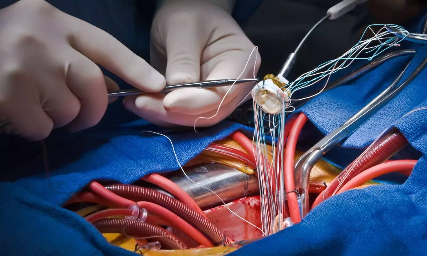
Young patients who have undergone the Ross procedure for aortic valve disease have shown excellent long-term survival, the majority without the need for additional surgery two decades later.

These findings, presented today at the 61st annual meeting of The Society of Thoracic Surgeons (STS), were the result of a 22-year study at the Narayana Institute of Cardiac Sciences in Bengaluru, India.

“The Ross operation can be performed safely with results comparable to mechanical valve replacement,” said the study’s lead author, cardiac surgeon Varun Shetty, MBBS, DNB (CVTS). “It has the added advantage of avoiding oral anticoagulation drugs and the risks associated with lifelong anticoagulation. In fact, most of our patients are not on any cardiac medications following one year after surgery.”

Dr. Shetty’s team followed 252 patients who underwent the Ross procedure, a heart valve replacement operation commonly used to treat younger patients with severe aortic valve disease. During the surgery, the malfunctioning aortic valve is replaced with the patient’s own healthy pulmonary valve-a technique called an autograft-and the pulmonary valve replaced with donor tissue.

“The main drawback of the Ross operation is failure of the autograft over time,” Dr. Shetty said. “We wanted to examine our outcomes, and we were pleasantly surprised to find that the freedom from re-intervention was 91% at 10 years and 87% at 20 years.”

Of the 252 patients in the study, 186 underwent a cylinder inclusion technique, or “mini root” replacement, while 25 received a Dacron-reinforced autograft. Three patients underwent the Ross-Konno procedure, in which the left-sided outflow root is enlarged to fit the pulmonary autograft. Twenty-five patients also received an ascending aorta replacement, two received hemiarch replacement, and four underwent septal myectomy-removal of abnormally thickened heart muscle.

At follow-up, Dr. Shetty’s team found that survival rates were 95.8% at 5 years, 94.3% at 10 years, 93.3% at 15 years, and 90.3% at 20 years. “Freedom from aortic regurgitation was 95.1%, 92.2%, 87.7%, and 84.5% at 5,10,15 and 20 years respectively,” the researchers reported.

Freedom from pulmonary regurgitation was lower—97% at 5 years, 83.6% at 10 years, 79.7% at 15 years, and 75.1% at 20 years.

“Individuals aged 18 years or younger exhibit notably higher rates of aortic regurgitation,” Dr. Shetty observed. “Risk factors for late autograft dysfunction include a dilated aortic annulus and ascending aorta.”

“Stabilization of the aortic annulus and pulmonary autograft will improve the re intervention rate in the future,” he added.

Because the Ross procedure uses living tissue, it not only can circumvent the need for anticoagulant medication, but also allows the valves to grow along with a growing child.

“The most important message from our study is that the Ross procedure offers excellent long-term survival rates with low risk of re-intervention even at 20 years,” Dr. Shetty said. “This makes the Ross operation an excellent substitute for mechanical valve replacement.”

Dr. Shetty’s team benchmarked their results with those recorded in the STS Congenital Heart Surgery Database (CHSD)-which contains more than 600,000 congenital heart surgery procedure records and currently has more than 1,000 participating physicians, including surgeons and anesthesiologists.

“Our study, performed at a single high-volume surgical center in India, demonstrates that the Ross operation can be performed with reproducible success across the globe,” Dr. Shetty said.

The CHSD is part of the STS National Database, one of the largest and most comprehensive clinical registries with nearly 10 million cardiothoracic procedures performed by 4,300+ surgeons. With over 95% of adult and congenital cardiac surgery procedures and a majority of lung cancer and esophageal cancer surgery in the U.S., the data and outcomes from the STS Database specialty registries provide true national benchmarks for clinical outcomes of cardiothoracic surgery.

Powered by WPeMatico

Prior Intravitreal Injections Increase Risk of Cataract Surgery Complications

According to a recent study published in the American Journal of Ophthalmology intravitreal injection (IVI) therapy has a greater risk of complications for patients undergoing cataract surgeries. The study was conducted by Cindy Z. and colleagues in the US.

Despite being effective and one of the commonest procedures to restore eyesight, cataract surgery might be impacted by IVI therapy, which is typically used to treat retinal conditions. A retrospective cohort study performed using the TriNetX database evaluated the link between IVI therapy before cataract surgery and the complications arising. Over 500,000 patients were followed for RD, anterior vitrectomy, retained lens, dislocated intraocular lens (IOL), secondary lens procedures, and endophthalmitis within defined post-operative intervals.

The study compared patients with IVI therapy in the 20 years before cataract surgery with those without any exposure to IVI as a control. Using propensity score matching (PSM), the researchers then balanced the baseline demographics, systemic health, and ocular co-morbid illnesses in the two groups that each contained 14,240 patients. Exclusion criteria included individuals requiring prior lensectomy and pars plana vitrectomy. The outcomes were measured up-to 14 days, up-to 30 days, and up-to 90 days after surgery, using robust statistical analyses.

Results

Complication Rates:

  • The overall complications were significantly greater within the 90-day period in the IVI group (3.3%, 447/13,719) compared with the control (2.4%, 340/13,945), with a relative risk of 1.34 (95% CI: 1.16–1.54, p<0.0005).

Retinal Detachment and Repairs:

  • The 30-day risk escalation of RD repair was demonstrated; RR 1.84 (95% CI: 1.27–2.66, p=0.001) and the 90-day risk escalation of RD repair occurs RR 2.05 (95% CI: 1.65–2.54, p<0.0005).

Anterior Vitrectomy in Certain groups:

  • There was no general association with anterior vitrectomy, save for DR patients in whom the risk rose to RR 1.24 (95% CI: 0.85–1.79, p=0.001) at 90 days.

Patients with Diabetes Among Medical Conditions:

  • Patients with DR and a prior IVI history showed higher rate risks for lensectomy, RD repair, and secondary lens procedures compared to those without a prior IVI (p<0.0005 for all).

Individuals with previous anti-VEGF therapy are at a remarkably higher risk of complications following cataract surgery, driven chiefly by higher rates of retinal detachment, with added risk for diabetic retinopathy. Inclusion of IVI history in preoperative assessments can facilitate a modification in surgical approaches, thus reducing postoperative risks and enhancing prognosis.

Reference:

Zhao, C. S., Chwialkowski, K., Wai, K. M., Mruthyunjaya, P., Rahimy, E., & Koo, E. B. (2025). Risk of cataract surgery complications in patients with prior intravitreal injection therapy. American Journal of Ophthalmology. https://doi.org/10.1016/j.ajo.2025.01.004

Powered by WPeMatico

Outdoor activities effectively reduce depression risk in older adults: Frontiers

A new study published in the recent issue of Frontiers in Psychology journal showed that promoting outdoor activities might be a useful public health tactic to lower the incidence of depression, especially for middle-aged and older individuals as well as members of particular ethnic groups. An estimated 300 million people worldwide suffer from depression, which is a major contributor to the global illness burden and economic difficulties.

Nearly 8.4% of individuals in the US alone face depression each year which reminds strongly the urgent need for efficient preventative and therapeutic measures. Outdoor activities have drawn attention among other therapies because of their potential to support mental health through special physiological and psychological processes. Increasing physical activity, boosting vitamin D production, lowering stress, and improving sleep quality are a few of them.

Prior research has shed important light on the advantages of outdoor activities, one such study showed that outdoor exercise was superior to indoor exercise in increasing vitality and optimism and that it considerably enhanced mood and psychological well-being. Individual variations in age and race may also influence how people react to outdoor activities.

This study uses data from the NHANES 2011–2018 to assess the association between outdoor time and depression in U.S. adults. This research aimed to offer solid evidence for incorporating outdoor activities into public health policies and tailoring treatments to fit the requirements of varied populations by concentrating on disparities across age and ethnic groups.

This study examined a total of 9,036 persons aged 20 and above using data from the NHANES 2011–2018 survey. The participants reported on their own as to how much time they spent outside and how depressed they were. The association between outdoor exercise and depression risk was evaluated using statistical analysis that took into consideration a number of demographic and lifestyle characteristics.

A 51% decreased incidence of depression was linked to spending more time outside. According to subgroup analysis, this connection was especially strong for non-Hispanic whites and non-Hispanic blacks, as well as persons 40 years of age and older. Overall, spending more time outside is substantially linked to a lower incidence of depression, especially for middle-aged and older persons, non-Hispanic white people, and Black people. These results highlight outdoor recreation as a useful and affordable way to improve mental health.

Source:

Liu, K., Guo, C., Xie, J., & Cheng, L. (2025). Outdoor activity time and depression risk among adults aged 40 years and older: a cross-sectional analysis of NHANES 2011–2018 data. Frontiers in Psychology, 16. https://doi.org/10.3389/fpsyg.2025.1506168

Powered by WPeMatico