Discovery explains kidney damage caused by blood pressure drugs
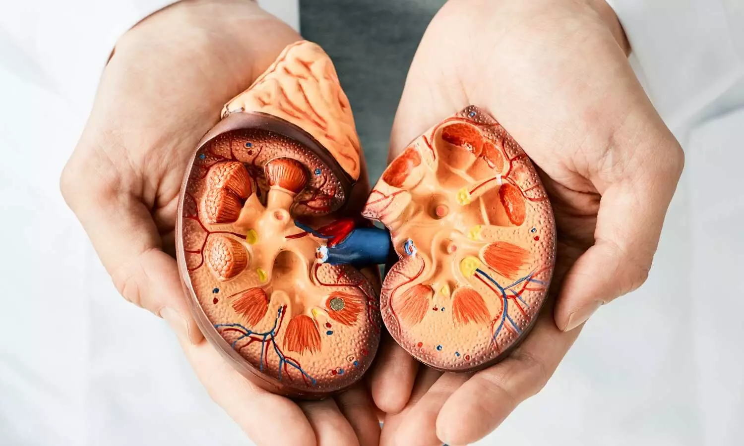
University of Virginia School of Medicine researchers have discovered how long-term treatment of high blood pressure with commonly prescribed drugs can destroy the kidney’s ability to filter and purify blood. The finding could open the door to better ways to manage high blood pressure and other vascular diseases.

The class of drugs, known as renin–angiotensin system (RAS) inhibitors, block the effects of the renin enzyme, relaxing blood vessels and allowing blood to flow more easily. They are widely used as first-line medications for hypertension (high blood pressure). But long-term use can take a terrible toll on the kidney, causing scarring and other dramatic physical changes that shift the organ’s focus from blood filtration to producing renin.

No longer able to clean the blood of impurities, the Frankensteined kidney becomes a “pathological neuro-immune endocrine organ,” as the UVA researchers describe it in a new scientific paper, that can cause serious health problems. But they say their discovery sets the stage for identifying ways to protect the kidney and better treat hypertension.

“The most commonly used and believed-to-be safe blood pressure medications may be damaging the kidneys,” said researcher R. Ariel Gomez, MD, of UVA’s Child Health Research Center. “We need to accurately understand the effects of long-term use of RAS inhibitors on the kidneys.”

Managing High Blood Pressure

High blood pressure affects more than 1.3 billion people around the world. The condition forces the heart to work too hard and can cause a host of other serious medical problems, such as stroke, myocardial infarction, kidney damage and vision loss.

The renin-angiotensin system (RAS) plays a crucial role in regulating blood pressure. Renin is a hormone enzyme that is produced by cells in the kidney which are stimulated when blood pressure drops.

RAS inhibitors are widely and effectively used for managing high blood pressure. They are quite safe when their use is supervised by a physician, but patients are routinely cautioned to contact their doctors if they notice signs of potential kidney damage such as reduced frequency of urination, swelling in the legs or feet or seizures.

The potential effects of chronic inhibition of RAS on the kidney are well known, but experts have been uncertain what causes these harmful changes. UVA’s new discovery offers answers: Excessive stimulation of renin-producing cells in the kidney causes the cells to revert to an invasive, embryonic state. In this state, these cells that line the tiny arteries in the kidney begin to grow too large. They start to secrete renin and substances that trigger other changes: New nerves grow like weeds; immature smooth muscle cells build up; scars form around the tiny blood vessels, called arterioles; and inflammatory cells infiltrate. The end result is “silent but serious” vascular disease, the researchers note in their new paper.

“Our 3D imaging clearly revealed that long-term RAS inhibition leads to hyperinnervation of renal arteries, together with arteriolar hypertrophy and immune inflammatory cell infiltration,” said researcher Manako Yamaguchi, PhD. “This neuro-immune-endocrine cooperation synergistically promotes increased production of renin to maintain blood pressure homeostasis, but, on the other hand, severe arteriolar hypertrophy reduces the blood filtration function of the kidney.”

By understanding what is causing the harmful changes in the kidney, scientists are now positioned to find ways to stop it. That could lead to better ways to treat high blood pressure without unwanted side effects, the researchers hope.

“Our next goal is to elucidate the whole picture of the interactions between renin cells, smooth muscle cells, nerves and inflammatory cells under RAS inhibition,” said researcher Maria Luisa S. Sequeira-Lopez, MD. “These findings may open new avenues for the prevention of adverse effects when treating hypertension.”

Reference:

Manako Yamaguchi, Lucas Ferreira de Almeida, Hiroki Yamaguchi, Xiuyin Liang, Transformation of the Kidney into a Pathological Neuro-Immune-Endocrine Organ, Circulation Research, https://doi.org/10.1161/CIRCRESAHA.124.32530

Powered by WPeMatico

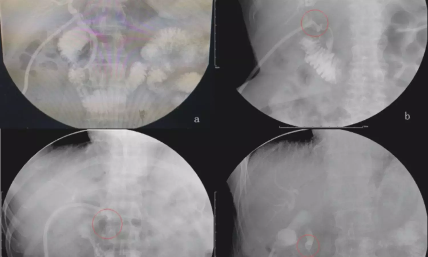
Routine Cholangiography during cholecystectomy associated with reduced bile duct injuries: Study

A recent ground-breaking study
found that routine cholangiography reduced the odds of bile duct injuries and
increased the chance of intraoperative recognition of major bile duct injuries
in individuals while undergoing cholecystectomy as per results that were
published in the journal JAMA Surgery.

Cholecystectomy is one of the most
common surgeries performed globally. Intraoperative cholangiography (IOC) is an
integral component of cholecystectomy and is performed either routinely or
selectively. Routine cholangiography (RC) is advocated to avoid systemic bile
duct injuries (BDI) during cholecystectomy and increase the quality of life. However,
it is associated with increased costs due to invasive testing and increased
postoperative imaging. Selective cholangiography (SC) is done in certain cases,
citing long operative times and costs as one of the factors. As there is an
ambiguity regarding the use of selective or routine cholangiography, researchers
conducted a comparative study to understand the perioperative outcomes of
cholecystectomy between surgeons who routinely vs selectively perform IOC.

A retrospective cohort study was
carried out from January 2015 through June 2023 within the Cleveland Clinic
Enterprise. About 18 hospitals and nine ambulatory surgery centers in 2 states Ohio
and Florida, were included. The study included adult patients who underwent
cholecystectomy for benign biliary disease. Routine cholangiography is defined
as more than 70% of cholecystectomies performed with IOC per surgeon over the
study period.

Findings:

  • A total of 134 surgeons performed 28,212
    cholecystectomies with 10,244 in the RC cohort.
  • The mean age of participants in the RC cohort
    was 52.71 years.
  • About 17,968 participants were included in the
    SC cohort.
  • The mean age of the participants was 52.33 years.
  • There were about 26 major BDIs (0.09%) and 105
    minor BDIs (0.34%).
  • RC was associated with decreased odds of major
    BDI and minor BDI compared with SC patients and surgeon characteristics were
    controlled.
  • The intraoperatively RC cohort was associated
    with more recognition of major BDIs than the SC cohort.
  • RC was not significantly associated with
    increased perioperative endoscopic retrograde cholangiopancreatography
    utilization or negative ERCP rate.

Thus, the study concluded that
routine cholangiography significantly reduced major and minor bile duct
injuries when compared to selective cholangiography. This underscores the
importance of incorporating routine cholangiography as a health system strategy
to reduce the risk of bile duct injuries in cholecystectomy.

Further reading: Gross A, Said
SA, Wehrle CJ, et al. Selective vs Routine Cholangiography Across a Health
Care Enterprise. JAMA Surg. Published online December 11,
2024. doi:10.1001/jamasurg.2024.5216.

Powered by WPeMatico

Tonsils or adenoids removal associated with the development of stress-related disorders: Study

A new study uncovered a concerning link between the surgical removal of tonsils or adenoids in early life and an increased risk of stress-related psychiatric disorders later in life. This study looked into the potential psychological repercussions of a common pediatric surgery which was published in Journal of American Medical Association.

The research analyzed data from Swedish nationwide population and health registries covered individuals born between 1981 and 2016. This  compared 2 groups where one had individuals who underwent tonsillectomy or adenoidectomy (exposed group) and other with those who did not (unexposed group). 

This population-matched cohort included a total of 83,957 exposed individuals and 839,570 unexposed counterparts, those who had the surgery exhibited a 43% higher risk of developing stress-related disorders. These included post-traumatic stress disorder (PTSD), acute stress reactions, and adjustment disorders. The hazard ratio (HR) for PTSD was even more pronounced at 1.55 which indicated a 55% increased risk when compared to unexposed individuals.

Also, in the sibling-matched cohort which consisting of 51,601 exposed individuals and 75,159 unexposed siblings, a 34% increased risk of stress-related disorders was observed which reinforced the findings from the larger cohort. PTSD risk in this cohort was also elevated (HR, 1.41).

The heightened risk was consistent across various demographic and clinical subgroups. Factors such as sex, age at surgery, time since surgery, parental educational attainment, and parental history of stress-related disorders did not significantly alter the results. The strongest association was seen in cases where surgery was performed due to adenotonsillar diseases or sleep and respiratory abnormalities.

This study highlights the need to explore the underlying mechanisms connecting early-life adenotonsillar conditions and their treatments to long-term mental health outcomes. It also raises questions about whether the stress of surgery or the health conditions prompting the procedure contribute to the observed risks.

While tonsillectomy and adenoidectomy are often necessary for managing severe medical conditions, the potential psychological effects should be factored into pre- and post-operative care by emphasizing the need for careful risk-benefit analyses and long-term monitoring of mental health outcomes in affected children.

Overall, this study opens the door for further investigation into the intersection of physical and mental health in pediatric populations by potentially reshaping how surgical interventions in children are approached.

Source:

Xiao, X., Yang, F., Yin, L., Isung, J., Ye, W., Mataix-Cols, D., Zhang, Z., Valdimarsdóttir, U., & Fang, F. (2024). Stress-Related Disorders Among Young Individuals With Surgical Removal of Tonsils or Adenoids. In JAMA Network Open (Vol. 7, Issue 12, p. e2449807). American Medical Association (AMA). https://doi.org/10.1001/jamanetworkopen.2024.49807

Powered by WPeMatico

Topical versus Systemic Antibiotics: Scaling and Root Planing Still Leads in Periodontal Therapy, Study Finds

Iran: A recent study explored the effects of local and systemic administration of metronidazole as adjunctive treatments for patients with chronic periodontitis. The findings, published in Clinical & Experimental Dental Research, revealed that all methods demonstrated comparable effectiveness, with scaling and root planing (SRP) remaining the gold standard for treating periodontal diseases.

While systemic metronidazole can penetrate deep into periodontal pockets, its use is often associated with side effects, including gastrointestinal disturbances and drug interactions. This led researchers to explore topical metronidazole in gel form as an alternative, given its targeted application and reduced systemic exposure. The findings suggest that the topical gel can be a safe and effective addition to SRP without the side effects associated with systemic delivery.

Chronic periodontitis is a progressive inflammatory disease that can lead to tooth loss if left untreated. The standard treatment for this condition is scaling and root planing considered the gold standard for managing periodontal diseases. However, using antibiotics as adjunctive therapies has gained attention for their potential to improve treatment outcomes.

Against the above background, Maryam Mehravani, Student Research Committee, Qazvin University of Medical Sciences, Qazvin, Iran, and colleagues sought to evaluate and compare the impacts of local versus systemic metronidazole in managing chronic periodontitis.

For this purpose, the researchers conducted a randomized clinical study involving 30 patients, each contributing three teeth for evaluation. Participants were divided into three groups: one received SRP alone, the second received adjunctive treatment with metronidazole tablets, and the third received adjunctive treatment with metronidazole gel.

Key parameters—bleeding on probing (BOP), pocket probing depth (PPD), and clinical attachment level (CAL)—were assessed at baseline and three months post-treatment.

The key findings were as follows:

  • There were significant improvements in BOP, CAL, and PPD levels across all treatments after three months compared to baseline.
  • There were no significant differences in BOP, CAL, and PPD levels between the three treatment groups post-treatment.

The findings showed that metronidazole gel, metronidazole tablets, and SRP treatment effectively improved clinical parameters such as CAL, PPD, and BOP. However, adding local or systemic metronidazole in patients with periodontitis beyond Stage II and Grade B showed no significant advantage over SRP alone.

“SRP remains the gold standard for periodontal disease treatment, and further research is necessary to validate these findings,” the researchers wrote.

“Systemic antibiotics can reach the depths of periodontal pockets but with notable side effects. As an alternative, a simpler form, such as a topical gel, may offer a more effective adjunctive therapy for treating chronic periodontitis,” they concluded.

Reference:

Mehravani, M., Houshyar, E., Jamalnia, S., & Gharaaghaji, R. (2024). Effects of Local and Systemic Metronidazole as Adjunctive Treatment in Chronic Periodontitis Patients. Clinical and Experimental Dental Research, 10(6), e70050. https://doi.org/10.1002/cre2.70050

Powered by WPeMatico

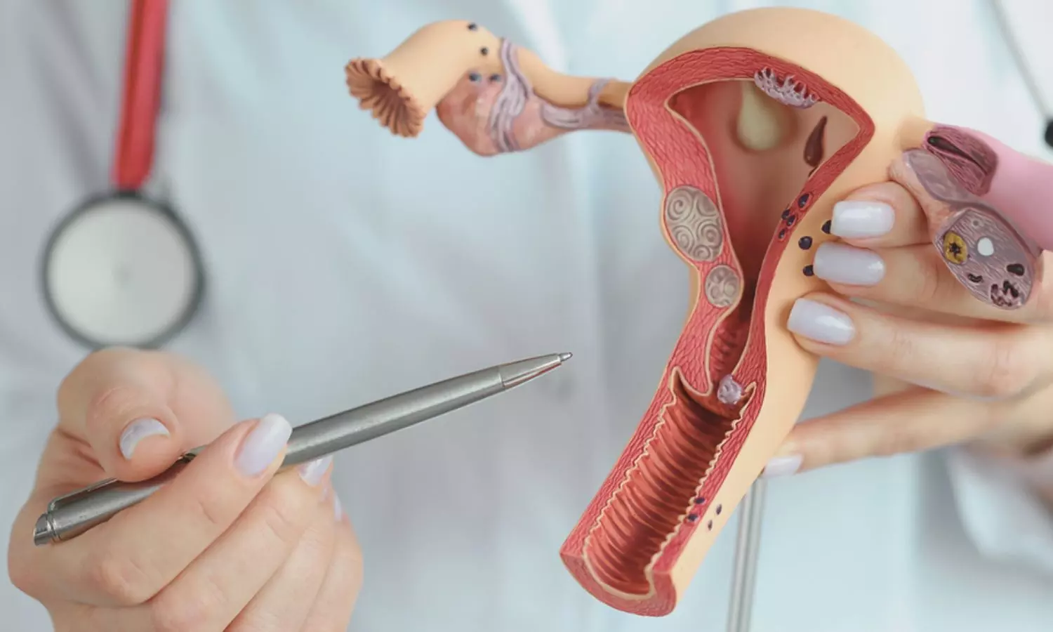
Vitamin D Deficiency Linked to Endometrial Polyps in Infertile Women: Study Highlights Nonlinear Correlation

China: A recent study published in Human Reproduction has shed light on the complex relationship between serum vitamin D levels and endometrial polyps (EPs) incidence in infertile women. It highlights a nonlinear correlation that could have significant implications for reproductive health.

“This study was the first to establish vitamin D deficiency as an independent risk factor for the incidence of endometrial polyps in infertile patients. Recognizing modifiable risk factors, such as vitamin D deficiency, could pave the way for developing new strategies to treat polyps or prevent their formation,” the researchers wrote.

Endometrial polyps are a common condition that can impact endometrial receptivity in women of reproductive age. Vitamin D influences cell proliferation, differentiation, apoptosis, angiogenesis, inflammation control, and immune modulation, apart from its well-known role in regulating calcium and phosphorus balance. Previous research has linked vitamin D levels to reproductive outcomes and highlighted an association between low vitamin D levels and the occurrence of colorectal and nasal polyps. However, limited evidence exists regarding the relationship between serum vitamin D levels and the incidence of EPs.

To address this gap, Xiqian Zhang, Center for Reproductive Medicine, Guangdong Women and Children Hospital, Guangzhou, Guangdong Province, China, and colleagues sought to investigate whether serum vitamin D levels are associated with EPs incidence in infertile women.

For this purpose, the researchers conducted a cross-sectional study at Guangdong Women and Children Hospital between January 2019 and October 2023, involving 3,107 infertile patients.

The participants included women who underwent hysteroscopy, of whom 642 were diagnosed with endometrial polyps, while 2,465 had a normal uterine cavity. Hysteroscopy findings were used to evaluate the risk of EPs, polyp size, the prevalence of multiple polyps, and the incidence of chronic endometritis (CE). Serum vitamin D levels were measured using chemiluminescence to determine total 25(OH)D concentrations.

Patients were categorized into two groups based on international guidelines for vitamin D deficiency: those with serum 25(OH)D levels below 50 nmol/L and those with levels equal to or above 50 nmol/L.

The study led to the following findings:

  • Among the participants, 23.8% were vitamin D deficient, with serum 25(OH)D levels below 50 nmol/L.
  • The incidence of endometrial polyps was significantly higher in the vitamin D-deficient group (<50 nmol/L) compared to those with sufficient levels (≥50 nmol/L), at 24.9% versus 19.3%.
  • There were no differences between the two groups in terms of polyp size, the proportion of multiple polyps, or the presence of chronic endometritis (CE).
  • After adjusting for confounding factors, having 25(OH)D levels ≥50 nmol/L was associated with a reduced risk of EPs (adjusted OR: 0.733).
  • Additional factors influencing polyp incidence included body mass index (BMI), type of infertility, CA125 levels, and CD138-positive plasma cells.
  • A positive linear relationship was identified between age and serum 25(OH)D levels through a linear regression model.
  • Subgroup analyses revealed a higher risk of EPs in the 25(OH)D <50 nmol/L group compared to the ≥50 nmol/L group in both younger patients (23.8% versus 19.1%) and older patients (28.0% versus 19.9%).
  • A smooth curve fitting model indicated a nonlinear correlation between 25(OH)D levels and the risk of EPs, with an optimal threshold of 51.8 nmol/L for 25(OH)D levels.
  • Subgroup analyses showed a nonlinear correlation between 25(OH)D levels and polyp risk in patients under 35 years. In contrast, there was a linear correlation in patients aged 35 years or older.

The authors emphasize that their findings should be interpreted with caution, as the study’s correlational nature does not establish causality. Furthermore, the strict inclusion and exclusion criteria may limit the generalizability of the results to broader populations, such as premenopausal women or those of different racial backgrounds.

“Further clinical trials and laboratory studies are necessary to explore the role of vitamin D in the development of EPs and to clarify the underlying mechanisms,” they concluded.

Reference:

Zhou, R., Zhu, Z., Dong, M., Wang, Z., Huang, L., Wang, S., Zhang, X., & Liu, F. (2024). Nonlinear correlation between serum vitamin D levels and the incidence of endometrial polyps in infertile women. Human Reproduction, 39(12), 2685-2692. https://doi.org/10.1093/humrep/deae241

Powered by WPeMatico

Study finds effectiveness of topical corticosteroid regimen for eosinophilic esophagitis

A new study published in the Clinical Gastroenterology and Hepatology journal examined the effectiveness of topical corticosteroid (tCS) regimens for eosinophilic esophagitis (EoE) has revealed that both once-daily and twice-daily dosing provide comparable outcomes. These findings could help simplify treatment protocols and reduce concerns about less frequent dosing regimens.

The research at the University of North Carolina involved a retrospective analysis of 522 patients with newly diagnosed EoE from the UNC EoE Clinicopathologic Database. The participants received either once-daily (n=122) or twice-daily (n=400) tCS regimens and underwent follow-up endoscopies with biopsies to assess treatment outcomes.

The key demographic differences emerged between the 2 groups. Patients on the twice-daily regimen were slightly older on average (28.8 years vs. 24.3 years) and reported higher rates of heartburn (40% vs. 25%). Despite these distinctions, treatment outcomes demonstrated no significant variation. The study evaluated several key measures of EoE treatment success, were both groups had similar rates of symptom improvement, with 78% for once-daily dosing and 76% for twice-daily dosing.

Defined as having fewer than 15 eosinophils per high-power field (eos/hpf) in post-treatment biopsies, histologic response rates were nearly identical which was 56% for once-daily dosing versus 58% for twice-daily. Post-treatment eosinophil levels averaged 20.8 for once-daily dosing and 25.6 for twice-daily, a statistically insignificant difference.

EoE Endoscopic Reference Scores were used to measure inflammation and structural changes, were similar in both groups (2.2 for once-daily and twice-daily). Also, a notable difference was the lower frequency of candida infections with once-daily dosing (2% vs. 8%) by suggesting it may have a better safety profile.

Multivariate analysis confirmed no significant difference in histologic response rates between the dosing regimens, with an adjusted odds ratio of 1.03. These findings indicate that both once-daily and twice-daily dosing are equally effective for managing EoE. This study provided reassurance to clinicians and patients that less frequent dosing does not compromise treatment efficacy. Also, the reduced risk of candida infections with once-daily dosing offers a strong advantage.

As EoE management continues to evolve, these results could inform simpler and more patient-friendly treatment regimens. Overall, the findings hold significant implications for clinical practice by highlighting that effective EoE treatment need not require the higher burden of twice-daily medication administration.

Source:

Reed, C. C., LaFata, S. S., Gee, T. S., Thel, H. L., Cameron, B. A., Xue, A. Z., Kiran, A., Ocampo, A. A., McCallen, J., Lee, C. J., Borinsky, S. A., Redd, W. D., Barlowe, T., Kaakati, R. N., Cotton, C. C., Eluri, S., & Dellon, E. S. (2024). Daily or Twice Daily Treatment with Topical Steroids Results in Similar Responses in Eosinophilic Esophagitis. In Clinical Gastroenterology and Hepatology. Elsevier BV. https://doi.org/10.1016/j.cgh.2024.10.016

Powered by WPeMatico

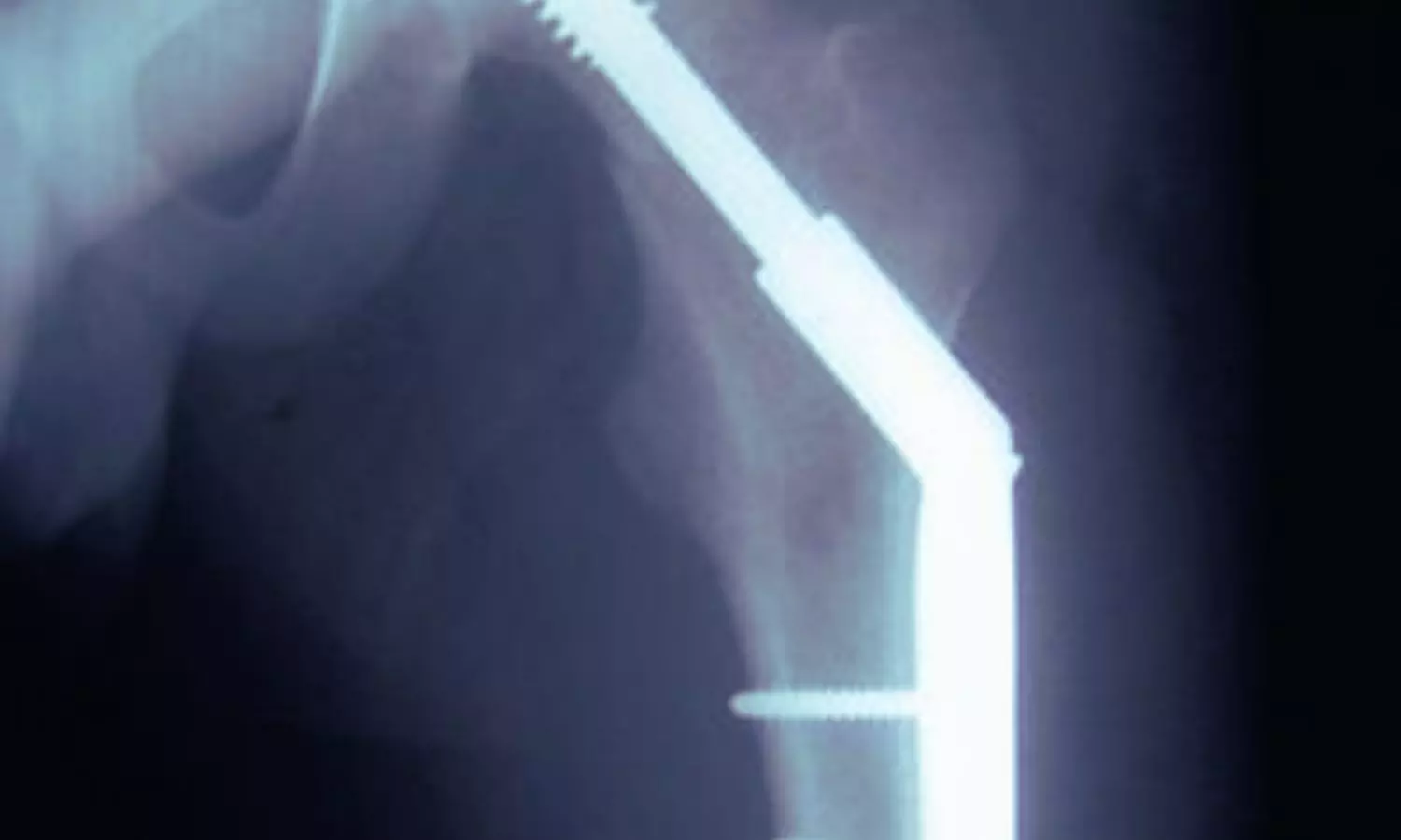
Rare Sliding Hip Screw Migration Poses Life-Threatening Risks to Pelvic Vessels: Two Cases Highlight

UK: Sliding hip screws (SHSs) are widely used for the surgical treatment of extracapsular neck or femur fractures, offering reliable stabilization and promoting healing. However, rare complications can arise, including the intrapelvic migration of an SHS, a life-threatening event that can compromise vital pelvic vessels. This has recently been highlighted in a report of two unusual cases linked to non-unions post-surgery, reported in BMJ Case Reports. 

Richard Laurence Donovan, Musculosketal Research Unit, University of Bristol Medical School, Bristol, UK, and colleagues reported two rare cases involving intrapelvic migration of the SHS lag screw caused by non-unions, posing a threat to the iliac vessels. They provided an up-to-date review of the literature, offering insights into the mechanisms by which a lag screw may migrate and proposing strategies to prevent this complication. 
Patient 1, an 80-year-old woman, presented with persistent pain in her left hip and difficulty mobilizing. She had undergone SHS fixation for a left-sided trochanteric neck of femur fracture five months earlier and spent weeks in a rehabilitation center with ongoing physiotherapy. Still, she continued to experience pain since her surgery. Her medical history included ischemic heart disease, a previous cardiac arrest, and atrial fibrillation (AF). Her mental status was intact, with a score of 10/10 on the abbreviated mental test, and her life expectancy remained good.
The patient was seen in the outpatient clinic two months post-surgery and showed progress in recovery. At her 18-month follow-up, a telephone consultation was held with her daughter due to her declining health. She had moved to a care home, mobilized short distances with a Zimmer frame, and was mostly housebound. Aside from a shuffling gait, she reported no hip complaints. Given her overall health and difficulties attending the clinic, she was discharged, and she has since passed away.
Patient 2, another woman in her 80s, was referred to a tertiary center with a three-week history of right-sided hip pain, which worsened over the last few days. She retained a small amount of discomfort with a straight leg raise. She had previously undergone SHS fixation for a right-sided trochanteric neck of femur fracture seven years ago. Fifteen months post-surgery, she experienced right medial thigh tenderness, with imaging revealing the failure of the most distal plate screw by breakage, managed without additional surgery. Her medical history included AF, congestive cardiac failure, abdominal aortic aneurysm, chronic kidney disease, hypertension, and coeliac disease. She lived independently with minimal assistance twice weekly, using a Zimmer frame for mobility but rarely leaving her home.
This patient was seen in the outpatient clinic two months post-surgery and was recovering well, mobilizing with a wheeled frame and reporting only occasional right hip ache. Radiographs confirmed no further lag screw migration. She preferred to avoid surgery as her symptoms were managed, and she was discharged with an open appointment.
The authors emphasize that orthopedic surgeons should remain aware of this rare but serious complication, as intrapelvic screw migration can pose a threat to the iliac vessels. They highlight the importance of using contrast-enhanced cross-sectional imaging to assess proximity to neurovascular structures. If necessary, collaboration with general or vascular surgeons should be considered to plan the surgical approach for intrapelvic screw retrieval.
“Adhering to manufacturer operative technique guides and avoiding skipped surgical steps are crucial. Additionally, surgeons should ensure guidewires do not penetrate the femoral head and that the femoral head and neck are not over-reamed,” they concluded.
Reference:
Donovan RL, Jiang M, Bassett J, et al. Rare intrapelvic migration of a sliding hip screw after hip fracture fixation. BMJ Case Reports CP 2024;17:e261689.

Powered by WPeMatico

Nocturnal Enuresis Significantly Prevalent in Children with Obstructive Sleep Apnea, reports study

A recent study published in the
Journal Children found that nocturnal enuresis is significantly prevalent in
children with obstructive sleep apnea (OSA). The study results suggest that
adenotonsillectomy can reduce nocturnal enuresis in these Children.

Obstructive sleep apnea (OSA) is
a breathing disorder characterized by repeated episodes of upper airway
obstruction during sleep, disrupting normal patterns. It is a sleep disorder
that peaks in children between 3-6 years of age. The breathing issues at night
can also affect dental occlusion and craniofacial development. Adenotonsillectomy
(AT) is the primary treatment for pediatric OSA. Nocturnal enuresis (NE) is intermittent
incontinence during sleep, and its prevalence decreases with age. Literature
shows that NE has a high prevalence in OSA and reduces when OSA is treated with
AT. However, there is limited data on the prevalence of NE in OSA children
compared to their healthy peers. Researchers conducted a study to determine the
prevalence of NE in children with apnea OSA, the effect of AT, and the width of
the arches and to compare them with control children without respiratory
problems.

The study included 298 children
within the age range of 2 to 12 years old. There were two age groups of
children aged 2–<5 years and ≥5–12 years. Participants were divided
into two main groups: an experimental group (n = 130) and a control group (n =
168). The experimental group was further divided into two subgroups: children
with OSA and NE (n = 51) and children with OSA but without NE (n = 79).
Children having at least one bedwetting incident per month were identified as
having NE. Arch widths were measured at the baseline and one year after. Polysomnography
was used to diagnose OSA, and the apnea-hypopnea index (AHI) was obtained. A Pediatric
Sleep Questionnaire (PSQ) was given to the parents to classify their children
into those with and without NE.

Findings:

  • This study included 130 subjects in the OSA
    group, while the control group consisted of 168 subjects.
  • There were no sexual differences in the occurrence
    of OSA
  • About 39.2% of patients in the OSA group experienced
    NE compared to 28% in the control group, which had a significant prevalence.
  • About 49% of the patients with both OSA and NE
    experienced a complete resolution of NE after surgery.
  • Both OSA groups had narrower arch widths than
    the control group (p = 0.012), with the NE group having the
    narrowest widths.
  • After the assessments before and after the
    surgery, children with OSA had a statistically significant prevalence of NE
    compared to the control group.
  • Patients with OSA, with and without NE, showed
    very similar AHI results, though those with NE had slightly higher AHI values.
  • Children with OSA exhibited narrower intercanine
    and intermolar widths in the upper and lower arches compared to the control
    group.

Thus, the study concluded that
nocturnal enuresis is more prevalent in children with OSA and should be
considered and evaluated as one of the first signs of breathing disorders. These
findings underscore the importance of recognizing nocturnal enuresis as one of
the potential clinical markers for OSA in pediatric patients.

Further reading: The Relationship
between Nocturnal Enuresis and Obstructive Sleep Apnea in Children. Doi: https://doi.org/10.3390/children11091148

Powered by WPeMatico

Sickle Cell Disease during pregnancy Significant Risk Factor for Maternal-Fetal Morbidity, finds study

Researchers have found that sickle cell disease (SCD) poses a significantly increased risk for maternal-fetal morbidities, especially in the last month of pregnancy. This is concluded by one of the largest studies into SCD pregnancies in India, undertaken at Kasturba Maternity Hospital, Gujarat. The study was conducted by Kapilkumar Dave and colleagues published in the International Journal of Obstetrics and Gynecology.

The study sought to quantify maternal-fetal risks associated with SCD and sickle cell traits and explore the epidemiology of maternal morbidity. The results are of utmost importance for critical insights into how SCD affects pregnancy outcomes and can provide actionable information on strategies to improve care in resource-limited settings.

This retrospective cohort study enrolled all pregnant women admitted to Kasturba Maternity Hospital from 2016 to 2021. In five years, there were 24,256 deliveries. The screening for SCD was done in these women, and pregnancy-related complications are documented meticulously. The analysis of risks was done with the help of Poisson and logistic regression models after accounting for confounding variables.

Among the 24,256 deliveries, 354 women (1.5%) had SCD, and 4216 women (17.4%) were identified as having the sickle cell trait.

Maternal Morbidity Risks:

Women with SCD had a significantly higher risk as compared to normal pregnancies :

  • Maternal death: AOR 13.7 (95% CI: 4.5-42.7).

  • Anemia: AOR 6.8 (95% CI: 4.5-10.2).

  • Severe anemia: AOR 4.3 (95% CI: 3.3-5.6).

  • Preterm delivery: AOR 4.5 (95% CI: 3.6-5.7).

  • Cesarean section: AOR 5.5 (95% CI: 4.7-7.0).

Fetal Outcomes:

  • Stillbirth: AOR 3.4 (95% CI: 2.3-5.3).

  • Low birth weight: AOR 3.1 (95% CI: 2.4-3.9).

Timing of Risk:

  • The greatest risks of maternal morbidities were in the last month of gestation.

SCD Severity:

  • Pregnant women with severe manifestations of SCD before conception were at increased risk for complications during pregnancy.

Maternal-fetal morbidities are increased by sickle cell disease, especially the risks of maternal death, anemia, preterm delivery, and poor fetal outcomes. This is a critical final month of gestation requiring high-level care for those women with SCD who had prior severe manifestations. This underscores the need for high-impact healthcare interventions.

Reference:

Dave K, Desai S, Desai T, Desai G. Adverse maternal and fetal outcomes among tribal pregnant women suffering from sickle cell disease: A retrospective cohort study in a community-based hospital situated in a tribal block of Gujarat, India. Int J Gynaecol Obstet. 2024 Nov 6. doi: 10.1002/ijgo.15999. Epub ahead of print. PMID: 39503239.

Powered by WPeMatico

AI tool analyzes placentas at birth for faster detection of neonatal, maternal problems, reveals study

A newly developed tool that harnesses computer vision and artificial intelligence (AI) may help clinicians rapidly evaluate placentas at birth, potentially improving neonatal and maternal care, according to new research from scientists at Northwestern Medicine and Penn State.

The study, which was published Dec. 13 in the print edition of the journal Patterns and featured on the journal’s cover, describes a computer program named PlacentaVision that can analyze a simple photograph of the placenta to detect abnormalities associated with infection and neonatal sepsis, a life-threatening condition that affects millions of newborns globally.

“Placenta is one of the most common specimens that we see in the lab,” said study co-author Dr. Jeffery Goldstein, director of perinatal pathology and an associate professor of pathology at Northwestern University Feinberg School of Medicine. “When the neonatal intensive care unit is treating a sick kid, even a few minutes can make a difference in medical decision making. With a diagnosis from these photographs, we can have an answer days earlier than we would in our normal process.”

Northwestern provided the largest set of images for the study, and Goldstein led the development and troubleshooting of the algorithms.

Alison D. Gernand, contact principal investigator on the project, conceived the original idea for this tool through her global health work, particularly with pregnancies where women deliver in their homes due to lack of health care resources.

“Discarding the placenta without examination is a common but often overlooked problem,” said Gernand, associate professor in the Penn State College of Health and Human Development (HHD) Department of Nutritional Sciences. “It is a missed opportunity to identify concerns and provide early intervention that can reduce complications and improve outcomes for both the mother and the baby.”

Why early examination of the placenta matters

The placenta plays a vital role in the health of both the pregnant individual and baby during pregnancy, yet it is often not thoroughly examined at birth, especially in areas with limited medical resources.

“This research could save lives and improve health outcomes,” said Yimu Pan, a doctoral candidate in the informatics program from the College of Information Sciences and Technology (IST) and lead author on the study. “It could make placental examination more accessible, benefitting research and care for future pregnancies, especially for mothers and babies at higher risk of complications.”

Early identification of placental infection through tools like PlacentaVision might enable clinicians to take prompt actions, such as administering antibiotics to the mother or baby and closely monitoring the newborn for signs of infection, the scientists said.

PlacentaVision is intended for use across a range of medical demographics, according to the researchers.

“In low-resource areas — places where hospitals don’t have pathology labs or specialists — this tool could help doctors quickly spot issues like infections from a placenta,” Pan said. “In well-equipped hospitals, the tool may eventually help doctors determine which placentas need further, detailed examination, making the process more efficient and ensuring the most important cases are prioritized.”

“Before such a tool can be deployed globally, core technical obstacles we faced were to make the model flexible enough to handle various diagnoses related to the placenta and to ensure that the tool can be robust enough to handle various delivery conditions, including variation in lighting conditions, imaging quality and clinical settings” said James Z. Wang, distinguished professor in the College of IST at Penn State and one of the principal investigators on the study. “Our AI tool needs to maintain accuracy even when many training images come from a well-equipped urban hospital. Ensuring that PlacentaVision can handle a wide range of real-world conditions was essential.”

How the tool learned how to analyze pictures of placentas

The researchers used cross-modal contrastive learning, an AI method for aligning and understanding relationship between different types of data — in this case, visual (images) and textual (pathological reports) — to teach a computer program how to analyze pictures of placentas. They gathered a large, diverse dataset of placental images and pathological reports spanning a 12-year period, studied how these images relate to health outcomes and built a model that could make predictions based on new images. The team also developed various image alteration strategies to simulate different photo-taking conditions so the model’s resilience can be evaluated properly.

The result was PlacentaCLIP+, a robust machine-learning model that can analyze photos of placentas to detect health risks with high accuracy. It was validated cross-nationally to confirm consistent performance across populations.

According to the researchers, PlacentaVision is designed to be easy to use, potentially working through a smartphone app or integrated into medical record software so doctors can get quick answers after delivery.

Next step: A user-friendly app for medical staff

“Our next steps include developing a user-friendly mobile app that can be used by medical professionals — with minimal training — in clinics or hospitals with low resources,” Pan said. “The user-friendly app would allow doctors and nurses to photograph placentas and get immediate feedback and improve care.”

The researchers plan to make the tool even smarter by including more types of placental features and adding clinical data to improve predictions while also contributing to research on long-term health. They’ll also test the tool in different hospitals to ensure it works in a variety of settings.

“This tool has the potential to transform how placentas are examined after birth, especially in parts of the world where these exams are rarely done,” Gernand said. “This innovation promises greater accessibility in both low- and high-resource settings. With further refinement, it has the potential to transform neonatal and maternal care by enabling early, personalized interventions that prevent severe health outcomes and improve the lives of mothers and infants worldwide.”

This research was supported by the National Institutes of Health National Institute of Biomedical Imaging and Bioengineering (grant R01EB030130). The team used supercomputing resources from the National Science Foundation-funded Advanced Cyberinfrastructure Coordination Ecosystem: Services & Support (ACCESS) program.

Powered by WPeMatico