Tranexamic acid may help control GI bleeding, prevent rebleeding in cirrhosis patients: Study

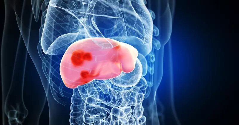
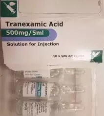
Tranexamic acid may help control GI bleeding, and prevent rebleeding in cirrhosis patients suggests a study published in the Hepatology.

Patients with Child-Turcotte-Pugh class B and C cirrhosis with upper gastrointestinal bleeding (UGIB) have systemic as well as localized (in the mucosa of the esophagus and stomach) fibrinolysis. The aim of this study was to evaluate the efficacy and safety of tranexamic acid in the treatment of acute UGIB in patients with cirrhosis. A total of 600 patients with advanced liver cirrhosis (Child-Turcotte-Pugh class B or C) presenting with UGIB were randomly allocated to either the tranexamic acid (n=300) or the placebo group (n=300). The primary outcome measure was the proportion of patients developing 5-day treatment failure. Failure to control bleeding by day 5 was seen in 19/300 (6.3%) patients in the tranexamic acid group and 40/300 (13.3%) patients in the placebo group (p=0.006). Esophageal endoscopic variceal ligation (EVL) site as a source of failure to control bleeding by day 5 among patients undergoing first-time esophageal EVL (excluding patients with a previous post-EVL ulcer as a source of bleed) was seen in 11/222 (4.9%) patients in the tranexamic acid group and 27/225 (1212.0%) patients in the placebo group (p=0.005). However, 5-day and 6-week mortality was similar in the tranexamic acid and placebo groups.

Tranexamic acid significantly reduces the failure to control bleeding by day 5 and failure to prevent rebleeding after day 5 to 6 weeks in patients with advanced liver cirrhosis (Child-Turcotte-Pugh class B or C) presenting with UGIB, by preventing bleeding from the EVL site.

Reference:

Kumar, Manoj1; Venishetty, Shantan1; Jindal, Ankur1; Bihari, Chhagan2; Maiwall, Rakhi1; Vijayaraghavan, Rajan1; Saggere Muralikrishna, Shasthry1; Arora, Vinod1; Kumar, Guresh3; Sarin, Shiv K.1. Tranexamic acid in upper gastrointestinal bleed in patients with cirrhosis: A randomized controlled trial. Hepatology 80(2):p 376-388, August 2024. | DOI: 10.1097/HEP.0000000000000817

Keywords:

Tranexamic, acid, control, GI bleeding, prevent, rebleeding, cirrhosis patients, study, Kumar, Manoj1; Venishetty, Shantan1; Jindal, Ankur1; Bihari, Chhagan2; Maiwall, Rakhi1; Vijayaraghavan, Rajan1; Saggere Muralikrishna, Shasthry1; Arora, Vinod1; Kumar, Guresh3; Sarin, Shiv K

Powered by WPeMatico

Ziresovir Shows Promise in Alleviating Bronchiolitis Symptoms in Hospitalized Infants with RSV: Study

China: In a promising development for pediatric care, a recent study has highlighted the efficacy of Ziresovir in treating infants hospitalized due to respiratory syncytial virus (RSV) infection. The research, published in the New England Journal of Medicine, provides vital insights into managing bronchiolitis, a common respiratory condition that can lead to severe complications in young children.

The researchers found that treatment with Ziresovir, an antiviral medication, significantly alleviated the signs and symptoms of bronchiolitis in infants and young children hospitalized with RSV infection, with no safety concerns reported.

Respiratory syncytial virus (RSV) is a major cause of serious illness in infants, and currently, there is no effective treatment available. However, results from a phase 2 trial indicate that ziresovir may be an effective option for treating infants hospitalized with RSV infection.

Against the above background, Xin Ni, Beijing Children’s Hospital, Beijing, China, and colleagues conducted a phase 3, multicenter, double-blind, randomized, placebo-controlled trial in China. The researchers enrolled participants aged 1 to 24 months hospitalized with RSV infection. They were randomly assigned in a 2:1 ratio to receive either ziresovir (doses ranging from 10 to 40 mg based on body weight) or a placebo, administered twice daily for five days.

The primary endpoint was the change in the Wang bronchiolitis clinical score from baseline to day three (defined as 48 hours after the first dose), with total scores ranging from 0 to 12, where higher scores indicated greater severity of symptoms. The intention-to-treat population included all participants with confirmed RSV infection who received at least one dose of either ziresovir or placebo, while the safety population encompassed all participants who received at least one dose of the treatment or placebo.

The following were the key findings of the study:

  • The intention-to-treat population included 244 participants, and the safety population included 302.
  • The reduction from baseline in the Wang bronchiolitis clinical score on day 3 was significantly greater with ziresovir than with placebo (−3.4 points versus −2.7 points).
  • The reduction in the RSV viral load at day 5 was greater in the ziresovir group than in the placebo group (−2.5 versus −1.9 log10 copies per milliliter).
  • Improvements were observed in prespecified subgroups, including participants with a baseline bronchiolitis score of at least eight and those six months of age or younger.
  • The incidence of adverse events related to the drug or placebo was 16% with ziresovir and 13% with placebo.
  • The most common adverse events that were assessed by the investigator as being related to the drug or placebo were diarrhea (in 4% and 2% of the participants, respectively), an elevated liver-enzyme level (in 3% and 3%, respectively), and rash (in 2% and 1%).
  • Resistance-associated mutations were identified in 15 participants (9%) in the ziresovir group.

“We found that ziresovir treatment reduced signs and symptoms of bronchiolitis in infants and young children hospitalized with RSV infection and there were no safety concerns,” the researchers concluded.

Reference:

Zhao S, et al “Ziresovir in hospitalized infants with respiratory syncytial virus infection” N Engl J Med 2024; DOI: 10.1056/NEJMoa2313551.

Powered by WPeMatico

Maternal Weight tied to Increased Atopy and Asthma Risk in kids of Women at Risk for Gestational Diabetes: Study

Estonia: The follow-up study group of the Gestational Diabetes Study (GDS) has shed light on the impact of maternal weight-related factors on the development of atopic diseases, including asthma, in children born to mothers at risk for gestational diabetes mellitus (GDM).

The study, conducted with a focus on children aged 1 to 5 years, found that among pregnant women at risk for GDM, maternal weight-related factors play a significant role in the development of atopic diseases, regardless of the GDM diagnosis. The findings published in BMC Pregnancy and Childbirth suggest that in addition to focusing on women with GDM, it is crucial to also pay attention to those at risk who do not develop the condition, as their children appear to be at a higher risk for atopic diseases.

Gestational diabetes mellitus is the most common metabolic disorder during pregnancy and is linked to negative outcomes in offspring, including an increased risk of developing atopic diseases in early childhood. However, research focusing specifically on women at risk for GDM—some of whom develop the condition while others do not—is limited. Additionally, there is a lack of information regarding adverse health outcomes in the children of these women. To fill this knowledge gap, Anu Bärenson, Tartu University Hospital, Children´s Clinic, Tartu, Estonia, and colleagues aimed to examine how various maternal health characteristics are associated with the development of these disorders in their offspring.

The follow-up study of the GDS took place at Tartu University Hospital in Estonia from 2014 to 2020 and involved 223 mother-child pairs. All participating women had at least one risk factor for GDM, although only some went on to develop the condition. Information regarding the relevant diagnoses was collected from Electronic Health Records. Allergen-specific IgE levels in the children’s serum were measured using ImmunoCAP Phadiatop Infant, with values of 0.35 kU/l or higher deemed positive. Statistical analysis was conducted using RStudio software (version 4.3.0).

The study led to the following findings:

  • Only the cases of GDM requiring using antidiabetic medications were associated with the development of asthma and/or allergic rhinitis at two years of age (aOR 4.68).
  • Maternal obesity (BMI > 30) was associated with offspring´s asthma or allergic rhinitis diagnosis at two years of age (aOR 3.15).
  • Maternal abnormal weight gain during pregnancy was associated with asthma and/or allergic rhinitis at five years of age (aOR 2.76).

The findings suggest that, in addition to women diagnosed with GDM who require antidiabetic medications, maternal weight-related factors play a significant role in the development of atopic and allergic diseases in the children of pregnant women at risk for GDM, irrespective of whether they actually develop the condition.

“Therefore, it is crucial to focus not only on mothers with GDM but also on those at risk who do not go on to develop it, as their children seem to face an even higher risk of allergic diseases,” they concluded.

Reference:

Bärenson, A., Tagoma, A., Varendi, H. et al. Atopy and asthma in children born to mothers at risk of gestational diabetes mellitus: a follow-up study. BMC Pregnancy Childbirth 24, 610 (2024). https://doi.org/10.1186/s12884-024-06819-y

Powered by WPeMatico

New Insights on Perioperative Cardiovascular Management in Noncardiac Surgery: AHA/ACC 2024 Guidelines Released

USA: The American College of Cardiology (ACC) and the American Heart Association (AHA) have issued an updated joint guideline for the cardiovascular management of adults undergoing non-cardiac surgery, incorporating a decade’s worth of new research since the previous guideline was released in 2014. 

The updates published in the Journal of the American College of Cardiology highlighted the most recent evidence regarding non-cardiac surgery and managing cardiovascular disease (CVD) risk factors before, during, and after the procedure.

The updated AHA/ACC guidelines provide comprehensive recommendations for managing cardiovascular disease (CVD) risk factors in patients scheduled for non-cardiac surgery. The key suggestions include appropriate cardiovascular testing, management of existing conditions, and guidelines on sodium-glucose cotransporter-2 (SGLT2) inhibitors for type 2 diabetes (T2D).

Annemarie Thompson, MD, MBA, chair of the guideline writing group and professor at Duke University Medical Center, emphasized the importance of summarizing evidence to aid clinicians. With around 300 million non-cardiac surgeries performed annually, the guidelines aim to enhance perioperative care and reduce cardiovascular complications.

Similar to the 2014 report, the updated guidelines include a perioperative algorithm to guide decision-making on blood pressure management and tailored strategies for patients with specific conditions, such as coronary artery disease and hypertrophic cardiomyopathy.

Healthcare professionals are encouraged to use targeted screenings to assess cardiac risk, with recommendations for emergency-focused cardiac ultrasound in cases of unexplained hemodynamic instability, when specialists are available. The guidelines also stress the importance of managing medications, advising that SGLT2 inhibitors should be paused 3 to 4 days before surgery to lower the risk of perioperative ketoacidosis.

In addition, the guidelines highlight the risks associated with glucagon-like polypeptide-1 (GLP-1) agonists, which may cause nausea and delayed gastric emptying, potentially increasing the risk of pulmonary aspiration during anesthesia.

Blood thinners can be safely paused before surgery and resumed postoperatively, with exceptions noted in the recommendations. The authors call for further research on myocardial injury after non-cardiac surgery (MINS), which affects about 20% of patients and is linked to worse outcomes.

The guidelines also address atrial fibrillation (AF) that may arise during or after surgery, urging close follow-up for patients with newly diagnosed AF to prevent stroke.

Developed with input from multiple medical societies, the guidelines emphasize a multidisciplinary approach to optimize care for patients with cardiovascular risks before, during, and after surgery. Thompson noted the need for collaborative efforts among specialists to improve patient outcomes in this growing population.

The following were the key takeaways from the guideline:

  1. A structured approach to perioperative cardiac assessment helps clinicians decide when to proceed with surgery or pause for further evaluation.
  2. Cardiovascular screening and treatment for patients undergoing non-cardiac surgery should follow the same guidelines as for non-surgical patients, ensuring timely interventions without unnecessary overscreening or overtreatment.
  3. Stress testing should be used selectively for patients undergoing non-cardiac surgery, particularly for those at lower risk, and only when testing is justified regardless of the surgery.
  4. Emphasizing a team-based approach is crucial for managing patients with complex anatomical or unstable cardiovascular conditions.
  5. New treatments for diabetes, heart failure, and obesity have important implications for perioperative care. SGLT2 inhibitors should be stopped 3 to 4 days before surgery to reduce the risk of perioperative ketoacidosis.
  6. Myocardial injury following non-cardiac surgery is a newly recognized condition that should be addressed, as it can have significant consequences for patients.
  7. Patients diagnosed with atrial fibrillation during or after non-cardiac surgery face a heightened stroke risk and require close monitoring to address reversible causes and consider long-term anticoagulation.
  8. Perioperative bridging of oral anticoagulants should be reserved for those at high risk for thrombotic complications and is not generally recommended for most patients.
  9. In cases of unexplained hemodynamic instability, emergency-focused cardiac ultrasound can be utilized for evaluation when clinical expertise is available, but it should not replace comprehensive transthoracic echocardiography.

Reference:

Thompson, A., Fleischmann, K. E., Smilowitz, N. R., De las Fuentes, L., Mukherjee, D., Aggarwal, N. R., Ahmad, F. S., Allen, R. B., Altin, S. E., Auerbach, A., Berger, J. S., Chow, B., Dakik, H. A., Eisenstein, E. L., Gerhard-Herman, M., Ghadimi, K., Kachulis, B., Leclerc, J., Lee, C. S., . . . Williams, K. A. (2024). 2024 AHA/ACC/ACS/ASNC/HRS/SCA/SCCT/SCMR/SVM Guideline for Perioperative Cardiovascular Management for Noncardiac Surgery: A Report of the American College of Cardiology/American Heart Association Joint Committee on Clinical Practice Guidelines. Journal of the American College of Cardiology. https://doi.org/10.1016/j.jacc.2024.06.013
Powered by WPeMatico

Study shows 0.25% lidocaine as effective as 0.5% Lidocaine for Dermatologic surgery: Study

Recent research found that 0.25% lidocaine is as effective as the commonly used 0.5% concentration in dermatological surgeries, with potential benefits of patient safety and effectiveness. The 0.25% dosage was satisfactory to the patients with an advantage of reduced risk of potential toxicity. The trial results were published in the journal Dermatologic Surgery. 

Lidocaine is a local anesthetic extensively used in dermatologic surgery. It is the staple anesthetic of choice for procedures such as Mohs surgery and standard excisions. Despite its widespread use, there is no formal standard was established for the optimal concentration of lidocaine to be used in these procedures. Most of the surgeons use 0.5% due to the lack of standardized concentration guidelines across clinics. However, recent literature has challenged this norm by suggesting that more dilute concentrations of lidocaine may be equally effective while offering additional benefits, including reduced toxicity risks and conservation of lidocaine supplies—critical during periods of shortage. As research showed that more dilute lidocaine concentrations may achieve similar anesthetic effects while minimizing risks, Yelena et al from Baylor College of Medicine, Houston, Texas conducted a study to evaluate the efficacy of 0.25% lidocaine in comparison to 0.5% lidocaine. They carried out a double-blind, randomized controlled trial to determine if the lower concentration could provide adequate anesthesia during dermatologic surgery, specifically in Mohs surgery and standard excisions, without compromising patient comfort or safety.

The trial involved 100 patients who were randomized to receive either 0.25% or 0.5% lidocaine for percutaneous anesthesia before undergoing cutaneous surgery. Objectivity was maintained by blinding both the patients and the surgeons to the concentration being administered. Postoperatively, patients responded to a survey that assessed their pain levels during the procedure, overall satisfaction with the anesthesia, and their willingness to undergo future dermatologic surgeries under the same conditions.

Findings:

  • The trial found no statistically significant differences between the two groups.
  • Patients who received 0.25% lidocaine reported similar pain levels, satisfaction rates, and willingness to undergo future procedures as those who received 0.5% lidocaine.
  • Additionally, there was no notable difference in the total volume of lidocaine used or the need for rescue doses of additional anesthetic.

Thus, the study concluded that 0.25% lidocaine is a safe and effective option for achieving anesthesia during Mohs surgery and standard excisions in dermatology. Lower concentration of the drug can provide comparable anesthetic efficacy to the standard 0.5% concentration. It also has the added advantage of patient safety by reducing the risk of toxicity. Furthermore, usage of the dilute solution for dermatological procedures could help conserve supplies, a significant consideration during times of lidocaine shortages. These results pave the way for optimized, high-value care in dermatologic surgery, prioritizing both patient safety and resource management.

Further reading: Dokic, Yelena MD; Mireles, Nabor Stephen BSA; Hu, Aileen Y. BS; Joshi, Tejas P. BS; Shimizu, Ikue MD; Ranario, Jennifer Song MD, MBA*. Efficacy of 0.25% Lidocaine Versus 0.5% Lidocaine in Dermatologic Surgery: A Double-Blind, Randomized Controlled Trial. Dermatologic Surgery ():10.1097/DSS.0000000000004319, July 12, 2024. | DOI: 10.1097/DSS.0000000000004319

Powered by WPeMatico

Adrenal gland adaptation in response to chronic intrauterine stress on Prenatal 2D Ultrasound and Doppler: Study

Fetal growth restriction (FGR) is defined as a pathological
in utero growth disorder primarily caused by factors related to the fetus, the
mother, or the placenta. Neonatal mortality rates are higher in FGR compared to
cases in which there is normal growth. Additionally, FGR is associated with
increased risks of both short- and long-term neonatal morbidities, such as
intraventricular hemorrhage, infections, respiratory distress, delayed brain
development, impaired endocrine function, and cardiovascular disease. The
principal cause of placenta-related FGR is insufficient remodeling of the
uterine spiral arteries that supply the placenta.

The maintenance of blood supply to vital organs such as the
brain, myocardium, and adrenal glands requires redistribution of fetal
circulation, primarily through the hypothalamus-pituitary-adrenal axis (HPA).
Glucocorticoid (GC) hormones, particularly cortisol, are crucial in managing
stress responses during fetal development and in regulating the growth and
maturation of fetal tissues and organs. In contrast, studies of placental
vascular diseases associated with FGR have reported elevated plasma cortisol
levels and decreased levels of adrenocorticotropic hormone (ACTH) in affected
fetuses compared to those with normal growth.

The fetal adrenal glands, appearing early at 28–30 days
postfertilization, are among the largest organs when the fetus is near term.
The fetal adrenal cortex undergoes rapid growth during the prenatal period and
divides into three zones: the fetal zone (FZ), the definitive zone (DZ), and
the transitional zone (TZ). The fetal adrenal medulla, however, is not recognizable
until delivery. The FZ is responsible for the synthesis of
dehydroepiandrosterone (DHEA) and dehydroepiandrosterone sulfate (DHEA-S),
which are crucial for facilitating placental estrogen production. Meanwhile,
the DZ and TZ, also known as the “neocortex,” are involved in the production of
cortisol and aldosterone during pregnancy.

The fetal adrenal glands comprise a highly vascularized
organ, which receives blood from several primary arteries: the superior adrenal
artery (SAA), middle adrenal artery (MAA), and inferior adrenal artery (IAA),
which originate from the inferior phrenic artery, abdominal aorta, and renal artery,
respectively. The superior and inferior portions of the DZ are primarily
supplied by the SAA and IAA, respectively, while the FZ is predominantly
supplied by the MAA. Theoretically, chronic fetal hypoxia and stress could
trigger the activation of the HPA axis, potentially affecting both the adrenal
vessels and the adrenal glands. The aim of this study was to compare the
differences in Doppler indices of the adrenal artery and adrenal gland sizes
between fetuses with growth restriction and those with normal growth.

A multicenter, cross-sectional study was conducted from
February to December 2023. Authors compared 34 FGR to 34 with normal growth in
terms of inferior adrenal artery (IAA) Doppler indices and adrenal gland
volumes.

The IAA peak systolic velocity (PSV) in the FGR group was 14
9±2 9 cm/s compared to 13 5±2 0 cm/s in the normal group, with a mean
difference of 1.4 cm/s (95% confidence interval [CI]: 0.27–2.65; p value =
0.017).

There were no significant differences between groups in
terms of IAA pulsatility index (PI), resistance index (RI), or
systolic/diastolic (S/D), with p values of 0.438, 0.441, and 0.658,
respectively. The volumes of the corrected whole adrenal gland and the
corrected neocortex were significantly larger in the FGR group, with p values
of 0.031 and 0.020, respectively.

The Doppler study of the IAA in fetuses with growth
restriction revealed a significant increase in PSV, while no changes were
observed in the PI, RI, and S/D compared to those with normal growth.
Additionally, both the corrected WAG volume and the corrected neocortex volume
were significantly enlarged in FGR.

Both increased IAA PSV and enlarged volumes of the corrected
WAG and neocortex were found in fetuses with FGR, suggesting significant
adrenal gland adaptation in response to chronic intrauterine stress.

Source: Suphawan Pattamathamakul,1 Chatuporn Duangkum , 1
Sukanya Chaiyarach; Wiley Journal of Pregnancy Volume 2024, Article ID 9968509,
10 pages https://doi.org/10.1155/2024/9968509

Powered by WPeMatico

Voice changes recorded on a smartphone can indicate flare up of COPD symptoms, reports research

Changes in people’s voices recoded on a smartphone can signal a serious flare up in symptoms of chronic obstructive pulmonary disease (COPD), according to a pilot study presented at the European Respiratory Society (ERS) Congress in Vienna, Austria.

COPD, which includes emphysema and chronic bronchitis, is a serious and long-term lung condition. According to the World Health Organisation, COPD is the third leading cause of death worldwide.

A flare-up in symptoms, such as difficulty breathing and coughing, is known as an exacerbation. During an exacerbation patients may need extra treatment or hospitalisation, and their risk of dying increases. However, if treatment can be given at a very early stage, these risks are far lower.

The study was presented by Ms Loes van Bemmel, a researcher in the department of respiratory medicine at Maastricht University Medical Centre in the Netherlands. She said: “After an exacerbation, patients report a lower quality of life, their lung function can decline, and patients are at an increased and prolonged risk for cardiovascular events such as heart attack and stroke. It’s vital to detect exacerbations as early as possible so appropriate treatment can be given. Unfortunately, it has proved difficult to detect exacerbations at their onset, since symptoms usually start when patients are at home.

“Patients and their families have told us that there are voice changes before and during exacerbations. We wanted to see if we could record patients’ speech at home and analyse these recordings for early signs of an exacerbation.”

Twenty-eight people with COPD took part in the research. Researchers asked them to record their voices via a smartphone app every day for 12 weeks. They recorded themselves saying “aah” for as long as they could manage with one breath, then either reading a short paragraph of a story or answering a question.

Participants also completed a daily questionnaire to gauge their COPD symptoms. During the study, there were 16 occasions when a patient experienced a flare-up of symptoms.

Researchers analysed the voice recordings and looked for changes that coincided with symptom flare-ups. They found that patients’ voices became higher pitched when an exacerbation was imminent. They also found more ‘jitter’ in patients voices when an exacerbation was beginning. Jitter is when a voice sounds breathy or hoarse.

Ms van Bemmel said: “There were clear differences between patients’ recordings on a normal day and on the first day of an exacerbation. This confirmed our hypothesis that speech changes significantly, even at the very beginning of an exacerbation.

“These are preliminary results, so our findings need to be validated in larger numbers of patients with COPD. If we are able to do this, it would pave the way for early detection and diagnosis of exacerbations in the home environment. This would enable patients to manage these events themselves at home.

“While every disease is different, speech analysis could potentially help in other respiratory diseases as well. We suspect there are speech biomarkers for many respiratory diseases.”

Ms van Bemmel and her colleagues are now planning research to build on their findings. In partnership with researchers at Radboud University Medical Centre in the Netherlands, the team will co-create a mobile app with people living with COPD. The SPEAK app will be used by patients to detect exacerbations via speech signals and give support for treating symptoms in the home. At the same time, the researchers are looking into ways to responsibly collect, store and analyse speech data so that the privacy of the speaker is maintained.

Professor Frits Franssen is Secretary of the ERS assembly on respiratory clinical care and physiology, Head of the Department of Respiratory Medicine at Maastricht University Medical Centre, Netherlands, and was not involved in the research. He says: “COPD is a common and serious condition. When symptoms flare up, it can lead to a long-term deterioration in health and can even be fatal. If we spot these exacerbations early and give treatment, we know that serious complications can often be averted. At the moment, that means the patient or their family deciding something is wrong then seeing a doctor for assessment and tests.

“This research is interesting because it suggests that the early signs of a COPD exacerbation can be picked up via changes in a patient’s voice recorded on a smartphone. This finding needs to be tested in a larger study. If it can be validated, this could lead to a quick and efficient system to alert a patient and their doctor that treatment is needed. Because it works via a smartphone, voice analysis could be used by anyone, at anytime and anywhere. This could ultimately save money, time and patients’ lives.”`

Reference:

Voice changes recorded on a smartphone can indicate a flare up of COPD symptoms, European Respiratory Society, Meeting:European Respiratory Society Congress 2024.

Powered by WPeMatico

Timing of dosing of BP medication makes no difference, reports research

Evening administration of blood pressure (BP)-lowering medications did not reduce the risk of cardiovascular events or death compared with morning administration, according to late-breaking research presented in a Hot Line session today at ESC Congress 2024.

“Evidence suggests that higher-than-normal levels of BP at night are associated with an increased risk of cardiovascular events. However, trials that have assessed the impact of administering BP-lowering medications at night have shown mixed results. In this meta-analysis, we gathered together all of the trial data and concluded that the timing of dosing does not affect outcomes,” explained study presenter, Professor Ricky Turgeon from the University of British Columbia, Vancouver, Canada.

A systematic review and meta-analysis was undertaken that included all parallel-group randomised controlled trials (RCTs) comparing night-time and morning administration of all BP-lowering medications. Studies had to have at least one cardiovascular outcome of interest, with follow-up of ≥500 patient-years per group and median follow-up ≥12 months. Trials were assessed using the Cochrane Risk of Bias 2 tool.

The primary endpoint was major adverse cardiovascular events (MACE, a composite of death from any cause, non-fatal myocardial infarction, non-fatal stroke and heart failure exacerbation). Secondary endpoints included individual components of MACE, all-cause hospitalisation and specific safety events (fractures, glaucoma-related events and worsening cognition).

Five RCTs were included with 46,606 patients – BedMed, BedMed-Frail, TIME, Hygia and MAPEC. The BedMed, BedMed-Frail and TIME trials were judged to be at overall low risk of bias, while there were some bias concerns with Hygia and MAPEC, particularly regarding the randomisation process.

Across the five trials, the incidence of MACE was not affected by evening vs. morning dosing (hazard ratio [HR] 0.71; 95% confidence interval [CI] 0.43–1.16). In a sensitivity analysis by risk of bias, the HR was 0.94 (95% CI 0.86–1.03) for MACE with evening vs. morning dosing in the three trials judged to have low bias and 0.43 (95% CI 0.26–0.72) in the two trials with bias concerns.

There was no difference in all-cause mortality for evening and morning dosing (HR 0.77; 95% CI 0.51–1.16). Similarly, all other secondary endpoints were not affected by evening vs. morning dosing, including for fractures, glaucoma events and cognitive events.

“Results from the meta-analysis provide conclusive evidence that there is no difference between evening and morning dosing. Patients should take their once-daily BP-lowering medications at whatever time best suits their preferences and circumstances,” concluded Professor Turgeon. 

Powered by WPeMatico

Throat problems could impair autonomic nervous system’s ability to regulate BP: JAMA

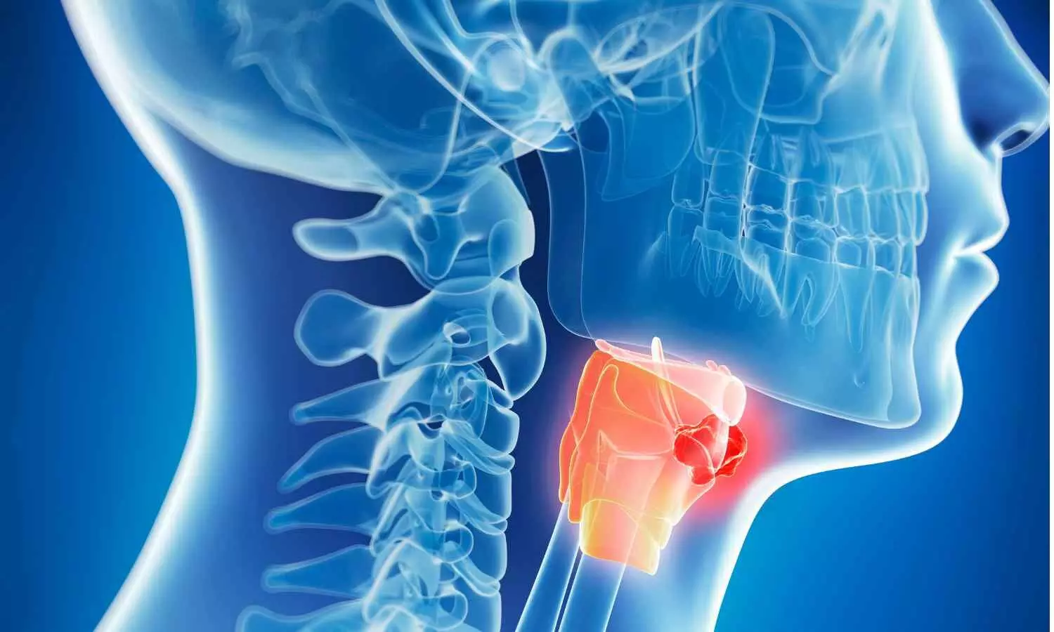
Patients with throat problems were less able to regulate their blood pressure in a new study led by the University of Southampton.

The study published in JAMA Otolaryngology is the first to observe reduced baroreflex sensitivity in patients with throat symptoms.

The baroreflex is a crucial part of the autonomic nervous system which detects changes in blood pressure and adjusts our heart rate and blood vessel tone accordingly to maintain stable blood pressure. It is what stops us from fainting when we stand up.

Researchers from the University of Southampton and University Hospitals of Dorset Foundation Trust believe the findings could be explained by the Vagas nerve (which controls the autonomic nervous system) prioritising protection of the airways over less urgent functions, such as blood pressure regulation.

“Our immediate survival depends on the throat being able to separate air and food passages each time we swallow,” says the lead author of the study Reza Nouraei, Professor of Laryngology and Clinical Informatics at the University of Southampton.

“The throat does this using delicate reflexes, but when these reflexes are disturbed, for example, due to a viral infection like Covid or exposure to reflux affecting nerves in this region, the control of this critical junction becomes compromised, giving rise to symptoms like the feeling of a lump in the throat, throat clearing and coughing.

“To compensate for a faulty throat, the autonomic control system must expend significant amounts of energy to maintain a safe airway. We found that in patients with a faulty throat, the heart, specifically a function called baroreflex, is less well controlled. This is one of the Peters that has been robbed to pay Paul.

“The problem with robbing this Peter is that it likely impacts long-term survival, as patients with reduced baroreflex function are more likely to die of a heart attack or stroke in years to come.”

The researchers compared the heart rates, blood pressure and baroreflex sensitivity of 23 patients admitted to Ear, Nose and Throat (ENT) surgery with aerodigestive (laryngopharyngeal) symptoms and 30 patients admitted to Gastroenterology with digestive (esophagogastric) symptoms at University Hospitals of Dorset NHS Foundation Trust.

Reflux was a common cause of symptoms in both groups – making up the majority of digestive group cases. Other causes like thinning of the vocal cord were present in the aerodigestive group.

The team found patients in the aerodigestive group had a higher resting heart rate, lower resting blood pressure, and lower baroreflex sensitivity, than those in the digestive group.

“Now, and especially since Covid which damages nerves, we are seeing more patients with throat symptoms,” says Professor Nouraei.

“Reduced baroreflex sensitivity impacts survival independent of other cardiovascular risks, so if the association we’ve discovered is confirmed by future studies, the need to make timely and accurate diagnoses and provide early and definitive treatments will become more pressing.”

The study adds to the increasing interest in the Vaus nerve and holistic health. As well as regulating blood pressure through the baroreflex, the Vagus nerve controls our heart rate, digestion, respiration, mood and a host of other bodily functions which affect our health and wellbeing.

Professor Nouraei says: “This study helps us to think about patients more holistically. As a clinician, if you can fix a problem in the throat that is potentially taking away bandwidth from the Vagus, then it frees up the nerve to give to the rest of the body.

“If there is a chance that throat problems can affect functions like the baroreflex, or have a wider impact on overall wellbeing, then they need more consideration.”

The researchers will now look at the long-term impacts of throat conditions on autonomic health and the effects of treatment.

Reference:

Nouraei SAR, Ayres L, Perring SJ. Baroreflex Sensitivity in Patients With Laryngopharyngeal Dysfunction—The Overwhelmed Vagus Hypothesis. JAMA Otolaryngol Head Neck Surg. Published online September 05, 2024. doi:10.1001/jamaoto.2024.2270

Powered by WPeMatico

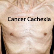
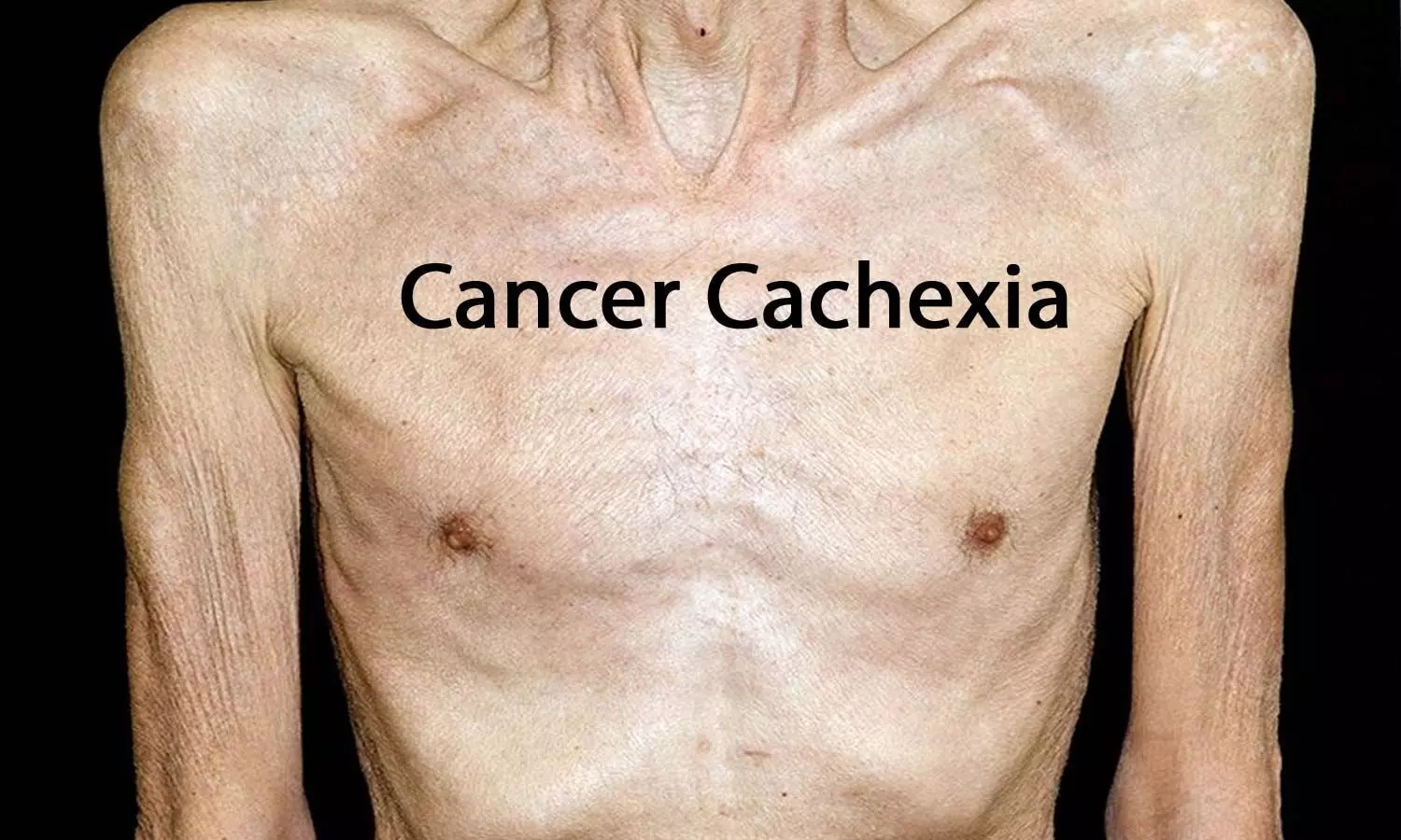
GDF-15 inhibition with ponsegromab improved cancer cachexia symptoms: NEJM

A new study by John Groarke and team found the cachexia decreased when ponsegromab was administered at escalating dosages to patients with cancer cachexia and raised circulating growth differentiation factor 15 (GDF-15) levels. The findings of this study were published in the New England Journal of Medicine.

One frequent cancer consequence that is linked to a higher risk of mortality is cachexia. In cancer cachexia, there is an increase in the amount of the circulating cytokine growth differentiation factor 15. Ponsegromab is a humanized monoclonal antibody that inhibits GDF-15 and was linked to better weight, appetite, and physical activity in addition to decreased serum GDF-15 levels.

And so, individuals with cancer cachexia and a high serum GDF-15 level (≥1500 pg per milliliter) were randomized in a phase 2 randomized, double-blind, 12-week trial in a 1:1:1:1 ratio to either receive ponsegromab at a dose of 100 mg, 200 mg, or 400 mg subcutaneously every 4 weeks for three doses, or receive a placebo. The main outcome measure at 12 weeks was the shift in body weight from the baseline. Digital assessments of physical activity, safety, and symptoms of hunger and cachexia were important secondary end goals.

The randomization was performed on a total of 187 subjects. Of these individuals, colorectal cancer accounted for 29%, pancreatic cancer for 32%, and non-small-cell lung cancer for 40%. At 12 weeks, there was a significant difference in weight increase between the ponsegromab groups and the placebo group. The median between-group difference was 1.22 kg for the 100-mg group, 1.92 kg for the 200-mg group, and 2.81 kg for the 400-mg group. Measures of hunger, symptoms of cachexia, and physical activity all showed improvements in the 400-mg ponsegromab group when compared to placebo. 70% of the patients in the ponsegromab group and 80% of the individuals in the placebo group had adverse events of any kind. Overall, the inhibition of GDF-15 with ponsegromab led to enhanced gain in weight and general activity level and lowered cachexia symptoms among patients with cancer cachexia and raised GDF-15 levels.

Source:

Groarke, J. D., Crawford, J., Collins, S. M., Lubaczewski, S., Roeland, E. J., Naito, T., Hendifar, A. E., Fallon, M., Takayama, K., Asmis, T., Dunne, R. F., Karahanoglu, I., Northcott, C. A., Harrington, M. A., Rossulek, M., Qiu, R., & Saxena, A. R. (2024). Ponsegromab for the Treatment of Cancer Cachexia. In New England Journal of Medicine. Massachusetts Medical Society. https://doi.org/10.1056/nejmoa2409515

Powered by WPeMatico