Menstrual Blood Loss may Determine Iron Status in Premenopausal Blood Donors: Study

Menstrual Blood Loss Determines Iron Status in Premenopausal Blood Donors suggests a study published in the Acta Obstetricia et Gynecologica Scandinavica.

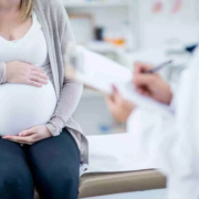
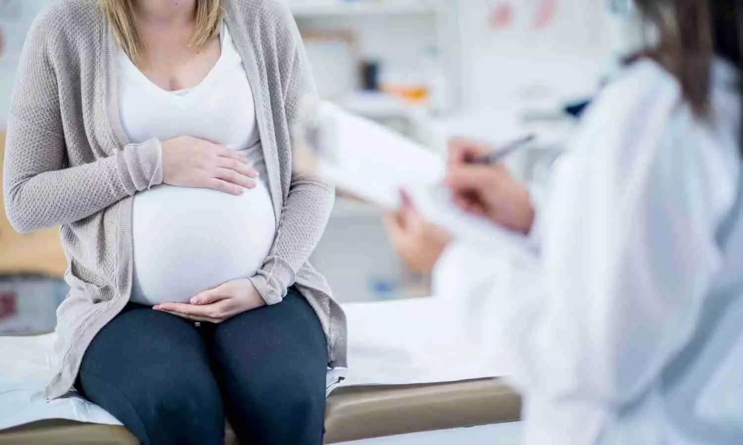
A study was done to prevent blood donors from developing iron deficiency (ferritin <15 μg/L) and subsequent anemia (hemoglobin <120 g/L), blood services rely on information about known risk factors, including the donor’s sex and age. For example, while Finnish women can donate whole blood with a minimum donation interval of 91 days, women in the 18 to 25-year-old age group are recommended to donate no more than once yearly. Menstrual blood loss is not accounted for in blood donation interval recommendations, despite being a known risk factor of iron deficiency.

We aim to investigate to what extent menstrual bleeding is associated with ferritin and haemoglobin levels in female blood donors and quantify the association of other menstruation-related variables not currently accounted for by blood services (i.e., use of hormonal contraception, heavy menstrual bleeding) with iron deficiency or anaemia. The study population comprised 473 premenopausal and 491 postmenopausal Dutch whole blood donors. Exclusion criteria were current pregnancy, BMI ≥50, ferritin ≥200, pictorial blood assessment chart (PBAC) ≥400, and age <18 or ≥70 years.

Menstrual blood loss was quantified using a PBAC, a semiquantitative method to evaluate the number of used menstrual products and the degree of staining. They identified predictors of log(ferritin)/haemoglobin and iron deficiency/anaemia using Bayesian linear and logistic regression models. They quantified the average percentage of variance in log(ferritin) and haemoglobin explained by the covariates. Results: Menstrual blood loss accounted for most of the explained variance in haemoglobin (8%) and second only to the number of days since last donation for ferritin (8%). Heavy menstrual bleeding (PBAC ≥150, OR = 3.56 [1.45-8.85], prevalence 13%) was associated with anaemia, and use of the levonorgestrel-releasing intrauterine device was negatively associated with iron deficiency (OR = 0.06 [0.01-0.44]). After statistical control for menstrual blood loss, age was not associated with iron status. Menstrual blood loss and blood donation were the most important determinants of iron status in premenopausal women. Thus, results suggest that accounting for menstrual blood loss in donation interval guidelines may benefit blood donors.

Reference:

Ekroos S, Karregat J, Toffol E, Castrén J, Arvas M, van den Hurk K. Menstrual blood loss is an independent determinant of hemoglobin and ferritin levels in premenopausal blood donors. Acta Obstet Gynecol Scand. Published online June 10, 2024. doi:10.1111/aogs.14890

Powered by WPeMatico

ADHD meds may help pregnant patients control opioid use disorder: Study

Opioid overdoses for pregnant patients are at an all-time high in the United States, even as overall numbers are improving. Attention deficit hyperactivity disorder (ADHD) is highly correlated with substance use disorders, yet treatment protocols to help expecting parents manage opioid use disorders and ADHD together are essentially nonexistent.

New research from Washington University School of Medicine in St. Louis may help change that. A study published in Nature Mental Health indicates that patients with opioid use disorders and ADHD who remain on their ADHD medications during their pregnancies are far more likely to adhere to treatment for opioid use, and far less likely to overdose, than are patients who stop taking ADHD medications.

This research is a step toward developing urgently needed resources and treatment guidelines, said Kevin Xu, MD, an assistant professor of psychiatry and the study’s first author, who is also the director for substance use disorders rotation for Washington University’s psychiatry residency program. “We have really never seen such rates of overdose among reproductive-age and pregnant people,” Xu said.

According to data from the National Institutes of Health (NIH), rates of overdose deaths doubled among pregnant and postpartum people in recent years (rising from 3.1 deaths per 100,000 in 2018 to 6.1 deaths per 100,00 in 2021). Opioid overdose accounts for about 10% of all pregnancy-related deaths. Despite research indicating that nearly one in four people diagnosed with ADHD also has a substance use disorder, there is little research available to physicians or their patients to guide how they can safely manage both conditions during pregnancy.

That lack of research struck psychiatry resident Tiffani Berkel, MD, PhD, as she was trying to advise patients navigating their pregnancies, which led her to approach Xu and propose this study.

“It’s very common for pregnant patients to ask their doctors, ‘Is this medication safe?’” Berkel said. “The physicians have to say, ‘We don’t know.’ That’s not very reassuring to a pregnant person. They have to do this risk-benefit analysis themselves.”

By examining de-identified prescription and Medicaid databases, Berkel, Xu and their co-authors focused on 168 pregnant patients who were receiving methadone or buprenorphine for opioid use disorder treatment and taking medications for ADHD. They analyzed how long the patients maintained their opioid use disorder treatments and how often they required emergency room care related to opioid use.

Because methadone clinics frequently require patients to stop taking any ADHD medications, there were not enough patients in that group to do a proper analysis. For patients treated with buprenorphine, the differences between patients who remained on their ADHD medications during their pregnancies and those who discontinued were striking: Those who continued with their ADHD medications stayed roughly two months longer on buprenorphine than did patients no longer taking ADHD medications. In line with that result, the researchers found there were fewer emergency room visits related to substance use disorder among patients taking ADHD medications: 41% of patients who continued their ADHD treatments went to the ER compared with 54% of those who stopped.

The reasons for the difference will need further investigation, but Berkel said one potential explanation is that ADHD drugs help control impulsivity and are therefore likely to improve patients’ ability to manage their substance use disorder treatments – with the additional benefit of improved attendance for regular pregnancy checkups as well.

Co-author Jeannie Kelly, MD, an associate professor of obstetrics and gynecology in the division of Maternal Fetal Medicine, said that research like this serves a real-world medical need to better understand the potential consequences of discontinuing ADHD treatments for patients with substance use disorders. Kelly treats patients in the Barnes Jewish Hospital, as do Xu and Berkel.

“Treatment of ADHD is a huge knowledge gap in obstetrics, and even more so in patients with substance use disorder,” Kelly said. “In obstetrics, a knowledge gap frequently leads to reluctance to treat because of unknown risks to the fetus. However, it’s also really important to discuss the risks of not treating, because untreated disease also can have huge implications for mom’s and baby’s health.”

Reference:

Xu, K.Y., Berkel, T.D.M., Martin, C.E. et al. Prescription psychostimulant use, admissions and treatment initiation and retention in pregnant people with opioid use disorder. Nat. Mental Health (2024). https://doi.org/10.1038/s44220-024-00270-w.

Powered by WPeMatico

Cranial Nerve Palsy Linked to Increased Stroke Risk in Young Adults: Study

Researchers have found that cranial nerve palsy (CNP) affecting the third, fourth, or sixth cranial nerve is associated with a higher risk of stroke, particularly among young adults. This association was strongest in individuals in their 30s, suggesting that early detection and monitoring could be crucial for preventing stroke in this demographic. A recent study was published in the Journal of The American Heart Association by Daye Diana Choi and colleagues.

CNP can impact ocular motor function, leading to symptoms such as double vision and difficulty with eye movement. While CNP is often considered a localized neurological issue, emerging evidence suggests that it may also be a marker for broader vascular concerns, including stroke. This study aimed to explore the relationship between CNP and stroke risk, with a focus on how age influences this association.

The cohort study included individuals diagnosed with third, fourth, or sixth CNP who participated in national health screenings within two years of diagnosis between 2010 and 2017. A control group, matched by sex and age at a 1:5 ratio, was used for comparison. Participants were followed until December 31, 2019. Using multivariable Cox proportional hazards regression analyses, the study assessed the risk of stroke associated with CNP, adjusting for covariates such as lifestyle, health behavior, underlying comorbidities, and Charlson comorbidity index scores.

The study included a significant cohort of individuals, with those diagnosed with ocular motor CNP demonstrating a higher risk of stroke compared to the control group (hazard ratio [HR], 1.23 [95% CI, 1.08-1.39]). Notably, the risk of stroke varied significantly with age:

  • 30s: The risk of stroke increased by 8.91 times (HR, 8.91 [95% CI, 1.63-48.66]).

  • 40s: The risk increased by 2.49 times (HR, 2.49 [95% CI, 1.39-4.45]).

  • 50s: The risk increased by 1.78 times (HR, 1.78 [95% CI, 1.31-2.42]).

  • 60s: The risk increased by 1.32 times (HR, 1.32 [95% CI, 1.08-1.62]).

  • 20s, 70s, and 80s: No significant increase in stroke incidence was observed in these age groups.

The findings underscore a pronounced age-related variation in stroke risk associated with CNP, with the most substantial risk observed in younger adults, particularly those in their 30s. This elevated risk highlights the importance of vigilant cardiovascular monitoring and preventive strategies in younger individuals diagnosed with CNP. The lack of significant risk increase in the very young (20s) and older age groups (70s and 80s) suggests that age-specific factors may modulate the vulnerability to stroke post-CNP diagnosis.

This cohort study establishes a clear association between ocular motor CNP and an elevated risk of stroke, particularly in younger adults. These findings emphasize the need for heightened awareness and preventive measures in patients with CNP to mitigate the risk of stroke.

Reference:

Choi, D. D., Cheon, D. Y., Park, K.-A., Han, K., Jung, J.-H., & Oh, S. Y. (2024). Age‐related risk of stroke following ocular motor cranial nerve palsy. Journal of the American Heart Association, 13(12). https://doi.org/10.1161/jaha.123.033437

Powered by WPeMatico

Study evaluates Ultrasound-Guided Upper Thoracic Erector Spinae Plane Block for Shoulder Surgery

The upper thoracic (T2) erector spinae plane block (UT-ESPB) has been suggested as a substitute for the interscalene brachial plexus block for pain management after shoulder surgery. Recent prospective exploratory study examined the efficacy of ultrasound-guided upper thoracic (T2) erector spinae plane block (UT-ESPB) for postoperative analgesia in patients undergoing proximal shoulder surgery under general anesthesia. The primary outcome was the incidence of phrenic nerve palsy, while secondary outcomes included block characteristics, postoperative pain scores, analgesic requirements, and quality of recovery.

A total of 43 patients were included in the study. The UT-ESPB was performed by injecting 0.4 mL/kg of 0.25% bupivacaine with 4 mg of dexamethasone at the T2 level. Diaphragmatic movements were assessed using ultrasound to detect phrenic nerve palsy.

The results showed that the incidence of phrenic nerve palsy was 0%. The sensory level achieved by the maximum number of patients at 30 minutes was C7-T5, and none of the patients had any motor block in the upper limb. Forty-two percent of patients did not require any rescue analgesia in the first 24 hours after surgery. In the remaining patients, the mean duration of analgesia was 724.2 ± 486.80 minutes, and the mean postoperative fentanyl requirement was 98.80 ± 47.02 μg. The median pain scores at rest and during movement were 2-3 and 3-4, respectively. The median quality of recovery score at 24 hours was 14 (15-14).

The authors concluded that the UT-ESPB resulted in a sensory loss from C7-T5 dermatomes without causing any weakness of the diaphragm or upper limb. However, the block was only moderately effective in terms of the total duration of analgesia, postoperative pain scores, analgesic requirement, and quality of recovery in patients undergoing proximal shoulder surgeries under general anesthesia. The authors suggested that further studies are required to establish the role of UT-ESPB due to the poor correlation between the sensory spread and clinical outcomes.

The study provides valuable insights into the use of UT-ESPB for shoulder surgery, highlighting its potential benefits in avoiding phrenic nerve palsy while also acknowledging the need for more research to optimize the analgesic efficacy of this regional anesthesia technique.

Key Points –

Here are the 3 key points from the research paper:

1. The study examined the efficacy of ultrasound-guided upper thoracic (T2) erector spinae plane block (UT-ESPB) for postoperative analgesia in patients undergoing proximal shoulder surgery under general anesthesia. The primary outcome was the incidence of phrenic nerve palsy, while secondary outcomes included block characteristics, postoperative pain scores, analgesic requirements, and quality of recovery.

2. The results showed that the incidence of phrenic nerve palsy was 0%, the sensory level achieved by the maximum number of patients at 30 minutes was C7-T5, and 42% of patients did not require any rescue analgesia in the first 24 hours after surgery. However, the block was only moderately effective in terms of the total duration of analgesia, postoperative pain scores, analgesic requirement, and quality of recovery.

3. The authors concluded that the UT-ESPB resulted in a sensory loss from C7-T5 dermatomes without causing any weakness of the diaphragm or upper limb, but further studies are required to establish the role of UT-ESPB due to the poor correlation between the sensory spread and clinical outcomes.

Reference –

Kumar D, Talawar P, Dhar M, Azam Q, Tripathy DK, Singla D, et al. The efficacy of ultrasound‑guided upper thoracic erector spinae plane block for postoperative analgesia in proximal shoulder surgery and its effect on phrenic nerve function: A prospective exploratory study. J Anaesthesiol Clin Pharmacol 2024;40:312‑7.

Powered by WPeMatico

Psoriasis potential risk factor for infective endocarditis among patients with permanent pacemaker: Study

Researchers have found that psoriasis significantly increases the risk of developing infective endocarditis (IE) in patients with permanent pacemakers (PPM). A recent study was published in the International Journal Of Dermatology by Chapman Wei and colleagues. This study highlights the need for heightened awareness and potential prophylactic measures to mitigate IE risk in this patient population.

Device-related infective endocarditis (IE) is a severe condition with high mortality rates. Identifying comorbidities that heighten IE risk is crucial for improving patient outcomes. Psoriasis, a chronic inflammatory skin condition, is increasingly recognized for its cardiovascular implications. However, its impact on IE risk in patients with PPM has not been well studied until now.

This study utilized the National Inpatient Sample database to analyze patients with PPM, distinguishing those with and without psoriasis. Demographic and comorbidity data were collected, and a 1:10,000 propensity matching for IE risk factors was performed. This approach aimed to identify independent associations between psoriasis and IE.

  • Out of 437,793 patients with PPM, 45 had psoriasis.

  • The study revealed a significantly higher rate of IE in psoriasis patients compared to those without psoriasis (4.4% vs. 0.6%; P < 0.01).

  • Multivariate analysis indicated that psoriasis patients had a 7.2-fold increased risk of developing IE (OR: 7.2 [1.7-30.2]; P < 0.01).

  • Further, post-match analysis showed an 8.3-fold increase in IE risk for psoriasis patients (OR: 8.3 [2.0-34.4]; P < 0.001).

The findings from this study demonstrate that psoriasis is a significant independent risk factor for IE in patients with PPM. This increased risk necessitates further research to corroborate these findings and explore potential prophylactic measures to prevent IE in this vulnerable population. Given the chronic inflammatory nature of psoriasis and its systemic effects, the association with heightened IE risk underscores the need for comprehensive cardiovascular care in psoriasis patients with PPM.

Psoriasis independently increases the risk of infective endocarditis in patients with permanent pacemakers. These findings suggest a need for further studies to confirm the association and develop targeted prophylactic strategies. Understanding the link between psoriasis and IE could lead to improved management and outcomes for patients with both conditions.

Reference:

Wei, C., Mustafa, N., Grovu, R., Wei, C., Rizvi, T., Bradu, S., & Mustafa, A. (2024). Impact of psoriasis on the risk of device‐related infective endocarditis in patients with permanent pacemakers: a propensity‐matched analysis. International Journal of Dermatology. https://doi.org/10.1111/ijd.17292

Powered by WPeMatico

Air pollution can increase cardiovascular risk for cancer patients, finds study

Modern therapies have extended the lives of many cancer patients; however, survivors often live with chronic health conditions, including cardiovascular disease. New research published in JACC: Cardio Oncology shows that air pollution plays a significant role in increasing cardiovascular disease and mortality in cancer patients and contributes to health disparities related to these conditions.

“The review underscores the critical need to consider environmental factors, especially air pollution, in cardio-oncology risk assessment and patient management,” said Xiaoquan Rao, MD, PhD, senior author of the study and a cardiologist at Tongji Hospital at the Tongji Medical College in Wuhan, China.

“By highlighting the significant role of air pollution in the cardiovascular health of cancer patients, our work aims to catalyze further research in this field and inform clinical practices and public health policies,” Rao said.

Rao noted that air pollution has been recognized as a significant risk factor for both cardiovascular diseases and cancer; however, little research has been done to study its effects, specifically in cardio-oncology or the overlap of both diseases. The new study was motivated by a need to fill that gap, Rao said.

Researchers reviewed papers published between 2000 and 2023 and found eight studies that directly explored the combined effect of air pollution on cardiovascular disease and cancer.

They found that exposure to PM2.5, or fine particulates in the air, was significantly associated with higher rates of both incidence and mortality of cardiovascular disease among cancer patients — and vice versa. Rao noted that air pollution exposure appears to impact multiple common risk factors shared by both cancer and cardiovascular disease, including inflammatory and oxidative stress pathways.

According to researchers, a surprising finding was that even short-term exposure to high pollution levels rapidly impacted cancer patients’ cardiovascular health.

“This suggests that even temporary deteriorations in air quality can have immediate adverse effects on vulnerable populations such as cardio-oncology patients,” Rao said.

The paper also highlights how air pollution contributes to health disparities worldwide. Disadvantaged populations are exposed to higher levels of air pollution, and cancer patients with a lower socioeconomic status face a higher risk of cardiovascular disease mortality linked to air pollution than the general public, researchers said.

“More research is needed, including clinical studies, to understand the impacts of air pollution on cardiovascular disease and cancer in greater detail,” Rao said.

Rao added that the findings can be used to better understand the risks of air pollution and help identify more vulnerable populations within cardio-oncology.

“This awareness is crucial for developing tailored air pollution exposure control measures and individualized patient management strategies aimed at mitigating cardiovascular disease risks among cancer patients,” Rao said.

Future research will focus on exploring air pollution-related health disparities across different types of cancer and cardiovascular diseases. Researchers also hope to evaluate the effectiveness of environmental interventions in reducing the impact of air pollution on cardio-oncology patients.

Reference:

Zhu, W, Al-Kindi, S, Rajagopalan, S. et al. Air Pollution in Cardio-Oncology and Unraveling the Environmental Nexus: JACC: CardioOncology State-of-the-Art Review. J Am Coll Cardiol CardioOnc. 2024 Jun, 6 (3) 347–362.https://doi.org/10.1016/j.jaccao.2024.04.003

Powered by WPeMatico

New, simple test detects rare fatal genetic heart condition, reveals research

A team of international researchers has revealed a new, simple clinical test to detect Calcium Release Deficiency Syndrome (CRDS), a life-threatening genetic arrhythmia that causes dangerously fast heartbeats and can lead to severe complications such as sudden cardiac arrest and death.

The new diagnostic method monitors for changes in electrocardiography (ECG) after a brief period of a fast heartbeat and a pause, which can occur naturally or be induced by artificially pacing the heart.

This research was co-led by Jason Roberts, a scientist at the Population Health Research Institute (PHRI), a joint institute of McMaster University and Hamilton Health Sciences, and Wayne Chen, a scientist and professor at the Libin Cardiovascular Institute and Hotchkiss Brain Institute at the University of Calgary’s Cumming School of Medicine, and published in JAMA.

While 60,000 cardiac arrests occur annually in Canada, CRDS remains undetectable with standard clinical tests, often resulting in the cardiac arrests being labeled as unexplained. This gap in understanding the underlying cause prevents the delivery of optimal care to survivors and vulnerable family members who may be affected by this genetic condition.

“This novel and simple diagnostic method, which can be performed using an electrocardiogram in a broad range of clinical settings, is hopefully an important step towards improving our evaluation of initially unexplained cardiac arrest (UCA),” said Roberts, co-principal investigator of the study.

Roberts and Chen led this multi-centre case-control study involving 68 study participants from 10 centres in seven countries. The participants were from four categories of heart conditions, including CRDS patients and UCA survivors. Accompanying studies from Chen’s lab revealed findings in genetic mouse models that mirrored those observed in humans. The mouse studies also provided insight into the underlying cellular mechanism responsible for this apparent ECG signature of CRDS.

“CRDS has been linked to many tragic incidents and heartbreaking stories affecting families. There have been numerous cases where patients experienced fainting episodes, but their tests showed no issues, which led doctors to believe the fainting was not due to a dangerous heart condition. A portion of these individuals, often young and otherwise healthy, subsequently suffered sudden cardiac arrests, and some did not survive,” said Roberts.

“This raised many questions in the medical community, most of which remained unanswered until Chen and his team established the existence of this rare genetic syndrome in 2021. However, the diagnosis has required specialized laboratory testing available only in research settings, making it inaccessible to most physicians and limiting the ability to care for potentially affected patients and their families.”

The study marks the initial stage of Roberts and Chen’s efforts in further exploring a diagnostic approach for CRDS and collecting additional data as part the ongoing PHRI DIAGNOSE CRDS trial. This trial represents a broader, international initiative aimed at strengthening their conclusions. Currently in the recruitment phase, DIAGNOSE CRDS aims to enroll 500 participants from 30 sites across 10 countries.

“We hope this test will help many families worldwide who have faced unexplained cardiac incidents or lost loved ones to them” added Roberts.

The team anticipates this simple pacing test will be incorporated into the routine diagnostic tests for initially unexplained cardiac arrest, providing hope for better outcomes and prevention of future tragedies.

“This is an important discovery because there is an urgent need for a clinical diagnostic test for CRDS,” says Chen. “This will allow us to identify individuals at risk, better understand the prevalence of CRDS and, hopefully, develop treatments for the condition.”

Reference:

Ni M, Dadon Z, Ormerod JOM, et al. A Clinical Diagnostic Test for Calcium Release Deficiency Syndrome. JAMA. Published online June 20, 2024. doi:10.1001/jama.2024.8599.

Powered by WPeMatico

Rare Case: Renal Sarcoidosis with Parotid and Uterine Complications

Japan: Renal sarcoidosis, a rare manifestation of sarcoidosis affecting the kidneys, has presented a challenging clinical scenario compounded by the involvement of the parotid glands and uterus in a recent case study. This complex interplay of symptoms underscores the intricate nature of the disease and highlights the importance of comprehensive management strategies.

The recent case study published in BMC Nephrology reported the first case of renal sarcoidosis complicated by parotid glands and uterus lesions. Pathological findings and the reactivity in Ga scintigraphy indicated the lesions’ presence in these organs.

Sarcoidosis is a systemic condition capable of impacting various organs. While pulmonary involvement is the most prevalent, renal manifestations are less common. Hirotaka Fukasawa, Iwata City Hospital, Iwata, Shizuoka, Japan, and colleagues reported a case of sarcoidosis with an exceptionally rare distribution, including renal lesions.

The case concerns a 51-year-old Japanese woman who was referred to the hospital due to bilateral parotid swelling and renal dysfunction without a history of previous renal issues. On physical examination, she exhibited bilateral parotid swelling, and lower leg edema, and reported hypermenorrhea. Laboratory tests revealed elevated serum creatinine (3.1 mg/dL), serum corrected calcium (10.6 mg/dL), serum angiotensin-converting enzyme (ACE) levels (44.9 U/L), and lysozyme levels (75 µg/mL). Urinary β2-microglobulin (β2-MG) levels were also elevated. Computed tomography (CT) scans showed swelling in both kidneys, parotid glands, and the uterus, confirmed by Ga scintigraphy, which revealed significant accumulation in these organs. Ocular screening for uveitis was negative.

During hospitalization, needle aspiration cytology of the parotid gland and endometrial cytology confirmed the presence of epithelioid cells indicative of sarcoidosis. Renal biopsy revealed tubulointerstitial nephritis with diffuse non-caseating epithelioid cell granulomas and multinucleated giant cells infiltrating the interstitial area and renal tubules. Immunohistochemical staining, including CD68, showed positive results in macrophages. Staining for 1-α hydroxylase (CYP27B1) and lysozyme also displayed remarkable positivity in multinucleated giant cells, and infiltrating cells, consistent with renal sarcoidosis diagnosis with parotid gland and uterine involvement.

Treatment commenced with prednisolone at 40 mg/day (0.8 mg/kg/day) from the 11th day of admission, resulting in rapid improvement of renal function, hypercalcemia, and reductions in urinary β2-MG, serum ACE, serum lysozyme, and serum 1,25(OH)2 vitamin D3 levels. A follow-up Ga scintigraphy showed the resolution of the previous organ accumulations. The patient was discharged on the 53rd day with a stable condition and maintained serum ACE levels within the normal range (7.2–18.4 U/L) for at least 30 months post-treatment.

In conclusion, the researchers documented a novel case of renal sarcoidosis with complications involving the parotid glands and uterus, an exceptionally rare distribution of lesions in sarcoidosis. Additionally, staining for 1-α hydroxylase and lysozyme revealed positive results in infiltrating cells, likely macrophages within renal granulomas. The presence of 1-α hydroxylase in these cells suggests a potential mechanism for hypercalcemia, while lysozyme positivity may serve as a supportive diagnostic marker for sarcoidosis in future cases.

Reference:

Kitamoto, S., Kaneko, M., Omata, K. et al. A case of renal sarcoidosis complicated by parotid gland and uterine lesions. BMC Nephrol 25, 198 (2024). https://doi.org/10.1186/s12882-024-03635-6

Powered by WPeMatico

Early Menopause and Hormone Therapy may Impact Lung Health: Insights from PLCO Trial Analysis

USA: A secondary analysis of data from the PLCO Trial has revealed intriguing insights into the relationship between early menopause, hormone therapy, and lung health outcomes. Published in the BMJ journal Thorax, the study underscores the potential impact of hormonal factors on respiratory health, adding a new dimension to understanding lung disease risks.

The researchers suggest that smoking with early natural menopause (early-M; <45 years of age) should be targeted for smoking cessation and lung cancer (LC) screening, irrespective of menopause type. The users of menopausal hormone therapy (MHT) had a lower likelihood of dying from lung cancer and respiratory diseases in ever smokers.

“Early menopause due to bilateral oophorectomy was associated with an increased risk of respiratory mortality,” the researchers reported. “That increased risk was observed both in ever smokers (HR 1.98) and never smokers (HR 1.91).”

Early natural menopause raises the risk of lung morbidities and mortalities in smokers. However, it is not known whether early-M due to surgery demonstrates similar effects and whether MHT protects against lung diseases. To fill this knowledge gap, Xiaochun Gai, Department of Internal Medicine, University of New Mexico School of Medicine, Albuquerque, New Mexico, USA, and colleagues aimed to assess the associations of early natural menopause and MHT with lung morbidities and mortalities using the prospective Prostate, Lung, Colorectal, and Ovarian (PLCO) trial.

For this purpose, the research team estimated the risk among 69,706 postmenopausal women in the PLCO trial, stratified by menopausal types and smoking status.

The study revealed the following findings:

  • Early M was associated with an increased risk of most lung disease and mortality outcomes in ever smokers with the highest risk seen for respiratory mortality (HR 1.98) in those with bilateral oophorectomy (BO).
  • Early-M was positively associated with chronic bronchitis, and all-cause, non-cancer, and respiratory mortality in non-smokers with natural menopause or BO, with the highest risk seen for BO— respiratory mortality (HR 1.91).
  • Ever MHT was associated with reduced all-cause, non-cancer, and cardiovascular mortality across menopause types irrespective of smoking status and was additionally associated with reduced risk of lung cancer, non-ovarian cancer, and respiratory mortality in ever smokers.
  • Among smokers, ever MHT use was associated with a reduction in HR for all-cause, non-cancer, and cardiovascular mortality in a duration-dependent manner.

As the scientific community continues to unravel the complex interplay between menopause, hormone therapy, and overall health, insights from studies like this underscore the importance of personalized healthcare approaches tailored to women’s unique hormonal profiles and medical histories. This research opens avenues for future investigations into optimizing respiratory health strategies for women transitioning through menopause.

Reference:

Gai X, Feng Y, Flores TM, et alEarly menopause and hormone therapy as determinants for lung health outcomes: a secondary analysis using the PLCO trialThorax Published Online First: 13 June 2024. doi: 10.1136/thorax-2023-220956

Powered by WPeMatico

Adjunctive systemic antibiotics fail to prevent additional surgical intervention after non-surgical peri-implantitis treatment: Study

Adjunctive systemic antibiotics fail to prevent additional surgical intervention after non-surgical peri-implantitis treatment suggests a study published in the Journal of Clinical Periodontology.

The aim of this retrospective long-term follow-up of a 3-month RCT was to assess whether non-surgical peri-implantitis treatment with adjunctive systemic antibiotics influenced the need for additional surgical treatment. Patients enrolled in an aftercare programme following non-surgical peri-implantitis treatment with or without systemic amoxicillin and metronidazole, were analysed. Data had previously been collected pre-treatment (T0) and 3 months after treatment (T1) and were additionally collected during subsequent aftercare visits, until the final assessment (T2). Primary outcome was the need for additional surgical peri-implantitis therapy during the aftercare programme, analysed via Kaplan–Meier analysis and Cox regression. Secondary outcomes involved clinical parameters, assessed using parametric and non-parametric tests. Results: Forty-five patients (22 AB− group, 23 AB+ group) were included. The mean follow-up time between T1 and T2 was 35.9 months (SD = 21.0). 73.9% of the AB+ group and 50.0% of the AB− group did not receive additional surgical therapy (log-rank test, p = .110). The adjusted Cox regression model did not provide a significant result for antibiotics (β = .441, 95% CI = 0.159–1.220, p = .115). Univariable regression analysis highlighted the influence of baseline peri-implant pocket depth on the need for surgical treatment (β = 1.446, 95% CI = 1.035–2.020, p = .031). Systemic amoxicillin and metronidazole administered during non-surgical peri-implantitis treatment do not seem to prevent the need for additional surgical therapy in the long term, during a structured aftercare programme.

Reference:

Hakkers, J., Vangsted, T. E., van Winkelhoff, A. J., & de Waal, Y. C. M. (2024). Do systemic amoxicillin and metronidazole during the non-surgical peri-implantitis treatment phase prevent the need for future surgical treatment? A retrospective long-term cohort study. Journal of Clinical Periodontology, 1–8. https://doi.org/10.1111/jcpe.14024

Keywords:

Adjunctive, systemic, antibiotics, fail, prevent, additional, surgical, intervention, non-surgical, peri-implantitis, treatment,study, Hakkers, J., Vangsted, T. E., van Winkelhoff, A. J., & de Waal, Y. C. M

Powered by WPeMatico