Families of Men with azoospermia and severe oligozoospermia more exposed to cancer risk: Study

A recent study published in the Human Reproduction found the familial multicancer patterns among men with azoospermia and severe oligozoospermia by offering valuable insights into the heterogeneity of cancer risk within these subfertility groups. The research utilized extensive population data to analyze cancer risks among families of subfertile men.

This study involved a retrospective cohort of 786 subfertile men including azoospermic and severely oligozoospermic individuals who were matched with fertile population controls. The research identified family members up to third-degree relatives for both subfertile men and controls with over 337,754 individuals over a period from 1966 to 2017.

The findings revealed distinct familial cancer patterns in the azoospermia and severe oligozoospermia cohorts that indicates variations in cancer risk not only between different types of subfertility but also within subfertility types. Also, this study identified significant increases in cancer risks in specific types among both cohorts when compared to control families.

In the azoospermia cohort, the increased risks were observed for bone and joint cancers, soft tissue cancers, uterine cancers, Hodgkin lymphomas, and thyroid cancer. The severe oligozoospermia cohort showed higher risk for colon cancer, bone and joint cancers and testis cancer, along with a decreased risk of esophageal cancer.

This analysis identified multiple clusters of familial multicancer patterns within both cohorts, with some expressing elevated cancer risks across various types. Certain clusters expressed increased odds of cancer diagnoses at young ages by including adolescent and young adult diagnoses, as well as pediatric cancer diagnoses.

Despite the strengths of the study, including comprehensive population data, the outcomes acknowledge limitations such as the lack of semen measures for fertile men and the absence of information on medical comorbidities and lifestyle factors. The implications of these findings are significant by offering a new opportunity for focused gene discovery and environmental risk factor studies.

Source:

Ramsay, J. M., Madsen, M. J., Horns, J. J., Hanson, H. A., Camp, N. J., Emery, B. R., Aston, K. I., Ferlic, E., & Hotaling, J. M. (2024). Describing patterns of familial cancer risk in subfertile men using population pedigree data. Human Reproduction, dead270. https://doi.org/10.1093/humrep/dead270

Powered by WPeMatico

Infection prevention and control for Ebola and Marburg disease: WHO guideline

Ten years on from the West African Ebola outbreak, the World Health Organization (WHO) updated its infection prevention and control guidelines for Ebola and Marburg disease.

The key recommendations are summarised in The BMJ today with an infographic and the full guideline is available on both the WHO website and the web based MAGICapp platform.

Case fatality rates for both Ebola and Marburg disease are high, averaging about 50% (and can range from 24% to 90%). Up to date, evidence based IPC guidelines are thus critical to ensuring a safe, systematic, and standardised approach during outbreaks.

The new guideline was developed by a group of experts and a patient representative supported by the WHO secretariat and reflects the knowledge and experience garnered from multiple outbreaks of Ebola and Marburg disease since 2014.

The updated guideline includes 11 new recommendations, 10 new good practice statements, and nine recommendations from previous guidelines to assist health and care workers implement effective infection prevention and control (IPC) measures and reduce the risk of Ebola and Marburg virus transmission.

The guideline development group prioritised 13 key questions and five background questions, and WHO commissioned systematic reviews to inform the formulation of the recommendations.

The group also carefully considered the balance between desirable and undesirable effects of interventions, the certainty of evidence, the evaluation of outcomes, resource use, the acceptability and feasibility of interventions to affected populations, and the impact of interventions on equity.

Although the recommendations pertain to all healthcare settings, some are also relevant to community settings-for example, when interacting in homes of individuals suspected or confirmed to have had Ebola or Marburg disease.

Updates include:

• Details of personal protective equipment (PPE) that should be worn for specific activities or risks (such as screening, triage, direct or indirect patient care, cleaning or other hygiene activities, and safe and dignified burial)

• Recommendations on the IPC ring approach (rapidly mobilising teams to enhance IPC activities in geographical “at risk” areas around infected individuals)

• A strong recommendation against spraying health and care workers with disinfectants such as chlorine who have direct or indirect contact with patients who have Ebola or Marburg disease during the removal of personal protective equipment

• Specific details of when to use single or double gloves (including heavy duty gloves) on the basis of activity risk, methods for glove disinfection, and changing of gloves between patients

The authors acknowledge that although the new guideline represents an advance, it also highlights the need for more evidence to inform effective IPC measures during outbreaks of filoviruses.

WHO calls for greater investment and engagement in research that will provide a stronger evidence base for IPC for Ebola and Marburg disease, and thus help direct future guidelines away from deep seated practices towards stronger evidence based practices.

Reference:

Willet V, Dixit D, Fisher D, Bausch D G, Ogunsola F, Khabsa J et al. Summary of WHO infection prevention and control guideline for Ebola and Marburg disease: a call for evidence based practice BMJ 2024; 384 :p2811 doi:10.1136/bmj.p2811.

Powered by WPeMatico

Acetazolamide ups rate of successful decongestion in acute decompensated heart failure, regardless of renal function: ADVOR trial

Belgium: Findings from the ADVOR trial have revealed the safety and efficacy of acetazolamide for treating congestion across the entire range of renal function; more pronounced beneficial effects are seen in patients with a lower estimated glomerular filtration rate (eGFR).

The researchers also revealed that acetazolamide treatment raises the risk of worsening renal outcomes during the treatment phase, which does not portend adverse outcomes as long as decongestion is achieved. The study findings were published online in the European Heart Journal.

Previous studies have shown that about 50% of patients with heart failure have chronic kidney disease (CKD), defined as eGFR< 60 mL/min/1.73 m2, which is linked with an impaired diuretic response. In such patients, achieving decongestion becomes a more arduous task. Additionally, worsening renal function (WRF), assessed by an increase in serum creatinine, is shown in up to 20%–40% of patients with acute decompensated heart failure (ADHF). Although highly prevalent, few trials have investigated diuretic strategies in patients with impaired renal function.

In the ADVOR trial in which 82% of patients had CKD, adding acetazolamide to standardized IV doop diuretics improved diuretic efficacy in patients with ADHF and volume overload, leading to a higher incidence of successful decongestion and shorter hospital stay.

In the ADVOR trial, acetazolamide improved decongestion in ADHF. There is no clarity on whether the beneficial effects of acetazolamide are consistent across the entire range of renal function. Additionally, limited information is available about the acetazolamide effect on renal function and the potential prognostic meaning of these treatment-induced alterations. Therefore, Wilfried Mullens, Hasselt University, Universiteitslaan, Diepenbeek, Belgium, and colleagues conducted a pre-specified subanalysis addressing these questions.

In the pre-specified analysis of the ADVOR trial, the researchers randomized 519 ADHF patients to IV acetazolamide or matching placebo on top of intravenous loop diuretics. The primary endpoints of diuresis, decongestion, clinical outcomes, and natriuresis are assessed according to baseline renal function. Renal function changes were evaluated between treatment arms.

The study led to the following findings:

  • The median estimated glomerular filtration rate (eGFR) was 40 mL/min/1.73 m²on admission.
  • Acetazolamide consistently increased the likelihood of decongestion across the entire spectrum of eGFR.
  • Natriuresis and diuresis were higher with acetazolamide, with a higher treatment effect for patients with low eGFR.
  • Acetazolamide was associated with a higher incidence of worsening renal function (WRF; rise in creatinine ≥ 0.3 mg/dL) during the treatment period (40.5% versus 18.9%), but there was no difference in creatinine after three months. This was not associated with a higher incidence of heart failure hospitalizations and mortality.
  • Decongestion at discharge was associated with a lower incidence of adverse clinical outcomes, irrespective of the onset of WRF.

“The findings showed that acetazolamide is linked to a higher rate of successful decongestion across the entire range of renal function; more pronounced effects regarding natriuresis and diuresis were seen in patients with a lower eGFR,” the researchers wrote.

“While worsening renal function occurred more frequently with acetazolamide, this was not associated with adverse clinical outcomes,” they concluded,

Reference:

Meekers E, Dauw J, Martens P, Dhont S, Verbrugge FH, Nijst P, Ter Maaten JM, Damman K, Mebazaa A, Filippatos G, Ruschitzka F, Tang WHW, Dupont M, Mullens W. Renal function and decongestion with acetazolamide in acute decompensated heart failure: the ADVOR trial. Eur Heart J. 2023 Oct 1;44(37):3672-3682. doi: 10.1093/eurheartj/ehad557. PMID: 37623428.

Powered by WPeMatico

Early initiation of ERT in Fabry disease can eliminate major organ damage and yield enhanced long-term benefits: Case Study

China: A recent article published in BMC Nephrology reports a case study of enzyme replacement therapy for Fabry disease (FD) presenting with proteinuria and ventricular septal thickening.

Fabry disease is an uncommon, X-linked, lysosomal storage disease that leads to defects in the glycosphingolipid metabolic pathway due to absent or deficient lysosomal α-galactosidase (α-Gal A) activity. This results in the accumulation of globotriaosylceramide (GL-3) within lysosomes in a wide range of cells, including cardiac, endothelial, corneal, and renal cells, and consequently, the progressive appearance of clinical symptoms in target organs.

As FD is a progressive condition, it is important to confirm a definitive diagnosis to have timely access to favourite monitoring, appropriate management, and supportive treatment. p1Q2EFCDXZSA

Enzyme replacement therapy (ERT), which involves the exogenous supplementation of α-Gal A enzyme has been successfully administered for treating Fabry disease.

Tianyang Ye, Department of Internal Medicine, The First Navy Hospital of Southern Theater Command, Zhanjiang, Guangdong, China, and colleagues report a case of a 37-year-old male with complaints of recurrent proteinuria and ventricular septal thickening.

A renal biopsy revealed vacuolization and foamy changes in podocytes, and the presence of zebra bodies and myelin-like bodies. α-Gal A activity of the white blood cells was very low, while the Lyso-GL-3 level was high. Additionally, genetic analysis revealed a gene variant c.902G > A p. Arg301Gln.

The patient was diagnosed with Fabry disease and subsequently received intravenous (IV) ERT with a dose of Agalsidase α (0.2 mg/kg, 17.5 mg every two weeks). During the 6-month follow-up, the values of proteinuria and ventricular septum thickness remain stable.

As FD is a progressive disease, the researchers suggest that initiating enzyme replacement therapy at an early age can effectively reduce the deposition of GL-3, attenuate the progressive clinical manifestations of FD, have the potential to eliminate major organ damage and provide greater long-term benefits.

“To our knowledge, FD exhibits a broad spectrum of phenotypic variability ranging from multiorgan involvement to diverse individual differences. Patients with FD diagnosis experience a multisystemic disorder characterized by life-threatening complications, such as progressive renal insufficiency, recurrent strokes, cardiomyopathy, and neuropathic pain, which can lead to Fabry crises,” the researchers wrote.

To conclude, the article reported a case of a 37-year-old male admitted with complaints of ventricular septal thickening and proteinuria. Subsequently, he received intravenous ERT with a dose of Agalsidase α.

“As Fabry disease is a progressive disorder, initiating ERT at an early age can effectively decrease the deposition of GL-3, attenuate the progressive clinical manifestations of FD, and provide greater long-term benefits,” the researchers wrote.

Reference:

Chen, Z., Yin, B., Jiao, J. et al. Case report: enzyme replacement therapy for Fabry disease presenting with proteinuria and ventricular septal thickening. BMC Nephrol 25, 61 (2024). https://doi.org/10.1186/s12882-024-03499-w

Powered by WPeMatico

What is effect of low-dose cadmium on airway epithelial cells in COPD patients?

Cigarette smoke exposure is associated with the development and severity of chronic obstructive pulmonary disease, or COPD, which is the third leading cause of death worldwide.

Cigarette smoke contains 2 to 3 micrograms of cadmium, a highly toxic metal and environmental pollutant, per cigarette. Burning tobacco releases cadmium oxide that can be adsorbed onto microparticles in smoke that travel deep into the lungs. Furthermore, the body is not able to remove cadmium, which accumulates in longtime smokers.

In a Scientific Reports study, University of Alabama at Birmingham researchers show how a low dose of cadmium produces a deleterious stress in lung epithelial cells, and their findings highlight potential therapeutic targets to be explored in cadmium-exposure and subsequent lung injury.

The research, led by Veena Antony, M.D., a professor in the UAB Department of Medicine, focuses on microRNA-381, and the expression of a chloride channel gene called ANO1 in lung tissue samples and airway epithelial cells. ANO1 helps produce mucus in the airway; but overproduction of mucus in chronic lung disease can lead to airway thickening and mucus blockage, adding to severity of the disease. Thus, overexpression of ANO1 can exacerbate COPD.

The UAB researchers compared lung tissue samples from nine “never” smokers, who had zero history of cigarette smoking, and lung tissue samples from 13 “ever” smokers with COPD who had a history of smoking that ranged from 15 to 25 pack years per person. One pack year is generally defined as smoking one pack of cigarettes a day for one year. The researchers found that “ever” smokers, in contrast to “never” smokers, had upregulated ANO1 expression in airway epithelial cells.

Similarly, airway epithelial cells in a bronchoalveolar lavage fluid from one non-COPD subject and one smoker with COPD showed greater ANO1 expression in the COPD-subject cells.

The researchers next tested the direct effect of very low doses of cadmium on normal human airway epithelial cells. These cells were grown on an air-liquid interface that allows the airway cells to differentiate normally. Two weeks of exposure to 0.5 or 1.0 micromolar cadmium chloride in the liquid layer increased expression of ANO1 12 to 14 times.

MicroRNAs have the ability to downregulate expression of a gene by direct interaction with that gene’s mRNA sequence. The UAB team used computer software analysis to identify microRNA-381 as the microRNA with most interaction with ANO1 mRNAs, suggesting that microRNA-381 is a negative regulator of ANO1. Some heavy metals are known to negatively regulate microRNAs.

Antony and colleagues used a synthetic inhibitor for microRNA-381 to inhibit the expression of microRNA-381 in primary human airway epithelial cells from subjects with COPD, and found that ANO1 expression was upregulated significantly. In contrast, adding a microRNA-381-mimic-a synthetic RNA that acts like microRNA-381 to increase the amount of negative regulation-to those cells decreased ANO1 expression. These results strengthened the premise of the UAB researchers that cadmium negatively regulates microRNA-381 expression to upregulate ANO1 expression in airway epithelial cells.

Lastly, researchers found that, even when primary human airway epithelial cells from subjects with COPD were also exposed to 1 micromolar cadmium chloride, the microRNA-381 inhibitor still upregulated ANO1 and the mimic still downregulated ANO1.

“Our observations from experiments involving low-dose cadmium-exposure of epithelial cells suggest that ANO1 is a direct target for miR-381, which is downregulated upon low-dose cadmium exposure,” Antony said. “Thus, cigarette-induced cadmium-toxicity may alter cellular homeostasis mechanisms at very low concentrations, and cadmium-exposure in a person with an existing pulmonary condition can have an additive or adverse effect with increased susceptibility toward infections and environmental allergens.

“This interaction of cadmium, microRNA-381 and ANO1 suggests that microRNAs may act as potential therapeutic targets to be explored further in cadmium-exposure and subsequent lung injury.”

Reference:

Singh, P., Li, F.J., Dsouza, K. et al. Low dose cadmium exposure regulates miR-381–ANO1 interaction in airway epithelial cells. Sci Rep 14, 246 (2024). https://doi.org/10.1038/s41598-023-50471-z.

Powered by WPeMatico

Diabetes independent risk factor for low back pain, disc degeneration: Study

A recent study published in the PNAS Nexus explored how diabetes affects the biomechanics of intervertebral discs that can offer crucial insights into spinal health. This study utilized advanced imaging techniques to investigate the behavior of collagen within the discs to uncover significant implications for both understanding disease mechanisms and developing potential treatments.

Synchrotron small-angle X-ray scattering was used to examine how alterations in collagen behavior contribute to changes in the ability of discs to resist compression. This research focused on comparing the discs of lean Sprague-Dawley rats to those of diabetic obese University of California Davis type 2 diabetes mellitus (UCD-T2DM) rats.

The research team identified two primary mechanisms discs from lean rats that enable resistance to compression at the nanoscale with the rotation of collagen fibrils within the annulus fibrosus and straightening and stretching of these fibrils. In discs from diabetic rats, both mechanisms were significantly impaired.

The study also revealed a 31% reduction in fibril rotation and a 30% decrease in collagen fibril strain in diabetic rat discs when compared to lean counterparts. This impairment was attributed to a stiffening of collagen fibrils that was evidenced by a 31% higher concentration of nonenzymatic cross-links in discs from diabetic rats.

Also, diabetic rat discs displayed earlier onset plastic deformations, like the fibril sliding and fibril-matrix delamination which indicates a compromised ability to withstand compression over time. These findings enables with understanding disc degeneration associated with aging and diabetes. The study paves way for targeted interventions to preserving spinal health by elucidating the key deformation mechanisms involved in whole-disc compression and highlighting how diabetes disrupts these processes.

Source:

Rosenberg, J. L., Schaible, E., Bostrom, A., Lazar, A. A., Graham, J. L., Stanhope, K. L., Ritchie, R. O., Alliston, T. N., Lotz, J. C., Havel, P. J., Acevedo, C., & Fields, A. J. (2023). Type 2 diabetes impairs annulus fibrosus fiber deformation and rotation under disc compression in the University of California Davis type 2 diabetes mellitus (UCD-T2DM) rat model. In D. Discher (Ed.), PNAS Nexus (Vol. 2, Issue 12). Oxford University Press (OUP). https://doi.org/10.1093/pnasnexus/pgad363

Powered by WPeMatico

Annual breast cancer screening beginning at 40 saves lives

Annual breast cancer screening beginning at age 40 and continuing to at least age 79 results in the highest reduction in mortality with minimal risks, according to a new study published today in Radiology, a journal of the Radiological Society of North America (RSNA).

Breast cancer is the second most common cause of cancer death for women in the U.S. Despite research demonstrating that consistent participation in screening mammography can reduce breast cancer deaths by 40%, only 50% or less of eligible women actually participate in annual screening.

“There is an ongoing debate over the recommendations for breast cancer screening, specifically about when to start and the frequency of screening,” said lead researcher Debra L. Monticciolo, M.D., professor of radiology at Dartmouth Geisel School of Medicine in Hanover, New Hampshire.

Dr. Monticciolo said a recommendation by the U.S. Preventive Services Task Force (USPSTF) in 2009 to screen every other year, or biennially, beginning at age 50 resulted in a nationwide decline in screening participation. The USPSTF drafted new recommendations in 2023, suggesting women participate in biennial screening between 40 and 74. The American College of Radiology, the Society of Breast Imaging and the National Comprehensive Cancer Network recommend annual screening for women at average risk for breast cancer beginning at age 40 and continuing as long as the woman is in good health.

In the study, Dr. Monticciolo and colleagues performed a secondary analysis of Cancer Intervention and Surveillance Modeling Network (CISNET) 2023 median estimates of breast cancer screening outcomes. CISNET modeling data gives researchers the opportunity to estimate the outcomes of screening at various frequencies and starting ages using U.S. data.

The researchers compared the benefits of screening, including mortality reduction, life years gained, breast cancer deaths averted, and its risks-including benign, or unnecessary, biopsies and recall rates-for four different scenarios: biennial screening of women 50-74 (the longstanding USPSTF recommendation), biennial screening of women 40-74 (the task force’s new draft recommendation), annual screening 40-74, and annual screening 40-79. CISNET does not offer modeling past age 79.

The review of CISNET estimates showed that annual screening of women 40-79 with either digital mammography or tomosynthesis showed a mortality reduction of 41.7%. Biennial screening of women 50-74 and 40-74 showed mortality reduction of 25.4% and 30%, respectively. Annual screening of women 40-79 years showed the lowest per mammogram false-positive screens (6.5%) and benign biopsies (0.88%) compared to other screening scenarios.

“The biggest takeaway point of our study is that annual screening beginning at 40 and continuing to at least age 79 gives the highest mortality reduction, the most cancer deaths averted, and the most years of life gained,” Dr. Monticciolo said. “There’s a huge benefit to screening annually until at least 79 and even more benefit if women are screened past 79.”

Dr. Monticciolo said that although the USPSTF uses CISNET modeling to formulate its recommendations, it refers to recall rates and benign biopsies as harms, rather than risks.

“To balance the harms and benefits of screening mammography, they’re willing to give up some mortality benefit to avoid women being recalled for additional imaging and benign biopsies,” she said.

According to the researchers’ analyses, the chance of a woman having a benign biopsy following annual screening is less than 1%, and all recall rates for screening mammography are under 10%. When screening is performed annually with tomosynthesis, the recall rate decreases to 6.5%.

“The risks of screening are non-lethal and manageable for most women,” she said. “But advanced breast cancer is often lethal. Breast cancer is easier to treat if it’s found earlier; we’re able to spare women extra surgeries and chemotherapy. It’s just a better idea to shift to early detection, and that’s what screening does.”

Dr. Monticciolo said she hopes that her study will add to the body of literature that supports annual screening beginning at age 40 as the best way to diagnose cancer early.

“This paper is important because it shows once again that there’s a tremendous increase in mortality benefit by screening annually between the ages of 40-79, and that the chances of experiencing harm are low on a per-exam basis,” she said. “It comes down to valuing women’s lives. I am hoping that primary care physicians see that risks of screening are manageable, and the benefits are tremendous. We need to do this for women.”

Reference:

Debra L. Monticciolo , R. Edward Hendrick, Mark A. Helvie, Outcomes of Breast Cancer Screening Strategies Based on Cancer Intervention and Surveillance Modeling Network Estimates, Radiology, https://doi.org/10.1148/radiol.232658.

Powered by WPeMatico

Repeated low-level red light therapy exhibits clinical significance as short-term alternative for childhood myopia control: Study

Egypt: Repeated Low-Level Red Light (RLRL) treatment for childhood myopia treatment demonstrated significant benefits over single-vision spectacles, recent research published in BMC Ophthalmology has revealed.

The systematic review and meta-analysis showed that RLRL treatment demonstrated significant benefits in terms of reducing axial length (AL), positively impacting subfoveal choroidal thickness (SFCT), and improving spherical equivalent refraction (SER) over a 3, 6, and 12-month follow-up.

“Repeated Low-Level Red Light exhibited clinical significance as a short-term alternative for myopia control with good user acceptability and no documented functional or structural damage,” the researchers wrote. However, the long-term effects of RLRL treatment and the rebound effect after cessation require further investigations.”

According to the researchers, this is the first systematic review and meta-analysis investigating only randomized controlled trials (RCTs) evidence supporting the efficacy of 650 nm RLRL for myopia control in the short term of 3, 6, and 12 months follow-up.

Myopia is the most prevalent form of refractive error that has a major adverse effect on visual function and causes blurring of vision. High myopia is linked to a significant risk of disorders that permanently impair vision, such as glaucoma, myopic maculopathy, retinal detachment, and staphyloma. As a result, myopia is a significant public health issue, and a strategy is urgently needed to stop its progression.

Mohamed Ashraf Youssef, Faculty of Medicine, Beni Suef University, Beni Suef City, Beni Suef, Egypt, and colleagues sought to determine if Repeated Low-Level Red Light treatment is beneficial in treating childhood myopia in terms of spherical equivalent refraction, axial length, and subfoveal choroidal thickness.

For this purpose, the researchers performed a systematic review of RLRL for myopia treatment in children compared to single vision spectacles (SVS). They employed a search strategy with keywords myopia and low-level light therapy, then they searched for online databases. The mean differences (MD) were used to evaluate the treatment effects.

The meta-analysis included five RCTs comprising 833 patients, 407 in the treatment group and 426 in the control group.

Based on the review, the researchers reported the following findings:

  • At a 3-month follow-up period, pooled studies show a statistical difference in AL between the RLRL and SVS group (MD = -0.16, SER (MD = 0.33), and SFCT (MD = 43.65).
  • At a 6 month follow-up period, pooled studies show a statistical difference in AL between the RLRL and SVS group (MD = -0.21), SER (MD = 0.46), and SFCT (MD = 25.07).
  • At a 12-month follow-up period, pooled studies show a statistical difference in AL between RLRL and SVS group (MD = -0.31) and SER (MD = 0.63).

In conclusion, the findings showed the clinical significance of RLRL for myopia control concerning SER, AL, and SFCT. It slowed down and reversed the myopia progression in a large proportion of children. RLRL therapy is an effective new alternative treatment for myopia control. However, there is a need to investigate the effect of long-term RLRL treatment and the rebound effect after cessation.

Reference:

Youssef, M.A., Shehata, A.R., Adly, A.M. et al. Efficacy of Repeated Low-Level Red Light (RLRL) therapy on myopia outcomes in children: a systematic review and meta-analysis. BMC Ophthalmol 24, 78 (2024). https://doi.org/10.1186/s12886-024-03337-5

Powered by WPeMatico

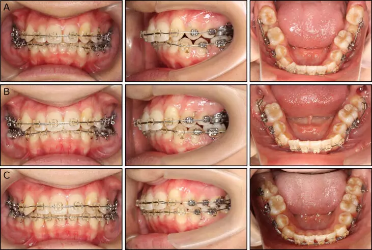
Bonding of second molars enhances the outcome of non-extraction fixed orthodontic treatment, finds study

Bonding of second molars enhances the outcome of non-extraction fixed orthodontic treatment, finds a study published in the Angle Orthodontist.

A study was done to compare orthodontic treatment (OT) outcomes in adolescents undergoing non-extraction fixed OT with or without bonding of second molars using the score of the American Board of Orthodontics Cast Radiograph Evaluation (C-R-Eval). This study included healthy adolescents with skeletal Class I or mild Class II/Class III malocclusion, normal or deep overbite (OB), and mild-to-moderate dental crowding (<5 mm) who underwent non-extraction fixed OT with (“bonded” group) or without (“not-bonded” group) bonding of second molars. Patient treatment records, pre- and posttreatment digital models, lateral cephalograms, and orthopantomograms were assessed. The evaluated outcomes included levelling of the curve of Spee (COS), OB, control of incisor mandibular plane angle (IMPA), number of emergency visits (related to poking wires and/or bracket failure of the terminal molar tubes), treatment duration, and C-R-Eval. Treatment variables were compared across time points and among groups. Results: The sample included 30 patients (mean age 16.07 ± 1.80 years) in the bonded group and 32 patients (mean age 15.69 ± 1.86 years) in the not-bonded group. The mean overall C-R-Eval score was significantly higher (P < .001) in the not-bonded group (25.25 ± 3.98) than in the bonded group (17.70 ± 2.97). There were no significant differences in mean changes of COS, OB, IMPA, or treatment duration among groups. The mean number of emergency visits was significantly higher in the bonded (3.3 ± 0.6) than the not-bonded group (1.9 ± 0.4) (P < .001). Bonding of second molars enhances the outcome of non-extraction fixed OT as demonstrated by the C-R-Eval without increasing treatment duration, irrespective of more emergency visits.

Reference:

Fares Alshuraim, Christopher Burns, Darren Morgan, Luay Jabr, Paul Emile Rossouw, Dimitrios Michelogiannakis; The second molar dilemma in orthodontics: to bond or not to bond?. Angle Orthod 2024; doi: https://doi.org/10.2319/071223-487.1

Keywords:

Bonding, second molars, enhances, outcome, nonextraction, fixed, orthodontic treatment, Fares Alshuraim, Christopher Burns, Darren Morgan, Luay Jabr, Paul Emile Rossouw, Dimitrios Michelogiannakis, Bonding of second molars, Cast-radiograph evaluation, Fixed orthodontic treatment, Treatment duration, Treatment outcomes, Angle Orthodontist

Powered by WPeMatico

Breakthrough Treatment for Polymorphic Light Eruption: Tofacitinib Shows Promise in Resistant Cases

A recent study has unveiled promising
results for individuals suffering from polymorphous light eruption (PMLE), a
sun-induced skin condition characterized by a delayed hypersensitivity
reaction. The study found that tofacitinib,
by inhibiting Janus kinase (JAK)1 and JAK3, may modulate the cytokine milieu in
PMLE, potentially reducing the expression of aberrant inflammatory T
lymphocytes. Thus, Researchers found that tofacitinib is
efficacious, in treating refractory cases of PMLE. The study, conducted on
seven patients who had previously failed multiple treatments, offers a glimmer
of hope for those struggling with the debilitating effects of this sun allergy.

The study results were published in the journal Dermatitis.

PMLE is recognized for its delayed
immune response to endogenous ultraviolet-induced skin antigens. Individuals
afflicted with this condition often experience itching and skin reactions after
sun exposure. Managing PMLE can be challenging, especially in cases where
standard treatments prove ineffective or lead to relapses.

In this groundbreaking study, seven patients
resistant to conventional therapies were given tofacitinib after providing
informed consent.

Findings:


  • The results were nothing short of
    remarkable.
  • Tofacitinib initiation led to a rapid reduction in itching, with a
    mean time of 3.1 ± 1.12 days.
  • Subsequently, clinical resolution was
    achieved in a surprisingly short period, with a mean time of 2.6 ± 1.1 weeks.


  • The duration of tofacitinib therapy
    varied among participants, ranging from 1 to 3 months, with an average duration
    of 2 ± 0.63 months.
  • Of significance, four out of seven
    patients experienced a recurrence within 5.5 weeks.

  • However, upon reinitiating
    tofacitinib, a prompt response was observed, signifying the medication’s
    potential in managing recurring PMLE episodes.


The mechanism behind tofacitinib’s
success in PMLE lies in its ability to inhibit JAK1 and JAK3, thereby
modulating the cytokine milieu associated with this sun allergy. By doing so,
the medication appears to reduce the expression of aberrant inflammatory T
lymphocytes, offering relief to patients who have struggled with the chronic
and often perplexing nature of PMLE.

This breakthrough presents a new
avenue for the treatment of sun-induced skin conditions, offering hope to
individuals who have found conventional therapies ineffective. The findings
open the door for further exploration into the role of JAK inhibitors in
managing other dermatological conditions with immune-mediated components.

While additional research is warranted
to fully understand the long-term effects and broader applicability of
tofacitinib in PMLE, this study marks a significant stride toward personalized
and effective treatments for those grappling with sun allergies and related
skin disorders.

Further reading: Tofacitinib: A Treatment Option for Recalcitrant Polymorphic Light Eruption and Its Mechanistic Rationale.  https://doi.org/10.1089/derm.2023.0360

Powered by WPeMatico