Long-Term Study reveals high Incidence of depression in patients after Stroke

Post-stroke depression (PSD) is a common complication affecting stroke survivors, but data on its long-term course and recurrence rates are limited. A recent prospective study from the U.K. provides valuable insights into the incidence and recurrence of PSD over an 18-year period, highlighting the importance of routine depression screening in stroke survivors. This study was published in the journal of Lancet Regional Health – Europe. The study was conducted by Liu L. and colleagues.

PSD can have significant implications for the recovery and quality of life of stroke survivors, yet it often goes unrecognized and untreated. Understanding the timing and recurrence patterns of depression following stroke is crucial for implementing effective interventions and support strategies.

Researchers analyzed data from the South London Stroke Register, including 3,864 stroke survivors assessed for depression over 18 years. Depression was measured using the Hospital Anxiety and Depression Scale, and patients were followed up to assess recurrence rates and long-term outcomes.

The key findings of the study were:

• The cumulative incidence of PSD over 18 years was 59.4%, with 87.9% of cases occurring within the first 5 years post-stroke.

• Within 3 months, 33.4% of PSD cases were identified, increasing to 54.6% within 1 year.

• Recurrence of depression was common, with 66.7% of patients experiencing recurrence, predominantly within 5 years of recovery.

• Patients with severe depression had lower recovery rates and higher recurrence rates compared to those with mild depression.

• Routine screening for depression within 5 years post-stroke is recommended, as depression can begin as early as 3 months post-stroke.

The study highlights the significant burden of PSD in stroke survivors and underscores the need for long-term monitoring and support. Early detection and intervention for depression can improve outcomes and enhance the overall well-being of stroke survivors.

Post-stroke depression is prevalent, with the majority of cases occurring within the first 5 years post-stroke. Routine screening for depression in stroke survivors is crucial for timely identification and management of this common complication. Long-term follow-up and support are essential to address the high recurrence rates observed in this population.

Reference:

Liu L, et al “Natural history of depression up to 18 years after stroke: a population-based South London Stroke Register study” Lancet Reg Health Eur 2024; DOI: 10. 1016/j.lanepe.2024. 100882.

Powered by WPeMatico

Even low levels of leisure time physical activity help to lower stroke risk, suggests new study

Even people whose physical activity levels fall short of recommended guidelines, but who manage to do some during their leisure time, are likely to have a lower risk of stroke than their sedentary peers, suggests a pooled data analysis of the available evidence, published online in the Journal of Neurology Neurosurgery & Psychiatry.

The effects are independent of age and sex, the findings show, prompting the authors to suggest that everyone should be encouraged to do whatever level of physical activity they can manage in their leisure time.

There’s no doubt that moderate to high levels of leisure-time physical activity curb stroke risk. But it’s not clear if even modest amounts confer protection, and if any such effects depend on age and sex, explain the authors.

While international guidelines recommend 150 minutes or more a week of moderate intensity physical activity or 75 minutes or more of vigorous intensity activity to lower the risk of cardiovascular disease, including stroke, not many adults achieve this, say the researchers.

To find out if lower levels of physical activity might still confer protection, the authors trawled research databases for relevant studies. They pooled the results of 15, involving 752,050 adults whose health had been monitored for an average of 10.5 years.

Each study assessed the potential impact of between 3-none, below target, and ideal-and 5-none, insufficient, low, moderate and intense-levels of leisure time physical activity on stroke risk.

The pooled data analysis of 5 studies assessing 3 levels of leisure time physical activity showed that, compared with no physical activity, the highest ‘ideal’ amount cut the risk of stroke by 29%, but that some ‘below target’ activity still reduced the risk by 18%.

Similar findings emerged for the pooled data analysis of the six studies reporting on 4, and the two reporting on 5 levels of leisure time physical activity. Compared with none, a moderate level of physical activity cut the risk of stroke by between 27% and 29%.

These effects were independent of sex and age, the analysis showed.

The authors acknowledge several limitations to their findings, chief among which were the variable definitions of different levels of activity used in the included studies and the reliance on subjectively assessed levels of physical activity.

Nevertheless, the authors conclude that recreational physical activity, even in small amounts could help ward off stroke in the long term.

“According to our results, all levels of [leisure time physical activity] can be beneficial for stroke prevention, including levels currently regarded as low or insufficient,” they write. “People should be encouraged to be physically active even at the lowest levels.”

Reference:

De Santis F, Romoli M, Foschi M, et alRisk of stroke with different levels of leisure-time physical activity: a systematic review and meta-analysis of prospective cohort studiesJournal of Neurology, Neurosurgery & Psychiatry Published Online First: 05 March 2024. doi: 10.1136/jnnp-2023-332457.

Powered by WPeMatico

Breakthrough Study Reveals Rapid Reduction of Gout-Related Crystal Deposits with Pegloticase Therapy

A new study found
that Monosodium urate (MSU) crystal deposits,
detectable with dual-energy CT (DECT), diminish rapidly during pegloticase
treatment, particularly when co-administered with methotrexate (MTX) for
uncontrolled gout. The volume of MSU crystals also slowed or stopped
even when SU was maintained at <6mg/dL with oral ULT.

The study findings were published in the journal Joint Bone Spine.

Monosodium urate (MSU) crystal deposits can be observed and measured
using dual-energy CT (DECT). Pegloticase effectively reduces serum urate (SU)
levels in uncontrolled gout patients, often with methotrexate (MTX) co-therapy
to enhance response and mitigate infusion reactions. However, limited
literature exists on DECT imaging during pegloticase+MTX treatment. Hence,
researchers conducted a study to address this gap by presenting DECT findings
from a larger cohort in a randomized controlled trial, investigating bone
erosion remodeling following MSU depletion with pegloticase therapy, and
exploring the impact of treatment duration. During the MIRROR RCT trial, patients were administered either
pegloticase (8mg every 2 weeks) along with methotrexate (MTX) orally at a dose
of 15mg/week, or pegloticase with a placebo (PBO).

A subgroup underwent DECT
imaging on Day 1 (the initial pegloticase infusion) and at Weeks 14, 24, and
52. Patients with paired baseline-Week 52 images were analyzed. Regions with
baseline MSU-crystal volume (VMSU) <0.5cm3 were excluded to minimize
artifact contributions. VMSU and bone erosion remodeling were evaluated.

Results:

  • Out of the eight patients included (six on MTX, two on PBO), five on MTX
    had undergone pegloticase therapy for 52 weeks, one on PBO for 42 weeks, and
    two on MTX and one on PBO for 6 weeks each.
  • Patients who discontinued pegloticase prematurely maintained serum urate
    (SU) levels <6mg/dL on allopurinol (n=2) or febuxostat (n=1).
  • By Week 52, VMSU had significantly decreased in both the pegloticase+MTX
    and pegloticase+PBO groups, with a more rapid reduction observed during
    pegloticase treatment.
  • Bone-erosion remodeling was noted in 29 out of 42 (69%) evaluated
    erosions, with 29 (69%) demonstrating a decrease in size, 4 (9.5%) exhibiting
    recortication, and 3 (7.1%) showing new bone formation.

Thus, the study concluded a swift reduction in monosodium urate (MSU) crystal volume
(VMSU) during pegloticase therapy, accompanied by concurrent bone remodeling
within a year. However, upon discontinuation of pegloticase, the rate of VMSU
reduction decelerated or halted, despite maintaining serum urate (SU) levels
below 6mg/dL with oral urate-lowering therapy (ULT).

Further reading: Examination of monosodium urate
crystal depletion and bone erosion remodeling during pegloticase treatment in
patients with uncontrolled gout: exploratory dual-energy computed tomography
findings from MIRROR RCT. Doi: 
https://doi.org/10.1016/j.jbspin.2024.105715                                  

Powered by WPeMatico

FDA approves injectable drug for three infections

The US Food and Drug Administration has approved injectable drug ceftobiprole medocaril sodium (Zevtera) for treatment of Staphylococcus aureus bacteremia (SAB) among adults. Ceftobiprole, a pyrrolidinone cephalosporin antibiotic, is active moiety of the prodrug ceftobiprole medocaril.

It is indicated for people with right-sided infective endocarditis, acute bacterial skin and skin structure infections (ABSSSI), and community-acquired bacterial pneumonia (CABP) in adult and pediatric patients 3 months to less than 18 years old. 

“The FDA is committed to fostering new antibiotic availability when they prove to be safe and effective, and Zevtera will provide an additional treatment option for a number of serious bacterial infections,” said Peter Kim, M.D., M.S., director of the Division of Anti-Infectives in the FDA’s Center for Drug Evaluation and Research. “The FDA will continue our important work in this area as part of our efforts to protect the public health.”

Zevtera’s efficacy in treating SAB was evaluated in a randomized, controlled, double-blind, multinational, multicenter trial. In the trial, researchers randomly assigned 390 subjects to receive Zevtera (192 subjects) or daptomycin plus optional aztreonam [the comparator] (198 subjects). The primary measure of efficacy for this trial was the overall success (defined as survival, symptom improvement, S. aureus bacteremia bloodstream clearance, no new S. aureus bacteremia complications and no use of other potentially effective antibiotics) at the post-treatment evaluation visit, which occurred 70 days after being randomly assigned an antibiotic. A total of 69.8% of subjects who received Zevtera achieved overall success compared to 68.7% of subjects who received the comparator.

Zevtera’s efficacy in treating ABSSSI was evaluated in a randomized, controlled, double-blind, multinational trial. In the trial, researchers randomly assigned 679 subjects to receive either Zevtera (335 subjects) or vancomycin plus aztreonam [the comparator] (344 subjects). The primary measure of efficacy was early clinical response 48-72 hours after start of treatment. Early clinical response required a reduction of the primary skin lesion by at least 20%, survival for at least 72 hours and the absence of additional antibacterial treatment or unplanned surgery. Of the subjects who received Zevtera, 91.3% achieved an early clinical response within the necessary timeframe compared to 88.1% of subjects who received the comparator.

Zevtera’s efficacy in treating adult patients with CABP was evaluated in a randomized, controlled, double-blind, multinational, multicenter trial. In the trial, researchers randomly assigned 638 adults hospitalized with CABP and requiring IV antibacterial treatment for at least 3 days to receive either Zevtera (314 subjects) or ceftriaxone with optional linezolid [the comparator] (324 subjects). The primary measurement of efficacy were clinical cure rates at test-of-cure visit, which occurred 7-14 days after end-of-treatment. Of the subjects who received Zevtera, 76.4% achieved clinical cure compared to 79.3% of subjects who received the comparator. An additional analysis considered an earlier timepoint of clinical success at Day 3, which was 71% in patients receiving Zevtera and 71.1% in patients receiving the comparator.

Given the similar course of CABP in adults and pediatric patients, today’s approval of Zevtera in pediatric patients three months to less than 18 years with CABP was supported by evidence from the CABP trial of Zevtera in adults and a trial in 138 pediatric subjects three months to less than 18 years of age with pneumonia.

For adults with SAB, the most common side effects of Zevtera included anemia, nausea, low levels of potassium in the blood (hypokalemia), vomiting, diarrhea, increased levels of certain liver tests (hepatic enzymes and bilirubin), increased blood creatinine, high blood pressure, low white blood cell count (leukopenia), fever, abdominal pain, fungal infection, headache and shortness of breath (dyspnea).

For adults with ABSSSI, the most common side effects of Zevtera included nausea, diarrhea, headache, injection site reaction, increased levels of hepatic enzymes, rash, vomiting and altered taste (dysgeusia).

For adults with CABP, the most common side effects of Zevtera included nausea, increased levels of hepatic enzymes, vomiting, diarrhea, headache, rash, insomnia, abdominal pain, vein inflammation (phlebitis), high blood pressure and dizziness. For pediatric patients with CABP, the most common side effects of Zevtera included vomiting, headache, increased levels of hepatic enzymes, diarrhea, infusion site reaction, vein inflammation (phlebitis) and fever.

Patients should not use Zevtera if they have a known history of severe hypersensitivity to ceftobiprole or any of the components of Zevtera, or other members of the cephalosporin antibacterial class.

Zevtera comes with certain warnings and precautions such as increased mortality in ventilator-associated bacterial pneumonia patients (an unapproved use), hypersensitivity reactions, seizures and other central nervous system reactions and Clostridioides difficile-associated diarrhea.

Zevtera was granted Priority Review, Fast Track and Qualified Infectious Disease Product designations for the CABP, ABSSSI and SAB indications.

The FDA granted the approval of Zevtera to Basilea Pharmaceutica International Ltd.

Powered by WPeMatico

Primary care strategy did not reduce hospitalizations at one year in kidney-dysfunction triad: ICD-Pieces study

USA: Using an electronic health record (EHR)-based algorithm plus practice facilitators embedded in primary care clinics did not reduce hospitalization at one year, according to a pragmatic trial involving patients with the triad of chronic kidney disease, hypertension, and type 2 diabetes.

“The hospitalization rate of patients in the intervention group at one year was about the same as that with usual care (20.7% vs 21.1%),” the researchers reported in the ICD-Pieces study published in the New England Journal of Medicine.

Patients with chronic kidney disease (CKD), type 2 diabetes (T2D), and hypertension (the kidney-dysfunction triad) are at high risk for multiple complications, end-stage kidney disease, and premature death. Despite the availability of effective therapies for these patients, there is a lack of results of large-scale trials examining the implementation of guideline-directed therapy to reduce death and complications risk in this population.

Miguel Vazquez, the University of Texas Southwestern Medical Center in Dallas, and colleagues conducted an open-label, cluster-randomized trial including 11,182 patients with the kidney-dysfunction triad being treated at 141 primary care clinics. They were assigned to receive an intervention that used a personalized algorithm (based on the patient’s EHR) to identify patients and practice facilitators to assist providers in delivering guideline-based interventions or to receive usual care.

The study’s primary outcome was hospitalization for any cause at 1 year. Secondary outcomes were readmissions, emergency department visits, dialysis, cardiovascular events, and death. Cardiovascular events included heart failure, acute coronary syndrome, and stroke, while cardiovascular procedures included cardiac catheterization and revascularization.

The researchers assigned 71 practices (enrolling 5690 patients) to the intervention group and 70 practices (enrolling 5492 patients) to the usual-care group.

The researchers reported the following findings:

  • The hospitalization rate at 1 year was 20.7% in the intervention group and 21.1% in the usual-care group (between-group difference, 0.4 percentage points).
  • The risks of emergency department visits, cardiovascular events, readmissions, dialysis, or death from any cause were similar in the two groups.
  • The risk of adverse events was also similar in the trial groups, except for acute kidney injury, which was observed in more patients in the intervention group (12.7% vs. 11.3%).

While the intervention missed the mark for reducing hospitalizations, Vazquez highlighted the importance of improving care for patients with the kidney-dysfunction triad.

“In this pragmatic trial involving patients with the kidney-dysfunction triad, using an EHR-based algorithm and practice facilitators embedded in primary care clinics did not translate into reduced hospitalization at 1 year,” the researchers concluded.

Reference:

Vazquez MA, et al “Pragmatic trial of hospitalization rate in chronic kidney disease” N Engl J Med 2024; DOI: 10.1056/NEJMoa2311708.

Powered by WPeMatico

Bariatric surgery linked to heart health improvements in people with severe obesity: Study

Bariatric surgery may result in significant cardiometabolic improvements, particularly among younger, female, or white people and those without comorbidities, according to new research published in the Journal of the Endocrine Society.

The United States has the highest obesity rates globally. In 2017-2018, about 40% of U.S. adults had obesity and 9% had severe obesity. The prevalence is particularly high among Black adults. Bariatric surgery is one method to help people with severe obesity lose a lot of weight and improve their health.

“Our study highlights how bariatric surgery not only leads to significant weight loss but also substantially improves heart health,” said study author Lei Wang, M.Phil., of Vanderbilt University Medical Center in Nashville, Tenn. “These health benefits include lower blood pressure, blood lipids and blood sugar, and an estimated 35% reduction in 10-year cardiovascular disease risk one year after surgery.”

“Additionally, 30% to 50% of people in our study experienced remissions of diabetes, hypertension and dyslipidemia,” added co-author Danxia Yu, Ph.D., of Vanderbilt University Medical Center. “Individuals who were younger, female, self-identified as white and had no history of cardiometabolic diseases tended to see greater post-surgery cardiometabolic improvements.”

The researchers analyzed over 7,800 people between the ages of 20 to 79 who underwent bariatric surgery during 1999-2022 at the Vanderbilt University Medical Center. Most study participants were women and white, but the study also includes a significant number of male patients and Black patients, who have been underrepresented in bariatric surgery research.

The researchers evaluated various cardiometabolic improvements, including blood pressure, cholesterol, glucose and HbA1c. They also measured remission rates of diabetes, hypertension and dyslipidemia, and the estimated 10-year cardiovascular disease risk.

Older, male or Black patients showed less reduction in 10-year heart disease risk and lower odds of diabetes, hypertension and dyslipidemia remission than younger, female or white patients. Patients with a history of diabetes, hypertension, dyslipidemia or cardiovascular disease showed less cardiometabolic improvements than those without.

“Our findings can help people with severe obesity experience better health outcomes and help us recognize which patients may require extra health management after surgery,” Wang said.

Reference:

Lei Wang, Michael T O’Brien, Xinmeng Zhang, You Chen, Wayne J English, Brandon Williams, Matthew Spann, Vance Albaugh, Xiao-Ou Shu, Charles R Flynn, Danxia Yu, Cardiometabolic Improvements After Metabolic Surgery and Related Presurgery Factors, Journal of the Endocrine Society, Volume 8, Issue 5, May 2024, bvae027, https://doi.org/10.1210/jendso/bvae027.

Powered by WPeMatico

lncreased fetal catecholamines associated with postnatal hypoglycemic episodes: JAMA

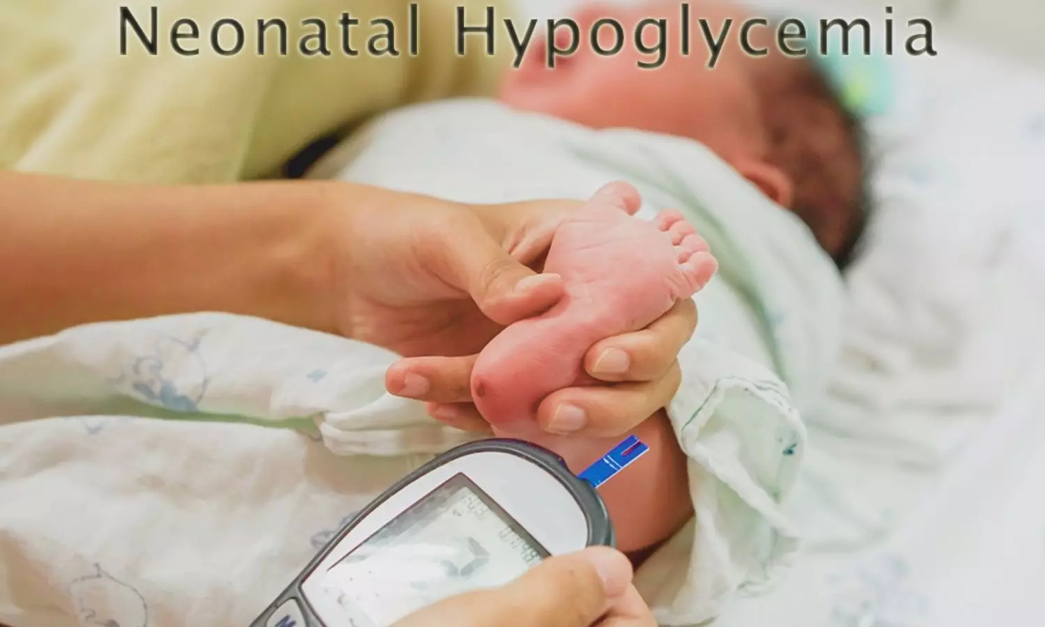
Neonatal hypoglycemia, a condition marked by low blood sugar levels in newborns, poses significant risks and can be particularly concerning for infants with perinatal stress or fetal growth restriction. Understanding the underlying mechanisms of this condition is crucial for effective management and prevention.

Perinatal stress and fetal growth restriction are known risk factors for neonatal hypoglycemia, but the precise pathomechanism remains unclear. In a recent study, researchers investigated whether elevated catecholamine concentrations in umbilical cord blood (UCB) and amniotic fluid (AF) are associated with neonatal hypoglycemia and whether they correlate with postnatal glycemic levels. This study was published in the JAMA Pediatrics. The study was conducted by Henrike H. and colleagues.

The key findings of the study were:

• The study involved 328 neonates, with 234 in the risk group and 94 in the control group.

• Growth-restricted neonates showed increased umbilical cord blood (UCB) concentrations of norepinephrine (21.10 vs 10.88 nmol/L), metanephrine (0.37 vs 0.12 nmol/L), and 3-methoxytyramine (0.149 vs 0.091 nmol/L) compared to controls.

• Neonates with perinatal stress had increased UCB concentrations of norepinephrine (22.55 vs 10.88 nmol/L), normetanephrine (1.75 vs 1.25 nmol/L), and 3-methoxytyramine (0.120 vs 0.091 nmol/L).

• UCB norepinephrine, metanephrine, and 3-methoxytyramine concentrations were positively correlated with the number of hypoglycemic episodes (rs = 0.146, p = 0.01; rs = 0.151, p = 0.009; and rs = 0.180, p = 0.002, respectively).

• These concentrations were also negatively correlated with the lowest measured blood glucose levels (rs = -0.149, p = 0.01; and rs = -0.153, p = 0.008).

• UCB catecholamine concentrations were negatively correlated with amniotic fluid C-peptide concentration (rs = -0.212, p = 0.005; rs = -0.182, p = 0.016; and rs = -0.183, p = 0.016).

• A p-value of less than 0.0083 was considered statistically significant due to multiple comparisons.

Neonates at risk for hypoglycemia exhibited elevated catecholamine concentrations in UCB, which correlated with postnatal hypoglycemic episodes and lower blood glucose levels. These findings suggest a link between perinatal stress, fetal growth restriction, and neonatal hyperinsulinemic hypoglycemia. Understanding these mechanisms could improve the management of newborns at risk for hypoglycemia.

Reference:

Hoermann, H., van Faassen, M., Roeper, M., Hagenbeck, C., Herebian, D., Muller Kobold, A. C., Dukart, J., Kema, I. P., Mayatepek, E., Meissner, T., & Kummer, S. (2024). Association of fetal catecholamines with neonatal hypoglycemia. JAMA Pediatrics. https://doi.org/10.1001/jamapediatrics.2024.0304

Powered by WPeMatico

Study finds high prevalence of hidden brain changes in people with heart disease

A new analysis involving over 13,000 people has found changes to blood vessels in the brain that can increase the risk of stroke and dementia are common in people with a range of heart conditions, regardless of whether they have experienced a stroke.

The new research, published today in Neurology®, the medical journal of the American Academy of Neurology, is the most comprehensive systematic review of ‘hidden’ brain changes in people with a range of heart conditions to date.

Lead author Dr Zien Zhou from The George Institute for Global Health said that identifying these changes could play an important role in choosing treatments for these patients.

“Although people with heart disease are two to three times more likely than the general population to have changes in their brain’s vascular system, they’re often overlooked, because these patients don’t routinely undergo brain imaging unless they have suffered a stroke,” he said.

“But it can make them more susceptible to the risk of brain bleeds from medications commonly used to treat or prevent blood clots – intracranial hemorrhage is a life-threatening complication with no proven treatment and a survival rate of less than 50 percent.”

Changes to blood vessels in the brain that can only be detected by brain imaging such as silent brain infarction (SBI) and cerebral small vessel disease (CSVD) are known to occur more commonly in older people or those who have hypertension. While not sufficient to cause obvious neurological symptoms, they can result in subtle neurological deficits and increase the longer-term risk of stroke or dementia.

To determine the prevalence of these hidden or covert cerebrovascular changes in adults with atrial fibrillation, coronary artery disease, heart failure or cardiomyopathy, heart valve disease, and patent foramen ovale (a hole in the heart), George Institute researchers conducted a meta-analysis of 221 observational studies published between 1988 and 2022.

The findings showed that in people with heart disease:

  • approximately one third had any form of SBI
  • a quarter had lacune (small cavities where neural tissue has died after a previous blockage or leakage from small arteries)
  • two-thirds had white matter lesions (damage to the protective coating around nerve fibres)
  • a quarter had evidence of asymptomatic microbleeds in the brain tissue, and
  • over one half had brain atrophy (a shrinking of the brain due to loss of neurons or connections between neurons).

The prevalence of these brain changes was generally the same between those with and without a recent stroke and there were no apparent sex differences in the results.

Dr Zhou said the study also confirmed that heart disease is one of the main causes of these changes that reflect brain ‘frailty’.

“While several potential mechanisms of the association between heart disease and hidden cerebrovascular injury have been proposed, the two conditions share common risk factors such as ageing, hypertension, type 2 diabetes, hyperlipidaemia, and smoking,” said Dr Zhou.

“It’s possible that a gradual decline in cardiac output in some patients with heart disease might affect how much blood is reaching the brain tissue, contributing to vascular changes and cognitive dysfunction in these patients,” he added.

“It’s also possible that hidden brain changes and cognitive dysfunction are a consequence of tiny blood clots traveling to the brain through the arterial circulation after forming in the heart.”

Dr Zhou said that more research was needed to look at the exact causes of these brain changes and the implications for managing these patients.

“We need to know whether performing an additional MRI in those considered for anticoagulation therapy – which is required for most people with heart disease – would be cost-effective in terms of preventing unwanted side effects,” he said.

“But refining the risks of brain clots and bleeds from anticoagulants and using this information to make the best treatment choice could improve treatment safety for people with heart disease.”

Reference:

Zien Zhou, Shoujiang You, Yuki Sakamoto, Ying Xu, Song Ding,  Wenyi Xu, Wenjie Li, Covert Cerebrovascular Changes in People With Heart Disease, Neurology, https://doi.org/10.1212/WNL.000000000020920

Powered by WPeMatico

Heart health declines rapidly after menopause, claims study

A woman’s cardiovascular risk can rise sharply after she goes through menopause, quickly catching up to men of a similar age and health profile, according to new findings presented at the American College of Cardiology’s Annual Scientific Session. Researchers said the study underscores the importance of recognizing and addressing early warning signs of heart disease risk in women as they lose the protective effects of estrogen after menopause.

“This is a unique study cohort of only post-menopausal statin users that signals that post-menopausal women may have risk of heart disease that is on par with males,” said Ella Ishaaya, MD, an internal medicine physician at Harbor-UCLA Medical Center in Torrance, California, and the study’s lead author. “Women are underscreened and undertreated, especially post-menopausal women, who have a barrage of new risk factors that many are not aware of. This study raises awareness of what those risk factors are and opens the door to indicating the importance of increased screening for coronary artery calcium (CAC).”

In the study, post-menopausal women underwent heart scans to assess their CAC score, a measure of plaque buildup-fat, calcium and other substances-in the heart’s arteries. CAC levels are assessed with a quick, non-invasive scan similar to an X-ray. A higher CAC score indicates a higher risk of a heart attack or other cardiac events.

Researchers analyzed data from 579 post-menopausal women who were taking statins to control their cholesterol and had undergone two CAC scans at least one year apart. Participants did not have heart disease at the time of the first scan. To compare CAC changes in men and women, each female participant was matched with a male of a similar profile in terms of age, race, statin use, blood pressure and diabetes status.

Researchers divided the participants into three groups with CAC levels of 1–99, 100–399, and 400 or higher at baseline. Between their first and second heart scan, women with baseline CAC of 1–99 saw their CAC rise by a median of eight points, double the median of four seen in their male counterparts. Similarly, women with baseline CAC of 100–399 saw their CAC rise by a median of 31 points, about double the median of 16 seen in males. There was no significant difference between sexes for those with baseline CAC of 400 or higher.

The findings suggest plaque buildup is accelerated in post-menopausal women compared to men, indicating that many women experience a steep rise in the risk of heart problems. Ishaaya said this is likely related to the drop in estrogen that women experience during menopause. Estrogen has long been known to have a protective effect on heart health, but researchers said many women and even many clinicians are not aware of what it means to lose that protection during menopause.

“After menopause, women have much less estrogen and shift to a more testosterone-heavy profile,” Ishaaya said. “This affects the way your body stores fat, where it stores fat and the way it processes fat; it even affects the way your blood clots. And all of those [changes] increase your risk for developing heart disease.”

Heart disease is the leading cause of death in both men and women, but women’s cardiovascular risk has traditionally been undertreated because women tend to develop heart disease at an older age than men and may experience different and sometimes more subtle symptoms.

Based on these results, researchers suggested post-menopausal women should talk to their doctor about heart disease risk factors and follow up on any recommended tests or monitoring. More women may benefit from heart scans when compared to the number of women currently receiving them, Ishaaya said.

Since all the women in the study were taking statins but many still saw a substantial rise in CAC, the results may also indicate that statins are not sufficient to keep plaque buildup in check for this population, Ishaaya said. Future studies could investigate the effectiveness of statins or other therapies in reducing plaque burden in post-menopausal women, she said.

ACC/American Heart Association guidelines recommend considering a heart scan to assess CAC when a person’s risk level is ambiguous or borderline based on standard risk factors. In the U.S. and many other countries, CAC scoring is most used to determine recommendations for statins for intermediate-risk and asymptomatic patients.

Reference:

Heart health declines rapidly after menopause, American College of Cardiology, Meeting: American College of Cardiology’s Annual Scientific Session.

Powered by WPeMatico

High Altitude Climbing Increases Risk of Cardiac Arrhythmia, reveals JAMA study

Exposure to high altitudes presents physiological challenges such as arterial hypoxemia, electrolyte imbalances, and periodic breathing, which can increase vulnerability to cardiac arrhythmias. Cardiac arrhythmias, including tachyarrhythmias and bradyarrhythmias, pose serious health risks, particularly in challenging environments such as high altitudes.

However, the incidence and characteristics of arrhythmias in individuals undertaking extreme altitude climbs like Mount Everest remain understudied. A recent study was published in the journal JAMA Cardiology. The study was conducted by Sherpa K. and colleagues. This prospective cohort study aimed to explore the incidence of tachyarrhythmias and bradyarrhythmias in healthy individuals during a high-altitude expedition to Mount Everest.

Healthy individuals participating in the expedition underwent baseline cardiac evaluations, including electrocardiogram and echocardiography, before ascending Mount Everest. Ambulatory rhythm recording was conducted both before and during the climb. The primary outcome was the incidence of a composite of supraventricular and ventricular tachyarrhythmias, as well as bradyarrhythmias.

The key findings of the study were:

  • 41 male individuals, with a mean age of 33.6 years, participated in the study.

  • 38.2% of participants experienced cardiac arrhythmias during the climb, with 45 primary endpoint-relevant events recorded.

  • Bradyarrhythmic events were more common, with 43 events documented in 13 individuals, compared to 2 ventricular tachycardias in 2 individuals.

  • Arrhythmias occurred more frequently at lower altitudes without supplemental oxygen, with 80% of events occurring in this setting.

  • The proportion of individuals with arrhythmias remained stable across altitudes, while event rates numerically increased between 5300 m and 7300 m before decreasing again at higher altitudes.

The study confirms a significant association between high-altitude exposure and the incidence of cardiac arrhythmias, with over one-third of healthy climbers experiencing arrhythmias during the ascent of Mount Everest. These findings underscore the importance of further research to explore the implications and management of cardiac rhythm disturbances in extreme altitude environments.

Understanding the prevalence and characteristics of cardiac arrhythmias in high-altitude climbers can inform risk assessment and management strategies for individuals undertaking similar expeditions. Additionally, these findings emphasize the need for medical interventions and monitoring to mitigate the risks associated with cardiac arrhythmias during high-altitude climbs.

Reference:

Sherpa, K., Sherpa, P. P., Sherpa, T., Rothenbühler, M., Ryffel, C., Sherpa, D., Sherpa, D. R., Sherchand, O., Galuszka, O., Dernektsi, C., Reichlin, T., & Pilgrim, T. (2024). Risk of cardiac arrhythmias among climbers on Mount Everest. JAMA Cardiology. https://doi.org/10.1001/jamacardio.2024.0364

Powered by WPeMatico